| 规格 | 价格 | 库存 | 数量 |
|---|---|---|---|
| 10mg |
|
||
| 25mg |
|
||
| 50mg |
|
||
| 100mg |
|
||
| 250mg |
|
||
| 500mg |
|
||
| Other Sizes |
|
| 靶点 |
Mycobacterium tuberculosis H37Rv( MIC50=2.7 nM )
|
|---|---|
| 体外研究 (In Vitro) |
体外活性:Q203 是一种咪唑并吡啶酰胺 (IAP) 化合物,通过靶向呼吸道细胞色素 bc1 复合物来阻断结核分枝杆菌的生长。它具有治疗结核病的潜力。 Q203 在低纳摩尔范围内抑制肉汤培养基中 MDR 和 XDR 结核分枝杆菌临床分离株的生长,并且在小鼠结核病模型中以低于 1 mg/kg 体重的剂量有效,这凸显了 Q203 的效力。 Q203 对参考菌株结核分枝杆菌 H37Rv 有活性,在肉汤培养基中的 MIC50 为 2.7 nM,在巨噬细胞内的 MIC50 为 0.28 nM。此外,Q203 显示出与每日一次给药兼容的药代动力学和安全性特征。总之,这些数据表明 Q203 是治疗结核病的有前途的新临床候选药物。激酶测定:Q203 对参考菌株结核分枝杆菌 H37Rv 有活性,在培养基中的 MIC50 为 2.7 nM,在巨噬细胞内的 MIC50 为 0.28 nM。细胞测定:
Telacebec (Q203; IAP6)对参考菌株结核分枝杆菌H37Rv有活性,最低浓度为抑制培养液中50%生物体(MIC50) 2.7 nM,巨噬细胞内MIC50为0.28 nM(图1b,c)。在三个中心采用四种不同的技术(见在线方法)测定培养液培养基中的MIC50,结果具有可比性。此外,我们通过琼脂板的CFU测定,证实了Q203在液体肉汤培养基中的活性。[1] 深入了解Telacebec (Q203; IAP6)并确定其分子靶点,我们选择了两种不同的IPA衍生物IPA04和IPA05的自发抗性突变体(Supplementary Fig. 5)。在确认了对Q203的稳定基因型抗性后(图3a),我们对来自独立生物重复的6个自发抗性突变体进行了全基因组测序。突变体显示Q203的MIC50持续增加了几个数量级,但仍然对标准抗结核药物敏感。我们在所有六个突变体中发现了细胞色素b亚基(qcrB,也称为细胞色素bc1复合体的Rv2196)中的单个氨基酸取代(图3b)。对另外18株独立的自发性耐药突变体(全部受试的18株)进行qcrB序列分析证实:Thr313突变为丙氨酸或异亮氨酸(图3b)与特拉西贝克(Q203;IAP6)耐药性相关。此外,通过同源重组在亲本结核分枝杆菌H37Rv中重新引入突变Ala313,获得了对Q203的抗性(图3a),表明这种取代直接和特异性地参与了对该化合物的抗性机制。在1 μM浓度下,对IPA04和IPA05的6个独立突变选择实验进行综合分析,结果表明,IPA04和IPA05的自发突变率为2.4 × 10⁻⁸,抗性突变体出现的概率较低。直接在Q203上选择的自发抗性突变体也对Q203具有高度抗性(补充图6),并且在qcrB中含有多态性T313A,而我们在两个泛易感和三个XDR临床分离株中未发现qcrB突变(补充图7)。最近,在由相关IPA衍生物选择的牛分枝杆菌卡介苗(BCG)突变体中发现了类似的qcrB多态性,该突变体在体外抑制结核分枝杆菌生长的活性低于Q203,且未优化用于临床。[1] 值得注意的是,在QP位点抑制剂的结合中起关键作用的几个残基(例如,stigmatellin)或涉及对此类抑制剂的抗性,或两者都位于ef区(补充图8),这表明Telacebec (Q203;IAP6)类似于作用于QP位点的非选择性抑制剂。考虑到细胞色素bc1在呼吸电子传递链中的关键作用,我们测试了Telacebec (Q203;IAP6 可能干扰结核分枝杆菌ATP合成。我们发现Telacebec (Q203; IAP6)触发了细胞内ATP的快速减少,IC50为1.1 nM(图3c)。在类似的实验条件下(见在线方法),莫西沙星或链霉素没有减少ATP池大小,而贝达喹啉有(IC50为27.7 nM)。最后,Q203能够在小于10 nM的IC50下干扰缺氧非复制结核中的ATP稳态(图3d)。在低浓度下快速抑制ATP合成强烈提示抑制细胞色素bc1活性是Q203[1]的主要作用方式。 |
| 体内研究 (In Vivo) |
Q203 显示出与每日一次给药兼容的药代动力学和安全性特征。 Q203的生物利用度为90%,终末半衰期为23.4小时。分布容积适中(5.27 l/kg体重),全身清除率低(4.03 mL/min/kg)。治疗 4 周后,在按每公斤体重 0.4、2 和 10 毫克 Q203 治疗的组中,观察到结核分枝杆菌 H37Rv 细菌载量分别减少了 90%、99% 和 99.9%
|
| 酶活实验 |
肝微粒体稳定性测定。[1]
将化合物(终浓度为2μM,溶于0.2%DMSO中)与0.5 mg mL-1人(200只,混合性别)、雄性狗、雄性大鼠或雄性小鼠肝微粒体在磷酸钾缓冲液中孵育。通过加入NADPH引发反应,并立即或在10、20、30或60分钟时停止,以精确估计清除率。采用带有电喷雾电离(ESI)的三重四极杆Quattro Premier质谱仪进行样品分析。样品通过捕集筒(Acquity BEH RP18 50 mm×2.1 mm,1.7μm,Waters,Milford,MA),然后通过分析柱。通过与0分钟时的初始量进行比较来计算剩余化合物的百分比。然后使用一级反应动力学计算半衰期。
CYP450抑制试验。[1] 该测定使用具有单个重组人细胞色素P450(rhCYP)同工酶的单个荧光探针底物,并根据先前发表的方法进行荧光检测36。用于每种同工酶的探针底物(在0.5%DMSO中)如下:CYP3A4为7-苄氧基-4-(三氟甲基)-香豆素,CYP2D6为3-[2-(N,N-二乙基-N-甲基铵)乙基]-7-甲氧基-4-甲基香豆素(AMMC),1A2和2C19为3-氰基-7-乙氧基香豆素(CEC),2C9为7-甲氧基-4-(三氟甲基)香豆素(MFC)。使用Victor3 V多标记板阅读器测量荧光。使用三倍连续稀释的八点浓度曲线测定IC50。 |
| 细胞实验 |
细胞毒性。[1]
如前所述,使用MTT(3-(4,5-二甲基噻唑-2-基)-2,5-二苯基溴化四唑)存活率测定法对人细胞系SH-SY5Y(脑)、HEK293(肾)和HepG2(肝)进行了细胞毒性测试。
结核分枝杆菌H37Rv ATP耗竭试验。[1] 将结核分枝杆菌H37Rv暴露于受试化合物24小时(需氧)或5天(厌氧),与等体积的BacTiter-Glo试剂混合,在黑暗中孵育10分钟。在Victor3 V多标签平板阅读器上记录发光。 |
| 动物实验 |
大鼠:Sprague Dawley大鼠用于药代动力学研究。化合物(Q203)的给药剂量为2 mg/kg体重,静脉注射;或10 mg/kg体重,口服。化合物(Telacebec (Q203; IAP6))配制于20% TPGS(d-α-生育酚聚乙二醇1000琥珀酸酯)中用于重复给药研究,配制于40% PEG400(pH 4)中用于单次给药研究。灌注前,使用1 mL注射器经下腔静脉采集血样。分别于给药后0.5、1、2、6、12、24和48小时从三只小鼠或大鼠中采集血样。血样在4℃下以3200g离心10分钟。离心后,收集血浆并冷冻保存,直至进行后续分析。化合物浓度采用液相色谱-质谱联用(LC-MS)法测定;
小鼠:研究了Telacebec (Q203; IAP6)在已建立结核病的小鼠模型中的疗效。在治疗14天和28天后,对感染小鼠肺部的细菌载量进行计数。Q203的给药剂量分别为0.4、2和10 mg/kg体重。贝达喹啉和异烟肼(INH)分别用作阳性对照,剂量分别为6.5和15 mg/kg体重。每组每个时间点使用5只小鼠。 药代动力学。[1] 使用BALB/c小鼠和Sprague Dawley大鼠进行药代动力学研究。化合物以2 mg/kg体重的剂量静脉注射或10 mg/kg体重口服给药。换言之,化合物[Telacebec (Q203; IAP6)]配制于20% TPGS(D-α-生育酚聚乙二醇1000琥珀酸酯)中用于重复给药研究,配制于40% PEG400(pH 4)中用于单次给药研究。灌注前,使用1 mL注射器经尾腔静脉采集血样。分别于给药后0.5、1、2、6、12、24和48小时从三只小鼠或大鼠中采集血样。血样在4℃下以3200g离心10分钟。离心后,收集血浆并冷冻保存,直至进一步分析。化合物浓度采用液相色谱-质谱联用(LC-MS)法测定。 在小鼠结核病模型中的体内疗效。[1] 急性模型按先前所述进行。简而言之,小鼠感染高剂量结核分枝杆菌H37Rv。感染后6天开始给药。药物[Telacebec (Q203; IAP6)]经口给药3天。通过菌落形成单位(CFU)计数测定感染小鼠肺部的细菌载量。对于已建立的小鼠模型,BALB/c小鼠经鼻内途径感染2 × 10²至2 × 10³ CFU的结核分枝杆菌H37Rv。感染后3周开始治疗。药物配制于20% TPGS中,每周5次,经口灌胃给药,持续28天。通过CFU计数测定感染小鼠肺部的细菌载量。对于组织病理学分析,将肺组织切片用10%中性福尔马林固定,石蜡包埋,并进行组织学处理。将切片(5 μm)进行苏木精-伊红染色。使用图像分析仪对组织切片进行形态学分析,以确定肉芽肿的大小和数量。 |
| 药代性质 (ADME/PK) |
Telacebec (Q203; IAP6) 在人、猴、大鼠和犬来源的微粒体和冷冻保存的肝细胞中的代谢稳定性较高(补充表 5),表明 Telacebec (Q203; IAP6) 在人体内可能具有良好的血药浓度。由于任何新的抗结核药物在临床上都需要与其他药物联合使用,因此避免药物相互作用至关重要。Telacebec (Q203; IAP6) 未抑制任何受试的细胞色素 P450 (CYP450) 同工酶,也未诱导人孕烷 X 受体 (hPXR) 活化(补充表 5)。此外,它既不是外排转运蛋白P-糖蛋白的底物,也不是其抑制剂(补充表5),表明其药物相互作用的可能性较低。[1]接下来,我们测定了Telacebec (Q203; IAP6)在小鼠体内的药代动力学特征(补充表6)。Telacebec (Q203; IAP6)的生物利用度为90%,末端半衰期为23.4小时。分布容积适中(5.27升/公斤体重),全身清除率较低(4.03毫升/分钟/公斤)。肺部药物浓度是血清药物浓度的2~3倍(补充表7),这对于抗结核药物而言是一个理想的特性。[15]鉴于其理想的药代动力学和安全性,我们评估了Telacebec (Q203; IAP6)的体内疗效。我们首先在急性结核病小鼠模型中评估了Telacebec (Q203; IAP6)16。结果显示,在10 mg/kg体重的剂量下,Telacebec (Q203; IAP6)可使细菌载量降低90%以上,其效果与贝达喹啉或异烟肼相当(图2a)。随后,我们在已建立的结核病小鼠模型中进一步评估了Telacebec (Q203; IAP6)。治疗4周后,我们观察到,在分别以0.4、2和10 mg/kg体重剂量治疗的组中,结核分枝杆菌H37Rv的细菌载量分别降低了90%、99%和99.9%(图2b)。与异烟肼相比,Telacebec (Q203; IAP6) 起效较慢;治疗的前两周细菌数量减少不到一个数量级,但在接下来的两周内减少了两个数量级以上。这种变化可能与其药代动力学特性或作用机制有关。值得注意的是,贝达喹啉也表现出类似的时效性疗效(图 2b)。我们还观察到,Telacebec (Q203; IAP6) 可减少肺部肉芽肿性病变的形成(图 2c-i)。在未经治疗的小鼠中,肺组织切片中含有多个结核性肉芽肿病灶(图 2c),主要由围绕肺泡内巨噬细胞的淋巴细胞组成(图 2f)。在异烟肼治疗组中,我们观察到肉芽肿病灶的体积缩小;然而,炎症病灶的数量与未治疗的对照组相当(图2d、g、i)。相比之下,我们仅在接受Telacebec(Q203;IAP6)治疗的小鼠肺部观察到数量有限的小肉芽肿灶(图2e-i)。值得注意的是,其他一些非常有效的药物,例如贝达喹啉,也对肺部病理具有显著的治疗作用[1]。
|
| 毒性/毒理 (Toxicokinetics/TK) |
鉴于结核病的成功治疗至少需要六个月,候选药物的安全性至关重要。为了评估Telacebec (Q203; IAP6)的细胞毒性,我们测定了三种真核细胞系中诱导细胞死亡的最低浓度。在浓度高达10 μM时,我们未在任何细胞系中观察到细胞毒性(图1d),这使得Telacebec (Q203; IAP6)的选择性指数>3700。我们使用hERG钾通道膜片钳技术检测Q203是否会因抑制hERG钾通道而导致QT间期延长。Q203未抑制hERG通道,表明其心脏毒性风险较低(补充表5)。此外,Q203 在迷你 Ames 诱变性试验和微核形成试验中均未显示出遗传毒性(补充表 5)。为了检测小鼠的急性毒性,我们给予小鼠高剂量 Q203,并观察 2 周。小鼠单次口服 1000 mg/kg 体重的 Q203 后耐受良好,未出现任何毒性临床症状。该剂量在 24 小时后血清中 Q203 浓度达到峰值 14.8 μg/ml,并在至少 10 天内维持在 >3 μg/ml 的水平(补充图 3)。此外,在大鼠长期给药研究中,每日以 10 mg/kg 体重的剂量给药 20 天,Q203 耐受性良好,未出现体重减轻(补充图 4)或毒性临床症状。这些数据表明,Q203 在长期暴露水平下具有良好的耐受性。[1]
|
| 参考文献 |
[1]. Nat Med.2013 Sep;19(9):1157-60.
|
| 其他信息 |
为应对结核病的流行以及多重耐药(MDR)和广泛耐药(XDR)结核病的蔓延,亟需新的治疗策略。这些疾病仍然是全球面临的严峻公共卫生挑战。目前最迫切的临床需求是发现能够缩短MDR和XDR结核病疗程且疗效与现有药物敏感性结核病疗法相当的强效药物。过去十年,人们发现了多种新型结核病治疗药物,其中一些目前正在进行临床试验。然而,鉴于候选药物在临床开发过程中的高失败率以及耐药性的出现,显然需要发现更多临床候选药物。本文报道了一类有前景的咪唑并吡啶酰胺(IPA)化合物,该类化合物通过靶向呼吸链细胞色素bc1复合物来抑制结核分枝杆菌的生长。优化的IPA化合物Q203在低纳摩尔浓度范围内即可抑制耐多药(MDR)和广泛耐药(XDR)结核分枝杆菌临床分离株在肉汤培养基中的生长,并且在结核病小鼠模型中,以低于1 mg/kg体重的剂量即可发挥疗效,这凸显了该化合物的效力。此外,Q203的药代动力学和安全性特征与每日一次给药方案相符。综上所述,我们的数据表明Q203是一种有前景的结核病治疗新候选药物。[1]
|
| 分子式 |
C43H44CLF3N4O8S2
|
|
|---|---|---|
| 分子量 |
901.41
|
|
| 精确质量 |
900.224
|
|
| 元素分析 |
C, 57.30; H, 4.92; Cl, 3.93; F, 6.32; N, 6.22; O, 14.20; S, 7.11
|
|
| CAS号 |
1566517-83-6
|
|
| 相关CAS号 |
1334719-95-7;1566517-83-6 (ditosylate);
|
|
| PubChem CID |
91617801
|
|
| 外观&性状 |
Typically exists as solid at room temperature
|
|
| tPSA |
184Ų
|
|
| 氢键供体(HBD)数目 |
3
|
|
| 氢键受体(HBA)数目 |
13
|
|
| 可旋转键数目(RBC) |
9
|
|
| 重原子数目 |
61
|
|
| 分子复杂度/Complexity |
1000
|
|
| 定义原子立体中心数目 |
0
|
|
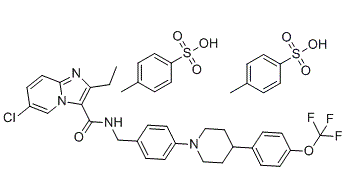
| SMILES |
ClC1C=CC2=NC(CC)=C(C(NCC3C=CC(=CC=3)N3CCC(C4C=CC(=CC=4)OC(F)(F)F)CC3)=O)N2C=1.S(C1C=CC(C)=CC=1)(=O)(=O)O.S(C1C=CC(C)=CC=1)(=O)(=O)O
|
|
| InChi Key |
CCGFTOLSNJBYDV-UHFFFAOYSA-N
|
|
| InChi Code |
InChI=1S/C29H28ClF3N4O2.2C7H8O3S/c1-2-25-27(37-18-22(30)7-12-26(37)35-25)28(38)34-17-19-3-8-23(9-4-19)36-15-13-21(14-16-36)20-5-10-24(11-6-20)39-29(31,32)33;2*1-6-2-4-7(5-3-6)11(8,9)10/h3-12,18,21H,2,13-17H2,1H3,(H,34,38);2*2-5H,1H3,(H,8,9,10)
|
|
| 化学名 |
6-chloro-2-ethyl-N-[[4-[4-[4-(trifluoromethoxy)phenyl]piperidin-1-yl]phenyl]methyl]imidazo[1,2-a]pyridine-3-carboxamide;4-methylbenzenesulfonic acid
|
|
| 别名 |
|
|
| HS Tariff Code |
2934.99.9001
|
|
| 存储方式 |
Powder -20°C 3 years 4°C 2 years In solvent -80°C 6 months -20°C 1 month |
|
| 运输条件 |
Room temperature (This product is stable at ambient temperature for a few days during ordinary shipping and time spent in Customs)
|
| 溶解度 (体外实验) |
|
|||
|---|---|---|---|---|
| 溶解度 (体内实验) |
注意: 如下所列的是一些常用的体内动物实验溶解配方,主要用于溶解难溶或不溶于水的产品(水溶度<1 mg/mL)。 建议您先取少量样品进行尝试,如该配方可行,再根据实验需求增加样品量。
注射用配方
注射用配方1: DMSO : Tween 80: Saline = 10 : 5 : 85 (如: 100 μL DMSO → 50 μL Tween 80 → 850 μL Saline)(IP/IV/IM/SC等) *生理盐水/Saline的制备:将0.9g氯化钠/NaCl溶解在100 mL ddH ₂ O中,得到澄清溶液。 注射用配方 2: DMSO : PEG300 :Tween 80 : Saline = 10 : 40 : 5 : 45 (如: 100 μL DMSO → 400 μL PEG300 → 50 μL Tween 80 → 450 μL Saline) 注射用配方 3: DMSO : Corn oil = 10 : 90 (如: 100 μL DMSO → 900 μL Corn oil) 示例: 以注射用配方 3 (DMSO : Corn oil = 10 : 90) 为例说明, 如果要配制 1 mL 2.5 mg/mL的工作液, 您可以取 100 μL 25 mg/mL 澄清的 DMSO 储备液,加到 900 μL Corn oil/玉米油中, 混合均匀。 View More
注射用配方 4: DMSO : 20% SBE-β-CD in Saline = 10 : 90 [如:100 μL DMSO → 900 μL (20% SBE-β-CD in Saline)] 口服配方
口服配方 1: 悬浮于0.5% CMC Na (羧甲基纤维素钠) 口服配方 2: 悬浮于0.5% Carboxymethyl cellulose (羧甲基纤维素) 示例: 以口服配方 1 (悬浮于 0.5% CMC Na)为例说明, 如果要配制 100 mL 2.5 mg/mL 的工作液, 您可以先取0.5g CMC Na并将其溶解于100mL ddH2O中,得到0.5%CMC-Na澄清溶液;然后将250 mg待测化合物加到100 mL前述 0.5%CMC Na溶液中,得到悬浮液。 View More
口服配方 3: 溶解于 PEG400 (聚乙二醇400) 请根据您的实验动物和给药方式选择适当的溶解配方/方案: 1、请先配制澄清的储备液(如:用DMSO配置50 或 100 mg/mL母液(储备液)); 2、取适量母液,按从左到右的顺序依次添加助溶剂,澄清后再加入下一助溶剂。以 下列配方为例说明 (注意此配方只用于说明,并不一定代表此产品 的实际溶解配方): 10% DMSO → 40% PEG300 → 5% Tween-80 → 45% ddH2O (或 saline); 假设最终工作液的体积为 1 mL, 浓度为5 mg/mL: 取 100 μL 50 mg/mL 的澄清 DMSO 储备液加到 400 μL PEG300 中,混合均匀/澄清;向上述体系中加入50 μL Tween-80,混合均匀/澄清;然后继续加入450 μL ddH2O (或 saline)定容至 1 mL; 3、溶剂前显示的百分比是指该溶剂在最终溶液/工作液中的体积所占比例; 4、 如产品在配制过程中出现沉淀/析出,可通过加热(≤50℃)或超声的方式助溶; 5、为保证最佳实验结果,工作液请现配现用! 6、如不确定怎么将母液配置成体内动物实验的工作液,请查看说明书或联系我们; 7、 以上所有助溶剂都可在 Invivochem.cn网站购买。 |
| 制备储备液 | 1 mg | 5 mg | 10 mg | |
| 1 mM | 1.1094 mL | 5.5469 mL | 11.0937 mL | |
| 5 mM | 0.2219 mL | 1.1094 mL | 2.2187 mL | |
| 10 mM | 0.1109 mL | 0.5547 mL | 1.1094 mL |
1、根据实验需要选择合适的溶剂配制储备液 (母液):对于大多数产品,InvivoChem推荐用DMSO配置母液 (比如:5、10、20mM或者10、20、50 mg/mL浓度),个别水溶性高的产品可直接溶于水。产品在DMSO 、水或其他溶剂中的具体溶解度详见上”溶解度 (体外)”部分;
2、如果您找不到您想要的溶解度信息,或者很难将产品溶解在溶液中,请联系我们;
3、建议使用下列计算器进行相关计算(摩尔浓度计算器、稀释计算器、分子量计算器、重组计算器等);
4、母液配好之后,将其分装到常规用量,并储存在-20°C或-80°C,尽量减少反复冻融循环。
计算结果:
工作液浓度: mg/mL;
DMSO母液配制方法: mg 药物溶于 μL DMSO溶液(母液浓度 mg/mL)。如该浓度超过该批次药物DMSO溶解度,请首先与我们联系。
体内配方配制方法:取 μL DMSO母液,加入 μL PEG300,混匀澄清后加入μL Tween 80,混匀澄清后加入 μL ddH2O,混匀澄清。
(1) 请确保溶液澄清之后,再加入下一种溶剂 (助溶剂) 。可利用涡旋、超声或水浴加热等方法助溶;
(2) 一定要按顺序加入溶剂 (助溶剂) 。
InvivoChem的所有产品仅用于作科学研究,不面向患者销售
Copyright 2020 InvivoChem LLC | All Rights Reserved 粤ICP备20063088号-1


 COA
COA
 463611831
