| 规格 | 价格 | 库存 | 数量 |
|---|---|---|---|
| 10 mM * 1 mL in DMSO |
|
||
| 1mg |
|
||
| 5mg |
|
||
| 10mg |
|
||
| 25mg |
|
||
| 50mg |
|
||
| 100mg |
|
||
| 250mg |
|
||
| 500mg |
|
||
| 1g |
|
||
| Other Sizes |
|

| 靶点 |
VEGFR2 (IC50 = 20 nM); FGFR1 (IC50 = 30 nM); PDGFRβ (IC50 = 510 nM)
|
|---|---|
| 体外研究 (In Vitro) |
体外活性:SU5402 抑制 VEGF、FGF、PDGF 依赖性细胞增殖,IC50 分别为 0.05 μM、2.80μM、28.4 μM。在 HUVEC 中,SU5416 以剂量依赖性方式选择性抑制 VEGF 驱动的有丝分裂,IC50 为 0.04 μM。在鼻咽上皮细胞中,SU5402 减弱 LMP1 介导的有氧糖酵解、细胞转化、细胞迁移和侵袭。在小鼠 C3H10T1/2 细胞中,SU 5402 减弱 FGF23 对细胞分化的影响 激酶测定:用工程基因感染草地贪夜蛾 (sf9) 细胞后,FGF-R1 和 Flk-1/KDR 的催化部分表达为 GST 融合蛋白杆状病毒。通过谷胱甘肽琼脂糖层析将受感染的 sf9 细胞裂解物中的 GST-FGFR1 和 GST-Flk1 纯化至同质。测定在 96 孔微量滴定板中进行,该板每孔用 2.0 μg 聚 Glu-Tyr 肽 (4:1) 的 0.1 mL PBS 包被过夜。将纯化的激酶在激酶测定缓冲液(100 mM Hepes pH 7.5、100 mM NaCl 和 0.1 mM 原钒酸钠)中稀释,并以每 0.05 mL 体积缓冲液 5 ng GST 融合蛋白添加到所有测试孔中。测试化合物用 4% DMSO 稀释并添加到测试孔中(0.025 mL/孔)。通过添加 0.025 mL 40 μM ATP/40 mM MnCl2 启动激酶反应,将板摇动 10 分钟,然后添加 0.025 mL 0.5 M EDTA 停止反应。最终 ATP 浓度为 10 μM,是实验确定的 ATP Km 值的两倍。阴性对照孔仅接受 MnCl2,不含 ATP。将板用 10 mM Tris pH 7.4、150 mM NaCl 和 0.05% Tween-20 (TBST) 洗涤三次。将兔多克隆抗磷酸酪氨酸抗血清以 1:10000 稀释度加入 TBST 中,孵育 1 小时。然后用TBST洗涤板3次。然后将与辣根过氧化物酶缀合的山羊抗兔抗血清添加至所有孔中1小时。用TBST洗涤板3次,并通过添加2,2'-偶氮双(3-乙基苯并噻唑啉-6-磺酸)(ABTS)来检测过氧化物酶反应。检测的颜色读数允许进行 20−30 分钟,并使用 410 nM 测试过滤器在 Dynatech MR5000 ELISA 板读取器上读取。细胞测定:用于体外生长的肿瘤细胞系(SF767T、SF763、EPH4-VEGF、C6、A375、A431、LNCAP、Calu-6、3T3Her2 和 488G2M2 细胞)在 37°C 的培养基中培养 5-10 小时。 %二氧化碳。 SU5416 在含有 DMSO (<0.5%) 的培养基中连续稀释,并在开始培养 1 天后添加到肿瘤细胞培养物中。 96 小时后使用磺基罗丹明 B 法测量细胞生长。使用四参数分析通过曲线拟合计算 IC50。
|
| 体内研究 (In Vivo) |
在小鼠中,SU5402(25 mg/kg,腹腔注射)通过抑制与肿瘤生长相关的血管生成过程来抑制一组肿瘤细胞系的皮下生长。
相比之下,在小鼠体内以无毒剂量全身给药SU5402可抑制来自各种组织来源的细胞的皮下肿瘤生长。SU5416的抗肿瘤作用伴随着从药物治疗动物身上切除的淡白色肿瘤的出现,这支持了该药物的抗血管生成特性。这些发现支持,对血管内皮生长因子受体酶活性的药理学抑制代表了一种限制多种肿瘤类型生长的新策略。[2] FGFR1抑制剂SU5402逆转MCT诱导的PH。[5] 为了证实与FGF2-siRNA治疗相关的肺血管改变和PH的减少与FGF2敲低有关,表明FGF2在PH中过量产生的关键作用,我们研究了选择性FGFR1抑制剂SU5402是否可以预防和/或逆转MCT在大鼠中诱导的PH。在MCT注射后第21至42天用SU5402治疗的大鼠中,第42天的评估显示,与用载体(生理盐水)治疗的大白鼠相比,PAP、RV/(LV+S)和远端动脉肌肉化明显降低(图8)。 |
| 酶活实验 |
基因组改变的杆状病毒感染草地贪夜蛾 (sf9) 细胞后,FGF-R1 和 Flk-1/KDR 的催化结构域表达为 GST 融合蛋白。使用谷胱甘肽琼脂糖层析,将感染的 sf9 细胞裂解物纯化至 GST-FGFR1 和 GST-Flk1 的同质性。在 96 孔微量滴定板中,每孔 0.1 mL PBS 中的 2.0 μg 聚谷氨酸-Tyr 肽 (4:1) 包被过夜,然后进行测定。使用激酶测定缓冲液(100 mM Hepes pH 7.5、100 mM NaCl 和 0.1 mM 原钒酸钠),以每 0.05 mL 体积缓冲液 5 ng GST 融合蛋白的比例将稀释的纯化激酶引入每个测试孔。将测试化合物用 4% DMSO 稀释后添加到测试孔中(0.025 mL/孔)。要启动激酶反应,请添加 0.025 mL 的 40 μM ATP/40 mM MnCl2。摇动板 10 分钟,然后添加 0.025 mL 0.5 M EDTA 以终止反应。 ATP 的终浓度为 10 μM,是实验测定的 ATP Km 值的两倍。阴性对照孔中添加 MnCl2 且不添加 ATP。使用 10 mM Tris pH 7.4、150 mM NaCl 和 0.05% Tween-20 (TBST) 对板进行三轮洗涤。将兔多克隆抗磷酸酪氨酸抗血清在 TBST 中的 1:10000 稀释液充满孔,持续一小时。然后用TBST洗板3次。然后,在一小时内,每个孔接受山羊抗兔抗血清和辣根过氧化物酶的缀合物。板的 3 次 TBST 洗涤后,添加 2,2'-azinobis(3-乙基苯并噻唑啉-6-磺酸) (ABTS) 以检测过氧化物酶反应。待测定的颜色读数显色 20 至 30 分钟后,使用 Dynatech MR5000 ELISA 酶标仪上的 410 nM 测试过滤器进行读取。
|
| 细胞实验 |
用于体外生长的肿瘤细胞系在 37°C、5–10% CO2 的培养基中生长。培养开始一天后,SU5402在含有 DMSO (<0.5%) 的培养基中连续稀释并添加到肿瘤细胞培养物中。采用磺基罗丹明B法测定96小时后细胞的生长情况。使用四参数分析和曲线拟合,确定 IC50。
蛋白质印迹分析: 总细胞裂解物(5-50µg蛋白质)通过10%或4-12%SDS-PAGE分离,并在免疫印迹前转移到PVDF膜上。 免疫荧光和免疫组织化学染色: 如前所述进行免疫荧光染色和免疫组织化学染色[3]。 细胞增殖试验: 用细胞增殖试剂CCK-8[3]进行细胞增殖试验。 软琼脂克隆试验: 软琼脂菌落形成试验如前所述[3]。 迁移和入侵检测: 分别使用CytoSelect 24孔伤口愈合检测试剂盒和CytoSelect24孔细胞侵袭检测试剂盒进行细胞迁移检测和Boyden腔侵袭检测。对于胶原凝胶侵袭试验,用I型胶原溶液制备胶原混合物[3]。 通过[3H]胸腺嘧啶掺入法评估SMC增殖。[5] 将PA-SMCs在含有15% FCS的DMEM中接种到24孔板中,密度为5 × 10^4细胞/孔,并使其贴壁。细胞在无血清培养基中进行48小时的生长停滞,然后用1 ml条件化的P-EC培养基处理。我们还测试了外源性PDGF(10 ng/ml)和FGF2(10 ng/ml)在有或无伊马替尼(10^-5 M)、EGF拮抗剂(10^-5 M)和SU5402(10^-5 M)的情况下对PA-SMC增殖的影响。在每种条件下,向每孔加入[3H]胸腺嘧啶(1 μCi/ml)。孵育24小时后,用PBS洗涤细胞两次,用冰冷的10%三氯乙酸处理,并溶解在0.1 N NaOH中(0.5 ml/孔)。对掺入的放射性进行计数,并以cpm/孔报告。 |
| 动物实验 |
小鼠:对9-12周龄的雄性ΔF508小鼠(CFTRtm1Eur,129/FVB背景)及其野生型同窝小鼠,连续一周腹腔注射DMSO或SU 5402(溶于DMSO,浓度为6 mg/mL),剂量为25 mg/kg体重。每日根据小鼠体重调整剂量。之后,吸入异氟烷进行麻醉,直至实验结束。为防止唾液腺受到胆碱能刺激,将50 μL胆碱能拮抗剂阿托品(1 mM)皮下注射至小鼠右侧颊部。注射侧颊部紧贴一小条滤纸,持续4分钟。然后在同一位置注射异丙肾上腺素(10 mM,37.5 μL),以诱发唾液的肾上腺素能分泌(时间 0)。在接下来的 30 分钟内,每 5 分钟更换一次滤纸条(预先在 Eppendorf 管中称重)。收集完成后,测量所有六条滤膜的重量,并将结果标准化为mg/g体重。
大鼠:成年雄性Wistar大鼠(200-250 g)皮下注射MCT(60 mg/kg),21天不进行任何治疗。为了评估FGFR1抑制剂SU 5402对已建立的肺动脉高压(PH)的潜在影响,体重200-250 g的成年雄性Wistar大鼠皮下注射MCT(60 mg/kg),21天不进行任何治疗,然后随机分为两组(每组10只动物):一组接受SU 5402(25 mg/kg/天)治疗,另一组从第21天到第42天不进行任何治疗。所有治疗均每日一次静脉(皮下)给药。 SU5402治疗对已建立的MCT肺动脉高压的影响。 [5] 为了评估FGFR1抑制剂SU5402对已建立的肺动脉高压(PH)的潜在影响,成年雄性Wistar大鼠(200-250 g)皮下注射MCT(60 mg/kg),21天后不进行任何治疗,然后随机分为两组(每组10只动物),一组从第21天至第42天接受SU5402(25 mg/kg/天)治疗,另一组接受溶剂对照。所有治疗均每日一次皮下注射。 |
| 参考文献 |
|
| 其他信息 |
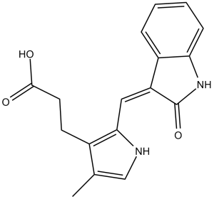
SU5402 是一种吲哚酮类化合物,其结构为 3-亚甲基吲哚酮,其中一个亚甲基氢原子被 3-(2-羧乙基)-4-甲基-1H-吡咯-2-基取代。它是成纤维细胞生长因子受体 1 酪氨酸激酶活性的 ATP 竞争性抑制剂,具有成纤维细胞生长因子受体拮抗剂的作用。它是一种单羧酸,属于吡咯类和吲哚酮类化合物。其功能与 3-亚甲基吲哚酮相关。
受体酪氨酸激酶 (RTK) 已被认为是治疗多种人类疾病的潜在靶点,包括癌症、炎症性疾病、心血管疾病(包括动脉再狭窄)以及肺、肝、肾纤维化疾病。已鉴定出三类在吡咯环C-3位连接丙酸官能团的3-取代吲哚啉-2-酮类化合物,它们是血管内皮生长因子(VEGF)、成纤维细胞生长因子(FGF)和血小板衍生生长因子(PDGF)受体酪氨酸激酶(RTK)的催化抑制剂。部分化合物能够抑制分离的血管内皮生长因子受体2(VEGF-R2)[胎肝酪氨酸激酶1(Flk-1)/激酶插入结构域受体(KDR)]、成纤维细胞生长因子受体(FGF-R)和血小板衍生生长因子受体(PDGF-R)酪氨酸激酶的活性,其IC50值达到纳摩尔级。因此,化合物 1 对 VEGF-R2 (Flk-1/KDR) 和 FGF-R1 酪氨酸激酶活性具有抑制作用,IC50 值分别为 20 nM 和 30 nM;而化合物 16f 对 PDGF-R 酪氨酸激酶活性具有抑制作用,IC50 值为 10 nM。本文讨论了这些化合物针对靶受体的结构模型和构效关系分析。采用溴脱氧尿苷 (BrdU) 掺入法测定了这些化合物的细胞活性。部分化合物表现出特异性和强效的细胞生长抑制作用。这些数据表明,这些化合物可用于抑制这些靶受体的功能。[1] 非角化性鼻咽癌 (NPC) 与 Epstein-Barr 病毒 (EBV) 感染密切相关。 EB病毒编码的潜伏膜蛋白1 (LMP1) 被认为在鼻咽癌 (NPC) 的发病机制中发挥重要作用,因为它能够激活多种细胞信号通路,这些通路共同促进细胞增殖、转化、血管生成和侵袭,并调节能量代谢。本研究发现,LMP1 可增加细胞对葡萄糖和谷氨酰胺的摄取,增强LDHA活性和乳酸生成,但降低丙酮酸激酶活性和丙酮酸浓度。此外,无论氧气供应情况如何,LMP1 均可增加PKM2、LDHA和FGFR1的磷酸化水平,以及PDHK1、FGFR1、c-Myc和HIF-1α的表达。综上所述,这些结果表明LMP1促进有氧糖酵解。关于FGFR1信号通路,LMP1不仅能增加FGFR1的表达,还能上调FGF2的表达,从而导致FGFR1信号通路的组成型激活。此外,两种FGFR1抑制剂(PD161570和SU5402)能够减弱LMP1介导的鼻咽上皮细胞的有氧糖酵解、细胞转化(增殖和非锚定依赖性生长)、细胞迁移和侵袭,表明FGFR1信号通路是LMP1介导的细胞生长转化中的关键通路。免疫组化染色显示,原发性鼻咽癌标本中普遍存在高水平的磷酸化FGFR1,且其表达与LMP1的表达呈正相关。此外,FGFR1抑制剂能够抑制鼻咽癌细胞的增殖和非锚定依赖性生长。我们目前的研究结果表明,LMP1介导的FGFR1激活促进有氧糖酵解和上皮细胞转化,从而提示FGF2/FGFR1信号通路激活参与了EBV驱动的鼻咽癌发病机制。[2] FGF23是一种骨源性激素,它通过抑制肾小管磷酸盐重吸收和降低循环中1,25(OH)2D和PTH水平来调节矿物质代谢。这些作用是通过FGF受体与其共受体Klotho结合和激活介导的,Klotho在肾脏远曲小管中表达。最近有报道称Klotho在骨骼组织中表达,这表明FGF23对参与骨骼发育和重塑的细胞具有直接的肾外作用,但其机制尚不明确。在本研究中,我们发现从Klotho基因敲除小鼠中分离的骨髓基质细胞形成的成骨细胞集落数量少于野生型小鼠,而脂肪细胞集落数量多于野生型小鼠。我们通过对小鼠C3H10T1/2细胞的实验,探讨了其潜在机制。我们发现Klotho表达较弱,且FGF23呈剂量依赖性地影响细胞谱系命运的决定。FGF23对细胞分化的影响可被FGF受体特异性酪氨酸激酶抑制剂SU5402减弱。我们的结果表明,FGF23直接影响骨髓基质细胞的分化。[4] 肺动脉高压(PH)是一种进行性、致命性肺部疾病,其特征是肺动脉平滑肌细胞(PA-SMC)增生,最终导致右心衰竭。肺内皮细胞(P-EC)来源的分子事件可能参与PH中PA-SMC的增生。因此,我们将培养的人肺动脉平滑肌细胞(PA-SMC)暴露于特发性肺动脉高压(IPH)患者或健康对照者的肺动脉内皮细胞(P-EC)条件培养基中,发现IPH P-EC条件培养基比对照P-EC条件培养基更能促进PA-SMC增殖。IPH P-EC培养基中FGF2水平高于对照组,而TGF-β1、PDGF-BB或EGF水平无明显差异。IPH和对照组PA-SMC之间FGF2诱导的增殖或FGF受体1型(FGFR1)mRNA水平无差异。使用siRNA敲低P-EC中的FGF2后,IPH P-EC培养基对PA-SMC生长的刺激作用降低了60%,对照P-EC培养基降低了10%。原位杂交显示,IPH患者肺血管内皮细胞重塑后,FGF2主要过度表达。重复静脉注射FGF2-siRNA可消除肺部FGF2的产生,从而预防并几乎逆转大鼠肺动脉高压模型。同样,使用SU5402进行FGFR1药理学抑制也可逆转同一模型中已建立的肺动脉高压。因此,内皮细胞FGF2在特发性肺动脉高压中过度产生,并促进特发性肺动脉高压中平滑肌细胞增生,这表明FGF2是治疗肺动脉高压的一个有前景的新靶点。[5] |
| 分子式 |
C17H16N2O3
|
|
|---|---|---|
| 分子量 |
296.32
|
|
| 精确质量 |
296.116
|
|
| 元素分析 |
C, 68.91; H, 5.44; N, 9.45; O, 16.20
|
|
| CAS号 |
215543-92-3
|
|
| 相关CAS号 |
SU 5402;215543-92-3
|
|
| PubChem CID |
5289418
|
|
| 外观&性状 |
Orange solid powder
|
|
| 密度 |
1.4±0.1 g/cm3
|
|
| 沸点 |
592.6±50.0 °C at 760 mmHg
|
|
| 熔点 |
>222ºC (dec.)
|
|
| 闪点 |
312.2±30.1 °C
|
|
| 蒸汽压 |
0.0±1.8 mmHg at 25°C
|
|
| 折射率 |
1.688
|
|
| LogP |
2.03
|
|
| tPSA |
82.19
|
|
| 氢键供体(HBD)数目 |
3
|
|
| 氢键受体(HBA)数目 |
3
|
|
| 可旋转键数目(RBC) |
4
|
|
| 重原子数目 |
22
|
|
| 分子复杂度/Complexity |
488
|
|
| 定义原子立体中心数目 |
0
|
|
| SMILES |
O=C(CCC1=C(NC=C1C)/C=C2C(NC3=C\2C=CC=C3)=O)O
|
|
| InChi Key |
JNDVEAXZWJIOKB-JYRVWZFOSA-N
|
|
| InChi Code |
InChI=1S/C17H16N2O3/c1-10-9-18-15(11(10)6-7-16(20)21)8-13-12-4-2-3-5-14(12)19-17(13)22/h2-5,8-9,18H,6-7H2,1H3,(H,19,22)(H,20,21)/b13-8-
|
|
| 化学名 |
3-[4-methyl-2-[(Z)-(2-oxo-1H-indol-3-ylidene)methyl]-1H-pyrrol-3-yl]propanoic acid
|
|
| 别名 |
|
|
| HS Tariff Code |
2934.99.9001
|
|
| 存储方式 |
Powder -20°C 3 years 4°C 2 years In solvent -80°C 6 months -20°C 1 month |
|
| 运输条件 |
Room temperature (This product is stable at ambient temperature for a few days during ordinary shipping and time spent in Customs)
|
| 溶解度 (体外实验) |
|
|||
|---|---|---|---|---|
| 溶解度 (体内实验) |
注意: 如下所列的是一些常用的体内动物实验溶解配方,主要用于溶解难溶或不溶于水的产品(水溶度<1 mg/mL)。 建议您先取少量样品进行尝试,如该配方可行,再根据实验需求增加样品量。
注射用配方
注射用配方1: DMSO : Tween 80: Saline = 10 : 5 : 85 (如: 100 μL DMSO → 50 μL Tween 80 → 850 μL Saline)(IP/IV/IM/SC等) *生理盐水/Saline的制备:将0.9g氯化钠/NaCl溶解在100 mL ddH ₂ O中,得到澄清溶液。 注射用配方 2: DMSO : PEG300 :Tween 80 : Saline = 10 : 40 : 5 : 45 (如: 100 μL DMSO → 400 μL PEG300 → 50 μL Tween 80 → 450 μL Saline) 注射用配方 3: DMSO : Corn oil = 10 : 90 (如: 100 μL DMSO → 900 μL Corn oil) 示例: 以注射用配方 3 (DMSO : Corn oil = 10 : 90) 为例说明, 如果要配制 1 mL 2.5 mg/mL的工作液, 您可以取 100 μL 25 mg/mL 澄清的 DMSO 储备液,加到 900 μL Corn oil/玉米油中, 混合均匀。 View More
注射用配方 4: DMSO : 20% SBE-β-CD in Saline = 10 : 90 [如:100 μL DMSO → 900 μL (20% SBE-β-CD in Saline)] 口服配方
口服配方 1: 悬浮于0.5% CMC Na (羧甲基纤维素钠) 口服配方 2: 悬浮于0.5% Carboxymethyl cellulose (羧甲基纤维素) 示例: 以口服配方 1 (悬浮于 0.5% CMC Na)为例说明, 如果要配制 100 mL 2.5 mg/mL 的工作液, 您可以先取0.5g CMC Na并将其溶解于100mL ddH2O中,得到0.5%CMC-Na澄清溶液;然后将250 mg待测化合物加到100 mL前述 0.5%CMC Na溶液中,得到悬浮液。 View More
口服配方 3: 溶解于 PEG400 (聚乙二醇400) 请根据您的实验动物和给药方式选择适当的溶解配方/方案: 1、请先配制澄清的储备液(如:用DMSO配置50 或 100 mg/mL母液(储备液)); 2、取适量母液,按从左到右的顺序依次添加助溶剂,澄清后再加入下一助溶剂。以 下列配方为例说明 (注意此配方只用于说明,并不一定代表此产品 的实际溶解配方): 10% DMSO → 40% PEG300 → 5% Tween-80 → 45% ddH2O (或 saline); 假设最终工作液的体积为 1 mL, 浓度为5 mg/mL: 取 100 μL 50 mg/mL 的澄清 DMSO 储备液加到 400 μL PEG300 中,混合均匀/澄清;向上述体系中加入50 μL Tween-80,混合均匀/澄清;然后继续加入450 μL ddH2O (或 saline)定容至 1 mL; 3、溶剂前显示的百分比是指该溶剂在最终溶液/工作液中的体积所占比例; 4、 如产品在配制过程中出现沉淀/析出,可通过加热(≤50℃)或超声的方式助溶; 5、为保证最佳实验结果,工作液请现配现用! 6、如不确定怎么将母液配置成体内动物实验的工作液,请查看说明书或联系我们; 7、 以上所有助溶剂都可在 Invivochem.cn网站购买。 |
| 制备储备液 | 1 mg | 5 mg | 10 mg | |
| 1 mM | 3.3747 mL | 16.8737 mL | 33.7473 mL | |
| 5 mM | 0.6749 mL | 3.3747 mL | 6.7495 mL | |
| 10 mM | 0.3375 mL | 1.6874 mL | 3.3747 mL |
1、根据实验需要选择合适的溶剂配制储备液 (母液):对于大多数产品,InvivoChem推荐用DMSO配置母液 (比如:5、10、20mM或者10、20、50 mg/mL浓度),个别水溶性高的产品可直接溶于水。产品在DMSO 、水或其他溶剂中的具体溶解度详见上”溶解度 (体外)”部分;
2、如果您找不到您想要的溶解度信息,或者很难将产品溶解在溶液中,请联系我们;
3、建议使用下列计算器进行相关计算(摩尔浓度计算器、稀释计算器、分子量计算器、重组计算器等);
4、母液配好之后,将其分装到常规用量,并储存在-20°C或-80°C,尽量减少反复冻融循环。
计算结果:
工作液浓度: mg/mL;
DMSO母液配制方法: mg 药物溶于 μL DMSO溶液(母液浓度 mg/mL)。如该浓度超过该批次药物DMSO溶解度,请首先与我们联系。
体内配方配制方法:取 μL DMSO母液,加入 μL PEG300,混匀澄清后加入μL Tween 80,混匀澄清后加入 μL ddH2O,混匀澄清。
(1) 请确保溶液澄清之后,再加入下一种溶剂 (助溶剂) 。可利用涡旋、超声或水浴加热等方法助溶;
(2) 一定要按顺序加入溶剂 (助溶剂) 。
NIH 3T3 Flk-1 cells (A) or NIH 3T3 platelet-derived growth factor β cells (B) grown to confluency were preincubated with SU5416 at concentrations ranging from 0.05 to 50 μm for 1 h at 37°C. Cancer Res. 1999 Jan 1;59(1):99-106. |
A375 cells (3 × 10⁶) were implanted subcutaneous into the hindflank region of female BALB/c nu/nu mice 8–12 weeks of age. Cancer Res. 1999 Jan 1;59(1):99-106. |
Rat C6 glioma cells were surgically implanted (0.5 × 10⁶ cells/animal) under the serosa of the colon in BALB/c nu/nu mice. Beginning 1 day after implantation, animals were treated once daily with a 50 μl i.p. bolus injection of either SU5416 at 25 mg/kg/day in DMSO or DMSO alone for 16 days. On day 16 after implantation, animals were euthanized, and their local tumors in the colon were first quantitated by measurement using venier calipers and then harvested. Cancer Res. 1999 Jan 1;59(1):99-106. |
 VEGFR2-IN-7
 SYHA1813
 VEGFR-2-IN-38
 BHEP
BHEP
InvivoChem的所有产品仅用于作科学研究,不面向患者销售
Copyright 2020 InvivoChem LLC | All Rights Reserved 粤ICP备20063088号-1


 COA
COA


 463611831
