| 规格 | 价格 | 库存 | 数量 |
|---|---|---|---|
| 1mg |
|
||
| 5mg |
|
||
| 10mg |
|
||
| 25mg |
|
||
| Other Sizes |
|

| 靶点 |
Human FP Receptor:0.4 nM (Ki); Human FP Receptor:0.53 nM (EC50); EP3 Receptor:67 nM (IC50)
Tafluprost acid targets the human prostanoid FP receptor, showing high affinity with a Ki of 0.4 nM and an EC50 of 0.53 nM. The FP receptor is a G protein-coupled receptor that mediates the effects of prostaglandin F2α. Activation of the FP receptor in the eye increases uveoscleral outflow of aqueous humor, leading to a reduction in intraocular pressure. |
|---|---|
| 体外研究 (In Vitro) |
在3T3-L1前脂肪细胞分化的早期和晚期,他氟前列素酸(10、100 nM)能有效抑制脂肪生成[2]。在野生型小鼠的原代脂肪细胞中,他氟前列素酸(100 nM)可减少脂肪生成,但在FP基因敲除小鼠的原代脂肪细胞中则无此作用[2]。人脐静脉内皮细胞(HUVEC)暴露于他氟前列素酸(10⁻⁴ M)6小时后,增殖和迁移受到刺激[4]。HUVEC管状结构的形成也受到刺激(-4 M,4-18小时)[4]。他氟前列素酸对人前列腺素FP受体具有高亲和力,Ki值为0.4 nM,EC50值为0.53 nM。它是FP受体的强效激动剂,其激活可导致房水流出增加。特定的体外活性数据与其作为前列腺素类似物的作用相符。
|
| 体内研究 (In Vivo) |
滴眼后4-8小时,Taf/T-FDC的降眼压效果与Lat/T-FDC几乎相当。Taf/T-FDC和Lat/T-FDC的降眼压峰值均出现在滴眼后8小时,两者之间无显著差异。在滴眼后24-30小时,二者的降眼压效果出现差异,Taf/T-FDC的降眼压效果显著优于Lat/T-FDC。Taf/T-FDC的降眼压效果可持续至滴眼后30小时,而Lat/T-FDC的降眼压效果在滴眼后28小时几乎消失。滴注Taf/T-FDC后,房水中噻吗洛尔浓度高于滴注Lat/T-FDC后(Cmax:3870 ng/mL vs 1330 ng/mL;AUCinf:3970 ng·h/mL vs 1250 ng·h/mL)。滴注Taf/T-FDC和Lat/T-FDC后,房水中他氟前列素酸/AFP-172和拉坦前列素酸的浓度分别与滴注他氟前列素和拉坦前列素单方制剂后的浓度相似。在所有评估时间点,无论未稀释还是稀释10倍,Taf/T-FDC对人角膜上皮细胞的细胞毒性作用均显著低于Lat/T-FDC[3]。
他氟前列素酸是他氟前列素的活性形式,也是其体内降眼压作用的来源。他氟前列素以滴眼液形式给药,并在眼内水解为他氟前列素酸。该酸形式随后作用于FP受体,增加房水流出,降低眼内压。他氟前列素已被批准用于治疗青光眼和高眼压症。 |
| 酶活实验 |
为了评估新型前列腺素F2α衍生物AFP-168(他氟前列素)的药理学特性,我们检测了其受体结合亲和力、降低眼压(IOP)的效果、对房水动力学的影响以及对黑色素生成的刺激作用。我们通过在器官浴槽中测量肌肉收缩、抑制血小板聚集以及与放射性标记配体的竞争性结合,确定了AFP-168羧酸衍生物他氟前列素酸/AFP-172的受体结合谱。在眼压测量研究中,我们使用了眼压正常的食蟹猴和激光诱导眼压升高的食蟹猴,并使用气动眼压计测量眼压。为了研究房水动力学,我们对眼压正常的猴子采用了眼压测量法(Goldmann压平式眼压计)、荧光光度法、双水平恒压灌注法以及同位素稀释和累积技术。我们测定了培养的B16-F0黑色素瘤细胞培养基和细胞体内的黑色素含量。Tafluprost酸/AFP-172对FP受体的亲和力(Ki:0.4 nM)是拉坦前列素羧酸PhXA85(Ki:4.7 nM)的12倍。单次滴注0.0025%的AFP-168可显著降低眼压正常和眼压升高猴子的眼压(分别为3.1 mmHg和11.8 mmHg,p < 0.01),而0.005%的拉坦前列素也可显著降低眼压(分别为2.1 mmHg,p < 0.01和9.5 mmHg,p = 0.059)。在眼压正常的猴子中,每日一次滴注0.001%、0.0025%或0.005%的AFP-168,持续5天,不仅在滴眼后数小时内显著降低眼压,而且在滴眼后24小时的药物谷浓度时也显著降低眼压。0.005%的拉坦前列素也能降低眼压,但在药物谷浓度时未观察到降低眼压的效果。 AFP-168 降低眼压的主要机制是使葡萄膜巩膜外流增加 65% (p < 0.05),并且与其他前列腺素类似,也增加了总房水流出率(增加 33%,p < 0.05)。在培养的 B16-F0 黑色素瘤细胞中,他氟前列素酸/AFP-172 (100 μM) 不刺激黑色素生成,而 PhXA85 (100 μM) 则有此作用。这些研究结果表明,AFP-168 对前列腺素 FP 受体具有高亲和力,在眼压正常和眼压升高的猴子中均具有强效的降眼压作用,且其作用优于拉坦前列素,并且对黑色素瘤细胞的黑色素生成刺激作用较小[1]。
可使用放射性配体结合试验评估他氟前列素酸与人前列腺素 FP 受体的结合亲和力,该试验使用表达重组 FP 受体的细胞膜制备物。竞争性结合实验中,使用递增浓度的他氟前列素酸与固定浓度的放射性标记 FP 受体配体(例如,[³H]前列腺素 F2α)进行竞争。在过量未标记配体存在的情况下测定非特异性结合。 |
| 细胞实验 |
为促进3T3-L1前脂肪细胞分化为成熟脂肪细胞,对其进行了处理。在分化的早期和晚期(第0、2和7天),分别向细胞中加入1至1000 nM的拉坦前列素酸(LAT-A)、曲伏前列素酸(TRA-A)、他氟前列素酸(TAF-A)、比马前列素(BIM)、比马前列素酸(BIM-A)、乌诺前列酮(UNO)或前列腺素F2α(PGF2α)。在第10天,使用油红O染色检测细胞内脂质。将照片上染色区域的面积与对照组进行比较。所有实验均采用盲法进行。接下来,使用FP受体敲除小鼠和野生型小鼠的原代培养脂肪细胞进行类似的实验。结果:在第0天或第2天添加前列腺素(PGs)时,LAT-A、TAF-A、BIM-A和PGF2α在10 nM和100 nM浓度下均显著抑制脂肪生成(第0天P < 0.01,第2天P < 0.05),而TRA-A仅在100 nM浓度下抑制脂肪生成。比马前列素和UNO在任何浓度下均未影响脂肪生成。在第7天添加前列腺素时,100 nM LAT-A、BIM-A或PGF2α显著抑制脂肪生成(P < 0.05)。在小鼠原代脂肪细胞培养中,LAT-A、TAF-A、BIM-A、TRA-A 和 PGF2α 显著抑制野生型脂肪细胞的脂肪生成(P < 0.05),但在 FP 基因敲除小鼠脂肪细胞中,任何一种前列腺素类似物均未抑制脂肪生成。结论:前列腺素类似物可能通过刺激 FP 受体抑制脂肪生成。尽管这些发现需要在与眼眶脂肪更密切相关的模型系统中进行进一步分析,但PG类似物可能通过抑制脂肪生成直接导致眼眶脂肪减少[2]。
在有或无FP受体拮抗剂(10 nM AL-8810)的情况下培养的HUVECs暴露于浓度递增的10⁻⁷、10⁻⁶、10⁻⁵、10⁻⁴和10⁻³ M的他氟前列素酸/AFP-172(他氟前列素的游离酸)。细胞增殖实验中,细胞数量通过微孔板读数仪使用CellTiter96® Aqueous One Solution细胞增殖检测试剂盒(Promega)获得。内皮细胞迁移采用BD Biocoat™血管生成系统和FluoroBlok™ 24孔插入板进行评估。使用BioTek FLx800荧光酶标仪对荧光标记的侵袭性血管内皮细胞进行定量检测。采用BD Biocoat血管生成系统,在Matrigel基质96孔板上评估内皮毛细血管样管的形成。采用实时定量逆转录-聚合酶链式反应(RT-PCR)检测血管内皮生长因子(VEGF)、环氧合酶-2(COX-2)和内皮型一氧化氮合酶(eNOS)的基因表达。采用免疫荧光染色和Western blot法检测COX-2蛋白。统计分析采用Student's t检验。[4] 在体外,使用表达重组FP受体的细胞评估他氟前列素酸在FP受体上的功能活性。将细胞用浓度递增的他氟前列素酸处理,并使用钙敏感荧光染料(例如 Fluo-4 AM)测量细胞内钙动员(FP 受体激活的下游信号)。测定钙动员的 EC50 值。 |
| 动物实验 |
在研究 A 中,分别于滴眼后 4 小时和 8 小时评估了 Taf/T-FDC 和 Lat/T-FDC 对眼压正常猴子的降眼压效果;在研究 B 中,分别于滴眼后 12 小时、14 小时、16 小时和 18 小时评估了 Taf/T-FDC 和 Lat/T-FDC 对眼压正常猴子的降眼压效果;在研究 C 中,分别于滴眼后 24 小时、26 小时、28 小时和 30 小时评估了 Taf/T-FDC 和 Lat/T-FDC 对眼压正常猴子的降眼压效果。在 Sprague Dawley 大鼠单次滴注 Taf/T-FDC 或 Lat/T-FDC 后,采用液相色谱-串联质谱法测定了噻吗洛尔、他氟前列素酸/AFP-172(他氟前列素的活性代谢形式)和拉坦前列素酸(拉坦前列素的活性代谢形式)的浓度,以评估药物向眼内的渗透情况。采用 3-(4,5-二甲基噻唑-2-基)-5-(3-羧基甲氧基苯基)-2-(4-磺基苯基)-2H-四唑法分析了 SV40 转化的角膜上皮细胞暴露于 Taf/T-FDC 或 Lat/T-FDC 1-30 分钟后的细胞毒性。评估了每种 FDC 的未稀释溶液和 10 倍稀释溶液[3]。
在动物研究中,通常不直接使用他氟前列素酸;而是将他氟前列素(前药)作为滴眼液给药。在青光眼或高眼压动物模型中,将他氟前列素局部滴眼,并使用眼压计测量眼内压。评估他氟前列素降低眼内压的疗效,其药理作用是由活性酸形式引起的。 |
| 药代性质 (ADME/PK) |
Taf/T-FDC 和他氟前列素单药治疗后,他氟前列素酸的浓度曲线相似,在 0.25–0.5 小时达到 Cmax,半衰期为 0.43–0.45 小时。Lat/T-FDC 和拉坦前列素单药治疗后,拉坦前列素酸的浓度曲线也相似,在 0.5 小时达到 Cmax,半衰期为 0.35–0.40 小时。[3] 他氟前列素酸的分子量为 410.45,分子式为 C22H28F2O5。它是一种具有二氟化结构的前列腺素类似物。标准参考文献中未详细列出其具体的理化性质,例如溶解度和 logP 值。
|
| 毒性/毒理 (Toxicokinetics/TK) |
他氟前列素酸的毒性特征与他氟前列素一致,后者作为滴眼剂使用时通常耐受性良好。常见不良反应包括眼部刺激、结膜充血和睫毛生长改变。由于眼部给药后全身吸收率低,因此全身毒性极小。
|
| 参考文献 |
|
| 其他信息 |
环氧合酶 (COX) 是花生四烯酸转化为前列腺素的关键酶。COX 有两种同工酶:COX-1 和 COX-2。COX-1 是一种管家基因,在大多数组织中表达;而 COX-2 是一种可诱导的早期中间基因,在炎症细胞中诱导表达,其作用与血管生成和肿瘤发生有关。在本研究中,10⁻⁴ M 的他氟前列素酸/AFP-172 仅刺激了三种基因中的一种——COX-2 的表达。在预先用 FP 受体拮抗剂处理的人脐静脉内皮细胞 (HUVEC) 中,他氟前列素酸/AFP-172 诱导的 COX-2 蛋白表达被阻断。这些结果表明,tafluprost acid/AFP-172 通过 FP 受体诱导 HUVEC 中 COX-2 的表达,这与 PGF2α 在子宫内膜腺癌中的作用类似。尽管 COX-2 促进血管生成的作用已广为人知,但 COX-2 介导的血管生成的具体机制仍不清楚。与 COX-1 抑制剂不同,COX-2 抑制剂被认为是抑制血管生成和肿瘤发生的潜在治疗药物。在本研究中,COX-2 抑制剂通过降低 HUVEC 的增殖、迁移和管状结构形成能力,消除了 Tafluprost acid/AFP-172 的促血管生成作用。这些结果提示 Tafluprost acid/AFP-172 通过与 COX-2 信号通路相互作用促进 HUVEC 的血管生成。COX-2 和 VEGF 似乎通过相互依赖的双基因表达通路参与血管生成。尽管已知COX-2可通过与VEGF系统相互作用来调节血管生成,但我们使用RT-PCR检测发现,AFP-172处理组和对照组之间VEGF-A的表达水平没有差异(图5)。因此,tafluprost/AFP-172的促血管生成作用与VEGF-A和eNOS无关。在眼部血管生成过程中,缺氧可通过与VEGF、COX-2和NOS系统相互作用来刺激血管生成。因此,我们可以推断tafluprost/AFP-172的促血管生成机制并非依赖于缺氧诱导的眼部血管生成,而是依赖于COX-2介导的、通过FP受体进行的前列腺素生物合成。在实际临床应用中,10⁻⁴ M远高于tafluprost/AFP-172的治疗浓度。此外,从角膜渗入玻璃体的眼药水量约为到达玻璃体剂量的1/104。由于一滴0.0015%的tafluprost眼药水含有约2.5 μg的tafluprost,因此玻璃体中可能含有250 pg的tafluprost。考虑到tafluprost的分子量(452.5),预计单次临床给药后,0.6 × 10⁻¹² M的tafluprost酸/AFP-172可到达玻璃体腔。基于我们观察到浓度为 10⁻⁴ M 的 tafluprost 酸/AFP-172 可刺激 HUVEC 细胞的血管生成,我们假设治疗剂量的 tafluprost 可能无法刺激伴有前段和后段病理性血管生成疾病(例如新生血管性青光眼、角膜炎、年龄相关性黄斑变性 (AMD) 和糖尿病视网膜病变)的青光眼患者的血管生成。然而,在某些方面,我们的体外实验结果不能直接应用于患者的治疗,尤其是不适用于伴有后段血管生成疾病(例如 AMD 和糖尿病视网膜病变)的患者。首先,眼部血管生成除了与眼内皮细胞相关外,还与其他眼部组织相关,例如视网膜色素上皮和脉络膜。其次,尽管之前的研究已利用HUVEC细胞探索了眼部病理生理学和发病机制,但HUVEC细胞与眼内皮细胞之间的相似性仍不明确。然而,由于10⁻⁴ M的Tafluprost酸/AFP-172浓度仍比tafluprost的药物浓度高约10,000倍(考虑到tafluprost滴眼液的治疗浓度为0.6 × 10⁻⁸ M),tafluprost似乎不会刺激前节血管生成过程,例如新生血管性青光眼、角膜感染和角膜移植。我们的结果与拉坦前列素在大鼠角膜模型中的血管生成作用截然不同。这可能是由于所用药物化合物和实验模型的差异造成的。他氟前列素酸/AFP-172 是他氟前列素的活性羧酸形式,与拉坦前列素不同,他氟前列素在15位碳原子上有两个氟原子,而非羟基。我们的体外实验结果可能与动物实验(例如,拉坦前列素研究中使用的大鼠角膜血管生成试验)中获得的体内实验结果有所不同。总之,我们证明他氟前列素酸/AFP-172 通过诱导HUVECs细胞上COX-2蛋白的表达发挥其血管生成作用。对于临床应用,特别是对于伴有眼部新生血管的青光眼患者,需要开展进一步研究,包括体内研究,以充分探索他氟前列素的血管生成机制。
他氟前列素酸(CAS# 209860-88-8)也称为他氟前列素游离酸和AFP 172。它是他氟前列素的活性形式,他氟前列素是一种前列腺素类似物,用于治疗青光眼和眼高压。他氟前列素酸对人前列腺素FP受体具有高亲和力,其Ki和EC50值分别为0.4 nM和0.53 nM。它被用作他氟前列素分析方法开发的参考标准。 |
| 分子式 |
C22H28F2O5
|
|---|---|
| 分子量 |
410.46
|
| 精确质量 |
410.19
|
| 元素分析 |
C, 64.38; H, 6.88; F, 9.26; O, 19.49
|
| CAS号 |
209860-88-8
|
| PubChem CID |
9978917
|
| 外观&性状 |
Colorless to light yellow ointment
|
| 密度 |
1.3±0.1 g/cm3
|
| 沸点 |
575.9±50.0 °C at 760 mmHg
|
| 闪点 |
302.1±30.1 °C
|
| 蒸汽压 |
0.0±1.7 mmHg at 25°C
|
| 折射率 |
1.579
|
| LogP |
2.8
|
| tPSA |
86.99
|
| 氢键供体(HBD)数目 |
3
|
| 氢键受体(HBA)数目 |
7
|
| 可旋转键数目(RBC) |
11
|
| 重原子数目 |
29
|
| 分子复杂度/Complexity |
557
|
| 定义原子立体中心数目 |
4
|
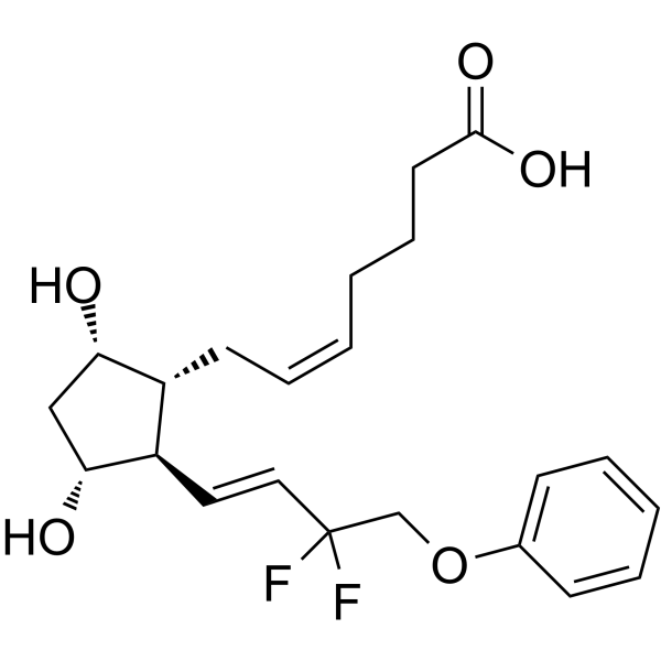
| SMILES |
C(=C/C[C@@H]1[C@@H](/C=C/C(COC2=CC=CC=C2)(F)F)[C@@H](C[C@@H]1O)O)/CCCC(=O)O
|
| InChi Key |
KIQXRQVVYTYYAZ-VKVYFNERSA-N
|
| InChi Code |
InChI=1S/C22H28F2O5/c23-22(24,15-29-16-8-4-3-5-9-16)13-12-18-17(19(25)14-20(18)26)10-6-1-2-7-11-21(27)28/h1,3-6,8-9,12-13,17-20,25-26H,2,7,10-11,14-15H2,(H,27,28)/b6-1-,13-12+/t17-,18-,19+,20-/m1/s1
|
| 化学名 |
(Z)-7-[(1R,2R,3R,5S)-2-[(E)-3,3-difluoro-4-phenoxybut-1-enyl]-3,5-dihydroxycyclopentyl]hept-5-enoic acid
|
| 别名 |
UNII-WTV8EPZ396; Tafluprost acid; 209860-88-8; AFP-172; Tafluprost (free acid); WTV8EPZ396; UNII-WTV8EPZ396; 5-Heptenoic acid, 7-[(1R,2R,3R,5S)-2-[(1E)-3,3-difluoro-4-phenoxy-1-buten-1-yl]-3,5-dihydroxycyclopentyl]-, (5Z)-; 5-Heptenoic acid, 7-((1R,2R,3R,5S)-2-((1E)-3,3-difluoro-4-phenoxy-1-buten-1-yl)-3,5-dihydroxycyclopentyl)-, (5Z)-;
|
| HS Tariff Code |
2934.99.9001
|
| 存储方式 |
Powder -20°C 3 years 4°C 2 years In solvent -80°C 6 months -20°C 1 month 注意: 本产品在运输和储存过程中需避光。 |
| 运输条件 |
Room temperature (This product is stable at ambient temperature for a few days during ordinary shipping and time spent in Customs)
|
| 溶解度 (体外实验) |
DMSO : ~100 mg/mL (~243.64 mM)
|
|---|---|
| 溶解度 (体内实验) |
注意: 如下所列的是一些常用的体内动物实验溶解配方,主要用于溶解难溶或不溶于水的产品(水溶度<1 mg/mL)。 建议您先取少量样品进行尝试,如该配方可行,再根据实验需求增加样品量。
注射用配方
注射用配方1: DMSO : Tween 80: Saline = 10 : 5 : 85 (如: 100 μL DMSO → 50 μL Tween 80 → 850 μL Saline)(IP/IV/IM/SC等) *生理盐水/Saline的制备:将0.9g氯化钠/NaCl溶解在100 mL ddH ₂ O中,得到澄清溶液。 注射用配方 2: DMSO : PEG300 :Tween 80 : Saline = 10 : 40 : 5 : 45 (如: 100 μL DMSO → 400 μL PEG300 → 50 μL Tween 80 → 450 μL Saline) 注射用配方 3: DMSO : Corn oil = 10 : 90 (如: 100 μL DMSO → 900 μL Corn oil) 示例: 以注射用配方 3 (DMSO : Corn oil = 10 : 90) 为例说明, 如果要配制 1 mL 2.5 mg/mL的工作液, 您可以取 100 μL 25 mg/mL 澄清的 DMSO 储备液,加到 900 μL Corn oil/玉米油中, 混合均匀。 View More
注射用配方 4: DMSO : 20% SBE-β-CD in Saline = 10 : 90 [如:100 μL DMSO → 900 μL (20% SBE-β-CD in Saline)] 口服配方
口服配方 1: 悬浮于0.5% CMC Na (羧甲基纤维素钠) 口服配方 2: 悬浮于0.5% Carboxymethyl cellulose (羧甲基纤维素) 示例: 以口服配方 1 (悬浮于 0.5% CMC Na)为例说明, 如果要配制 100 mL 2.5 mg/mL 的工作液, 您可以先取0.5g CMC Na并将其溶解于100mL ddH2O中,得到0.5%CMC-Na澄清溶液;然后将250 mg待测化合物加到100 mL前述 0.5%CMC Na溶液中,得到悬浮液。 View More
口服配方 3: 溶解于 PEG400 (聚乙二醇400) 请根据您的实验动物和给药方式选择适当的溶解配方/方案: 1、请先配制澄清的储备液(如:用DMSO配置50 或 100 mg/mL母液(储备液)); 2、取适量母液,按从左到右的顺序依次添加助溶剂,澄清后再加入下一助溶剂。以 下列配方为例说明 (注意此配方只用于说明,并不一定代表此产品 的实际溶解配方): 10% DMSO → 40% PEG300 → 5% Tween-80 → 45% ddH2O (或 saline); 假设最终工作液的体积为 1 mL, 浓度为5 mg/mL: 取 100 μL 50 mg/mL 的澄清 DMSO 储备液加到 400 μL PEG300 中,混合均匀/澄清;向上述体系中加入50 μL Tween-80,混合均匀/澄清;然后继续加入450 μL ddH2O (或 saline)定容至 1 mL; 3、溶剂前显示的百分比是指该溶剂在最终溶液/工作液中的体积所占比例; 4、 如产品在配制过程中出现沉淀/析出,可通过加热(≤50℃)或超声的方式助溶; 5、为保证最佳实验结果,工作液请现配现用! 6、如不确定怎么将母液配置成体内动物实验的工作液,请查看说明书或联系我们; 7、 以上所有助溶剂都可在 Invivochem.cn网站购买。 |
| 制备储备液 | 1 mg | 5 mg | 10 mg | |
| 1 mM | 2.4363 mL | 12.1815 mL | 24.3629 mL | |
| 5 mM | 0.4873 mL | 2.4363 mL | 4.8726 mL | |
| 10 mM | 0.2436 mL | 1.2181 mL | 2.4363 mL |
1、根据实验需要选择合适的溶剂配制储备液 (母液):对于大多数产品,InvivoChem推荐用DMSO配置母液 (比如:5、10、20mM或者10、20、50 mg/mL浓度),个别水溶性高的产品可直接溶于水。产品在DMSO 、水或其他溶剂中的具体溶解度详见上”溶解度 (体外)”部分;
2、如果您找不到您想要的溶解度信息,或者很难将产品溶解在溶液中,请联系我们;
3、建议使用下列计算器进行相关计算(摩尔浓度计算器、稀释计算器、分子量计算器、重组计算器等);
4、母液配好之后,将其分装到常规用量,并储存在-20°C或-80°C,尽量减少反复冻融循环。
计算结果:
工作液浓度: mg/mL;
DMSO母液配制方法: mg 药物溶于 μL DMSO溶液(母液浓度 mg/mL)。如该浓度超过该批次药物DMSO溶解度,请首先与我们联系。
体内配方配制方法:取 μL DMSO母液,加入 μL PEG300,混匀澄清后加入μL Tween 80,混匀澄清后加入 μL ddH2O,混匀澄清。
(1) 请确保溶液澄清之后,再加入下一种溶剂 (助溶剂) 。可利用涡旋、超声或水浴加热等方法助溶;
(2) 一定要按顺序加入溶剂 (助溶剂) 。
InvivoChem的所有产品仅用于作科学研究,不面向患者销售
Copyright 2020 InvivoChem LLC | All Rights Reserved 粤ICP备20063088号-1


 463611831
