| 规格 | 价格 | 库存 | 数量 |
|---|---|---|---|
| 10mg |
|
||
| 25mg |
|
||
| 50mg |
|
||
| 100mg |
|
||
| 250mg |
|
||
| 500mg |
|
||
| 1g |
|
||
| 5g |
|
||
| Other Sizes |
|
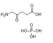
描述:5-氨基-4-氧代戊酸磷酸酯(氨基乙酰丙酸磷酸酯)是一种经FDA批准的局部用药,用于治疗光化性角化病。它是血红素生物合成的中间体,也是四吡咯的通用前体。
| 靶点 |
Endogenous Metabolite
|
|---|---|
| 体外研究 (In Vitro) |
研究人员正在探索5-氨基乙酰丙酸盐酸盐(ALA,一种非荧光前药)能否刺激恶性胶质瘤细胞生成荧光卟啉,从而用于术中肿瘤识别和切除。中位随访时间为35.4个月(95% CI 1.0-56.7)。139例接受5-氨基乙酰丙酸治疗的患者中,90例(65%)所有增强肿瘤均被完全切除;而131例接受白光治疗的患者中,仅有36例(36%)达到此效果(两组差异为29% [95% CI 17-40],p < 0.0001)。接受 5-氨基乙酰丙酸 (5-ALA) 治疗的患者 6 个月无进展生存率高于接受白光治疗的患者(41.0% [32.8-49.2] vs 21.1% [14.0-28.2];组间差异 19.9% [9.1-30.7],p= 0.0003,Z 检验)[1]。研究表明,单独使用 5-ALA 不足以在不增加术后神经功能恶化风险的情况下实现完全切除。此外,对于功能性 III 级胶质瘤,术中磁共振成像 (iMRI) 联合功能性神经导航明显优于 5-ALA 切除术 [2]。
|
| 体内研究 (In Vivo) |
尽管关于恶性胶质瘤细胞减灭术的争论仍在继续,但普遍认为肿瘤切除范围的扩大能够提高患者的总体生存率。然而,术中难以区分正常组织和病变组织,这阻碍了肿瘤切除范围的最大化。在此背景下,本研究探讨了两种成熟的肿瘤可视化方法:5-氨基乙酰丙酸(5-ALA)荧光引导手术和整合功能性神经导航的术中磁共振成像(iMRI),并将其作为一种双重术中可视化(DIV)方法。研究纳入了37例根据影像学表现疑似恶性胶质瘤(WHO III级或IV级)的患者。其中21例实验序列显示,5-ALA技术实现了肿瘤完全切除,并经iMRI证实。另有14例5-ALA技术显示肿瘤完全切除的序列未能被iMRI证实,iMRI检测到了残留肿瘤。进一步分析表明,这些残留肿瘤可被归类为功能性II级肿瘤(邻近重要脑区)。对于位于重要功能区附近的恶性胶质瘤亚组,荧光引导切除联合高场磁共振成像术中评估显著提高了肿瘤切除率,从 61.7% 提高到 100%;单独使用 5-氨基乙酰丙酸 (5-ALA) 不足以在不增加术后神经功能恶化风险的情况下实现肿瘤全切除。此外,对于功能性 III 级胶质瘤,术中磁共振成像联合功能性神经导航显著优于 5-ALA 切除技术。切除率可从 57.1% 提高到 71.2%,且不引起术后神经功能缺损[2]。
|
| 细胞实验 |
本研究检测了97例食管鳞状细胞癌(ESCC)患者病理标本中GPX4和HMOX1的表达,并进行了预后分析。采用实时聚合酶链式反应(RT-PCR)、RNA微阵列和蛋白质印迹分析评估了5-氨基乙酰丙酸(5-ALA)在体外铁死亡中的作用。Ann Surg Oncol. 2021 Jul;28(7):3996-4006. https://pubmed.ncbi.nlm.nih.gov/33210267/
|
| 动物实验 |
双术中可视化(DIV)方案[2]
术前立即进行肿瘤体积测量。随后仅使用5-ALA信号进行肿瘤切除,若无可见信号则判定切除彻底。此判断始终由主刀医生完成。间歇性地投射功能性神经导航数据,以防止意外损伤功能性脑区。在每个切除阶段结束时,系统地检查肿瘤腔,以排除残留肿瘤。一旦检测不到5-ALA信号,则进行术中磁共振成像(iMRI)扫描。如果确认切除范围,则由主刀医生决定结束手术。否则,重新分割残留肿瘤体积,并根据神经导航继续切除。在所有这些情况下,一旦去除薄薄的“健康”脑实质层和/或随后优化观察角度,即可在后续手术中重新检测到5-ALA信号。重复上述步骤直至检测不到 5-ALA 信号,且术中磁共振成像 (iMRI) 证实无对比增强肿瘤。iMRI 检测到的额外切除组织也由经验丰富的神经病理学家进行分析,证实存在病理性胶质瘤细胞浸润。如果神经导航数据显示功能区域内仍存在 5-ALA,则有意终止相应方向的进一步手术。此外,本研究还使用了铁死亡抑制剂 ferrostatin-1 和脂质过氧化试剂来对抗经 5-ALA 处理的细胞系。最后,通过 5-ALA 对食管鳞状细胞癌 (ESCC) 皮下异种移植小鼠模型的影响,证实了 5-ALA 的作用。Ann Surg Oncol. 2021 Jul;28(7):3996-4006. https://pubmed.ncbi.nlm.nih.gov/33210267/ |
| 药代性质 (ADME/PK) |
吸收
口服生物利用度为50-60%。### 外用凝胶 在一项纳入12名患有轻度至中度日光性角化病(AK)且面部或前额至少有10处AK病变的成年受试者的试验中,评估了氨基乙酰丙酸(ALA)和原卟啉IX(PpIX)的药代动力学(PK)。受试者在封闭回路条件下接受单次涂抹一管ALA(2克),持续3小时,随后进行光动力疗法(PDT),总覆盖面积为20平方厘米。基线血浆ALA和PpIX浓度的平均值±标准差分别为20.16±16.53 ng/mL和3.27±2.40 ng/mL。在大多数受试者中,ALA涂抹后3小时内血浆ALA浓度升高至2.5倍。基线校正后,ALA(n=12)的平均±标准差浓度-时间曲线下面积(AUC0-t)和最大浓度(Cmax)分别为142.83±75.50 ng·h/mL和27.19±20.02 ng/mL。达到Cmax的中位时间(Tmax)为3小时。### 外用溶液 两项人体药代动力学(PK)研究在患有轻度至中度上肢日光性角化病的受试者中进行,受试者一侧上肢至少有6处病灶,另一侧上肢至少有12处病灶。单剂量方案包括两次将ALA溶液(每次含354 mg ALA HCl)直接涂抹于病灶部位,随后进行3小时的封闭治疗,之后进行光疗。第一项PK研究纳入了29名受试者,并评估了ALA的PK参数。基线校正后的ALA最大浓度(Cmax)平均值±标准差为249.9±694.5 ng/mL,达峰时间(Tmax)中位数为给药后2小时。ALA的平均暴露量(以浓度-时间曲线下面积(AUCt)表示)为669.9±1610 ng·hr/mL。ALA的平均消除半衰期(t1/2)为5.7±3.9小时。在14名受试者中进行了第二次药代动力学(PK)研究,并测定了ALA和PpIX的PK参数。在50%(7/14)的受试者中,至少50%的样本中基线校正后的PpIX浓度为负值,因此无法可靠地估计AUC。经基线校正后,ALA 和 PpIX 的 Cmax 平均值 ± 标准差分别为 95.6 ± 120.6 ng/mL 和 0.95 ± 0.71 ng/mL。ALA 和 PpIX 的达峰时间 (Tmax) 中位数分别为给药后 2 小时和 12 小时。ALA 的平均 AUCt 为 261.1 ± 229.3 ng·hr/mL。ALA 的平均半衰期 (t1/2) 为 8.5 ± 6.7 小时。### 口服溶液 在 12 名健康受试者中,服用推荐剂量的 ALA 溶液后,ALA 的绝对生物利用度为 100.0% ± 1.1,范围为 78.5% 至 131.2%。ALA 的血浆峰浓度中位数为 0.8 小时(范围 0.5–1.0 小时)。 排泄途径 在12名健康受试者中,给予推荐剂量的氨基乙酰丙酸(ALA)溶液后12小时内,母体ALA经尿液排泄率为34 ± 8%(平均值±标准差),范围为27%至57%。 分布容积 在健康志愿者中,静脉注射氨基乙酰丙酸的分布容积为9.3 ± 2.8 L,口服为14.5 ± 2.5 L。[11961050] 代谢/代谢物 外源性氨基乙酰丙酸(ALA)代谢为原卟啉IX(PpIX),但ALA代谢为PpIX的比例尚不清楚。PpIX的平均血浆AUC不到ALA的6%。局部用药后,药物在皮肤内原位合成原卟啉IX。半衰期:口服给药后的平均半衰期为0.70 ± 0.18小时,静脉给药后的平均半衰期为0.83 ± 0.05小时。生物半衰期:局部应用氨基乙酰丙酸溶液的平均消除半衰期(t1/2)为5.7 ± 3.9小时,口服溶液的平均半衰期为0.9 ± 1.2小时。在另一项涉及6名健康志愿者的药代动力学研究中,使用128 mg剂量,口服给药后的平均半衰期为0.70 ± 0.18小时,静脉给药后的平均半衰期为0.83 ± 0.05小时。 |
| 毒性/毒理 (Toxicokinetics/TK) |
毒性概述
基于推测的作用机制,局部应用氨基乙酰丙酸 (ALA) 溶液后,ALA 代谢为原卟啉 IX (PpIX) 而产生光敏作用。PpIX 会在应用氨基乙酰丙酸的皮肤部位积聚。当暴露于适当波长和能量的光时,积聚的 PpIX 会发生光动力反应,这是一个依赖于光和氧同时存在的细胞毒性过程。光吸收导致卟啉分子处于激发态,随后 PpIX 自旋向分子氧转移生成单线态氧,单线态氧可进一步反应生成超氧阴离子和羟基自由基。氨基乙酰丙酸用于光化性角化病病变的光敏化,并结合使用 BLU-UTM 蓝光光动力治疗仪 (BLU-U) 进行照射,构成了氨基乙酰丙酸光动力疗法 (PDT) 的基础。 妊娠期和哺乳期影响 ◉ 哺乳期用药概述 目前尚无关于哺乳期口服氨基乙酰丙酸的信息。为尽量减少婴儿接触,口服给药后应暂停哺乳24小时。由于全身吸收率极低,预计哺乳不会导致婴儿局部接触氨基乙酰丙酸。氨基乙酰丙酸诱导的光动力疗法已成功用于治疗各种乳头皮肤损伤。该疗法似乎有助于维持乳头解剖结构,从而促进母乳喂养。 ◉ 对母乳喂养婴儿的影响 截至修订日期,未找到相关的已发表信息。 ◉ 对母乳喂养和母乳的影响 截至修订日期,未找到相关的已发表信息。 给药途径 口服生物利用度为50-60%。 蛋白结合 在体外研究中,当氨基乙酰丙酸 (ALA) 的浓度达到推荐剂量下 ALA 溶液最大血浆浓度的约 25% 时,ALA 的平均蛋白结合率为 12%。 在一项随机对照多中心 III 期试验(全分析人群,n=270)中,接受 5-氨基-4-氧戊酸磷酸酯(口服,20 mg/kg 体重)治疗的患者与接受白光常规显微手术的患者相比,术后 7 天内报告的严重不良事件或任何器官系统类别不良事件的发生率无差异。5-氨基-4-氧戊酸磷酸酯 组 (n=139) 与白光组 (n=131) 中最常见的早期严重不良事件为:偏瘫(4 例 [3%] vs 2 例 [2%],p=0.7);失语症(3例[2%] vs 0例,p=0.1);惊厥(3例[2%] vs 1例[1%],p=0.6);以及硬膜外血肿(1例[1%] vs 1例[1%],p=1.0)。所有接受5-氨基-4-氧戊酸磷酸酯治疗的患者中报告的失语症均在基线时存在,但在术后加重。30天时,5-氨基-4-氧戊酸磷酸酯组有5例(4%)患者死亡,白光组有3例(2%)患者死亡(差异1.3% [95% CI -3.4至6.2])。 [1] 在安全性人群(n=289,其中158例在5-氨基-4-氧代戊酸磷酸酯组,131例在白光组)中,除术后24小时外,各组实验室指标均无显著差异。术后24小时,5-氨基-4-氧代戊酸磷酸酯组的γ-谷氨酰转肽酶中位数为正常值上限的0.93倍(范围0.18-9.71),而白光组为0.72倍(0.24-8.43)(p=0.05)。 5-氨基-4-氧戊酸磷酸酯组和白光组的丙氨酸氨基转移酶中位数分别为正常值上限的1.05倍(0.32-8.53)和0.84倍(0.18-7.36)(p=0.003),天冬氨酸氨基转移酶中位数分别为正常值上限的0.72倍(0.22-5.60)和0.53倍(0.11-3.56)(p<0.0001)。[1] 在III期临床试验中,两组患者术后6周的Karnofsky评分中位数均为90分(5-氨基-4-氧戊酸磷酸酯组患者评分范围为20-100分,白光组患者评分范围为10-100分)。 6个月时,5-氨基-4-氧代戊酸磷酸酯组中有28%(95% CI 19-36)的患者Karnofsky功能状态评分下降至60分或以下,而白光组中这一比例为31%(20-42)(log-rank检验p=0.5)。两组患者采用改良版NIH卒中量表评估的神经功能障碍程度无显著差异。[1] |
| 参考文献 |
[1]. Stummer, W., et al., Fluorescence-guided surgery with 5-aminolevulinic acid for resection of malignant glioma: a randomised controlled multicentre phase III trial. Lancet Oncol, 2006. 7(5): p. 392-401.
[2]. Eyupoglu, I.Y., et al., Improving the extent of malignant glioma resection by dual intraoperative visualization approach. PLoS One, 2012. 7(9): p. e44885. |
| 其他信息 |
5-氨基乙酰丙酸盐是5-氨基乙酰丙酸的单盐酸盐。它代谢生成原卟啉IX,一种可在皮肤中积聚的光敏化合物。它与蓝光照射联合使用,用于治疗面部或头皮的轻度至中度日光性角化病。它具有抗肿瘤药物、光敏剂、皮肤科药物和前药的双重作用。它含有5-氨基乙酰丙酸。
氨基乙酰丙酸盐是氨基乙酰丙酸(一种氨基酮)的盐酸盐形式,用于局部光敏治疗。氨基乙酰丙酸(ALA)是一种代谢前药,可转化为光敏剂原卟啉IX(PpIX),后者可在细胞内积聚。当暴露于适当波长(红光或蓝光)的光下时,PpIX 会催化单线态氧的生成,单线态氧是一种内毒素,可进一步反应生成超氧阴离子和羟基自由基。这会导致细胞毒性作用。 PpIX 是血红素合成过程中由琥珀酰辅酶A和甘氨酸产生的中间体化合物。它被用作光化学疗法治疗日光性角化病。 药物适应症 Gliolan 适用于成人患者,用于在恶性胶质瘤(WHO III 级和 IV 级)手术中可视化恶性组织。 用于治疗成人面部和头皮的轻度至中度日光性角化病(Olsen 1 级至 2 级;参见 5.1 节),以及癌变区域。本研究旨在探索使用5-氨基乙酰丙酸治疗不适合手术治疗的浅表性和/或结节性基底细胞癌成年患者的治疗方案,这些患者可能由于潜在的治疗相关并发症和/或美容效果不佳而无法接受手术治疗。 背景:5-氨基乙酰丙酸是一种非荧光前药,可导致荧光卟啉在恶性胶质瘤细胞内积聚——这一发现已被用于术中识别和切除这些肿瘤。本研究旨在评估使用5-氨基乙酰丙酸进行荧光引导切除对肿瘤根治性、无进展生存期、总生存期和并发症的影响。方法:322例年龄在23至73岁之间、疑似恶性胶质瘤且适合进行完全增强肿瘤切除的患者被随机分为两组:一组接受荧光引导下5-氨基乙酰丙酸(5-ALA,20 mg/kg体重)切除术(n=161),另一组接受常规白光显微手术(n=161)。主要终点为早期MRI(即术后72小时内进行的MRI检查)无增强的患者人数以及MRI评估的6个月无进展生存期。次要终点包括术后MRI的残余肿瘤体积、总生存期、神经功能缺损和毒性反应。我们报告了一项中期分析的结果,该分析纳入了270例患者(139例接受5-ALA治疗,131例接受白光治疗),排除了由中心审核员(不知晓治疗分配情况)评估的组织学和放射学结果不符合纳入标准的患者。根据方案,中期分析结果导致研究终止。主要终点和次要终点均采用意向性治疗分析方法在全分析人群中进行分析。本研究已在http://www.clinicaltrials.gov注册,注册号为NCT00241670。结果:中位随访时间为35.4个月(95% CI 1.0–56.7)。在接受5-氨基乙酰丙酸治疗的139例患者中,90例(65%)增强肿瘤被完全切除;相比之下,在接受白光治疗的131例患者中,仅有47例(36%)肿瘤被完全切除(组间比较:29% [95% CI 17–40],p<0.0001)。接受 5-氨基乙酰丙酸治疗的患者 6 个月无进展生存期长于接受白光治疗的患者(41.0% [32.8–49.2] vs 21.1% [14.0–28.2];组间差异:19.9% [9.1–30.7],p=0.0003,Z 检验)。两组患者术后 7 天内报告的严重不良事件或任何器官系统不良事件的发生率均无差异。结论:5-氨基乙酰丙酸衍生的肿瘤荧光可以更彻底地清除增强肿瘤,从而提高恶性胶质瘤患者的无进展生存期。[1] 背景:5-氨基乙酰丙酸 (5-ALA) 是一种天然氨基酸,因其具有肿瘤特异性代谢途径特征而被广泛用于癌症治疗。本研究表明,5-氨基乙酰丙酸(5-ALA)通过谷胱甘肽过氧化物酶4(GPX4)和血红素加氧酶1(HMOX1)诱导铁死亡,并在食管鳞状细胞癌(ESCC)中发挥抗肿瘤作用。方法:检测97例ESCC患者病理标本中GPX4和HMOX1的表达,并进行预后分析。采用实时荧光定量PCR(RT-PCR)、RNA芯片和Western blot分析评估5-ALA在体外铁死亡中的作用。此外,本研究还使用铁死亡抑制剂ferrostatin-1和脂质过氧化试剂检测了5-ALA处理的细胞系。最后,通过皮下异种移植食管鳞状细胞癌(ESCC)小鼠模型进一步证实了5-ALA的作用。结果表明,GPX4上调和HMOX1下调是食管鳞状细胞癌(ESCC)预后不良的因素。在KYSE30细胞的RNA芯片分析中,铁死亡是最常被诱导的通路之一;5-ALA处理抑制了GPX4的表达并上调了HMOX1的表达。这些结果通过RT-PCR和Western blotting实验得到了验证。此外,5-ALA可导致脂质过氧化增加,并在多种癌细胞系中发挥抗肿瘤作用,而这种作用可被铁死亡抑制剂ferrostatin-1抑制。体内实验表明,5-ALA抑制肿瘤组织中GPX4的表达并上调HMOX1的表达,从而导致肿瘤体积缩小。结论:5-ALA通过调节GPX4和HMOX1的表达诱导食管鳞状细胞癌(ESCC)细胞发生铁死亡。因此,5-ALA 可能是一种有前景的食管鳞状细胞癌(ESCC)新型治疗药物。参考文献:Ann Surg Oncol. 2021 Jul;28(7):3996-4006. 5-氨基-4-氧代戊酸磷酸酯(5-氨基乙酰丙酸)用于荧光引导下恶性胶质瘤切除术。在一项随机对照III期临床试验(NCT00241670)中,疑似恶性胶质瘤患者在麻醉诱导前2-4小时口服20 mg/kg体重的5-氨基乙酰丙酸。荧光引导手术使65%的患者在术后早期MRI检查中实现了增强肿瘤的完全切除(白光引导组为36%,p<0.0001),并提高了6个月无进展生存率(41.0% vs 21.1%,p=0.0003)。在5-氨基-4-氧戊酸磷酸酯组中,中位无进展生存期为5.1个月(95% CI 3.4-6.0),而白光组为3.6个月(3.2-4.4)。该研究得出结论,5-氨基-4-氧戊酸磷酸酯产生的肿瘤荧光能够实现更彻底的切除,从而改善无进展生存期。[1] 在一项前瞻性研究中,37例恶性胶质瘤患者接受了5-氨基-4-氧戊酸磷酸酯(麻醉前3小时口服20 mg/kg)治疗,用于荧光引导手术,并结合术中磁共振成像(iMRI)作为双重术中可视化(DIV)方法。对于功能性 II 级肿瘤(邻近重要脑区),单独使用 5-ALA 可达到 71.7% 的切除率,而联合术中磁共振成像 (iMRI) 可将切除率提高至 100% (p<0.002)。对于功能性 III 级肿瘤(位于重要脑区),单独使用 5-ALA 可达到 57.6% 的切除率,而联合术中磁共振成像 (DIV) 可将其提高至 71.2% (p<0.05)。该联合疗法显著提高了肿瘤全切除率,且未造成术后神经功能缺损,尤其适用于邻近重要脑区的肿瘤。[2] |
| 分子式 |
C5H9NO3.H3O4P
|
|---|---|
| 分子量 |
229.12508
|
| 精确质量 |
229.035
|
| 元素分析 |
C, 26.21; H, 5.28; N, 6.11; O, 48.88; P, 13.52
|
| CAS号 |
868074-65-1
|
| 相关CAS号 |
5451-09-2 (HCl);106-60-5 (free);868074-65-1 (phosphate);
|
| PubChem CID |
24737828
|
| 外观&性状 |
Typically exists as solid at room temperature
|
| tPSA |
167.96
|
| 氢键供体(HBD)数目 |
5
|
| 氢键受体(HBA)数目 |
8
|
| 可旋转键数目(RBC) |
4
|
| 重原子数目 |
14
|
| 分子复杂度/Complexity |
171
|
| 定义原子立体中心数目 |
0
|
| SMILES |
O=C(CCC(CN)=O)O.O=P(O)(O)O
|
| InChi Key |
XWNWBYZHOAIHTK-UHFFFAOYSA-N
|
| InChi Code |
InChI=1S/C5H9NO3.H3O4P/c6-3-4(7)1-2-5(8)9;1-5(2,3)4/h1-3,6H2,(H,8,9);(H3,1,2,3,4)
|
| 化学名 |
5-amino-4-oxopentanoic acid;phosphoric acid
|
| 别名 |
Aminolevulinic acid phosphate; 868074-65-1; 5-Aminolevulinic acid phosphate; Pentanoic acid, 5-amino-4-oxo-, phosphate (1:1); UNII-FM8DCR39GH; delta-Aminolevulinic acid phosphate; FM8DCR39GH; Aminolevulinic acid phosphate [INCI];
|
| HS Tariff Code |
2934.99.9001
|
| 存储方式 |
Powder -20°C 3 years 4°C 2 years In solvent -80°C 6 months -20°C 1 month |
| 运输条件 |
Room temperature (This product is stable at ambient temperature for a few days during ordinary shipping and time spent in Customs)
|
| 溶解度 (体外实验) |
May dissolve in DMSO (in most cases), if not, try other solvents such as H2O, Ethanol, or DMF with a minute amount of products to avoid loss of samples
|
|---|---|
| 溶解度 (体内实验) |
注意: 如下所列的是一些常用的体内动物实验溶解配方,主要用于溶解难溶或不溶于水的产品(水溶度<1 mg/mL)。 建议您先取少量样品进行尝试,如该配方可行,再根据实验需求增加样品量。
注射用配方
注射用配方1: DMSO : Tween 80: Saline = 10 : 5 : 85 (如: 100 μL DMSO → 50 μL Tween 80 → 850 μL Saline)(IP/IV/IM/SC等) *生理盐水/Saline的制备:将0.9g氯化钠/NaCl溶解在100 mL ddH ₂ O中,得到澄清溶液。 注射用配方 2: DMSO : PEG300 :Tween 80 : Saline = 10 : 40 : 5 : 45 (如: 100 μL DMSO → 400 μL PEG300 → 50 μL Tween 80 → 450 μL Saline) 注射用配方 3: DMSO : Corn oil = 10 : 90 (如: 100 μL DMSO → 900 μL Corn oil) 示例: 以注射用配方 3 (DMSO : Corn oil = 10 : 90) 为例说明, 如果要配制 1 mL 2.5 mg/mL的工作液, 您可以取 100 μL 25 mg/mL 澄清的 DMSO 储备液,加到 900 μL Corn oil/玉米油中, 混合均匀。 View More
注射用配方 4: DMSO : 20% SBE-β-CD in Saline = 10 : 90 [如:100 μL DMSO → 900 μL (20% SBE-β-CD in Saline)] 口服配方
口服配方 1: 悬浮于0.5% CMC Na (羧甲基纤维素钠) 口服配方 2: 悬浮于0.5% Carboxymethyl cellulose (羧甲基纤维素) 示例: 以口服配方 1 (悬浮于 0.5% CMC Na)为例说明, 如果要配制 100 mL 2.5 mg/mL 的工作液, 您可以先取0.5g CMC Na并将其溶解于100mL ddH2O中,得到0.5%CMC-Na澄清溶液;然后将250 mg待测化合物加到100 mL前述 0.5%CMC Na溶液中,得到悬浮液。 View More
口服配方 3: 溶解于 PEG400 (聚乙二醇400) 请根据您的实验动物和给药方式选择适当的溶解配方/方案: 1、请先配制澄清的储备液(如:用DMSO配置50 或 100 mg/mL母液(储备液)); 2、取适量母液,按从左到右的顺序依次添加助溶剂,澄清后再加入下一助溶剂。以 下列配方为例说明 (注意此配方只用于说明,并不一定代表此产品 的实际溶解配方): 10% DMSO → 40% PEG300 → 5% Tween-80 → 45% ddH2O (或 saline); 假设最终工作液的体积为 1 mL, 浓度为5 mg/mL: 取 100 μL 50 mg/mL 的澄清 DMSO 储备液加到 400 μL PEG300 中,混合均匀/澄清;向上述体系中加入50 μL Tween-80,混合均匀/澄清;然后继续加入450 μL ddH2O (或 saline)定容至 1 mL; 3、溶剂前显示的百分比是指该溶剂在最终溶液/工作液中的体积所占比例; 4、 如产品在配制过程中出现沉淀/析出,可通过加热(≤50℃)或超声的方式助溶; 5、为保证最佳实验结果,工作液请现配现用! 6、如不确定怎么将母液配置成体内动物实验的工作液,请查看说明书或联系我们; 7、 以上所有助溶剂都可在 Invivochem.cn网站购买。 |
| 制备储备液 | 1 mg | 5 mg | 10 mg | |
| 1 mM | 4.3643 mL | 21.8217 mL | 43.6433 mL | |
| 5 mM | 0.8729 mL | 4.3643 mL | 8.7287 mL | |
| 10 mM | 0.4364 mL | 2.1822 mL | 4.3643 mL |
1、根据实验需要选择合适的溶剂配制储备液 (母液):对于大多数产品,InvivoChem推荐用DMSO配置母液 (比如:5、10、20mM或者10、20、50 mg/mL浓度),个别水溶性高的产品可直接溶于水。产品在DMSO 、水或其他溶剂中的具体溶解度详见上”溶解度 (体外)”部分;
2、如果您找不到您想要的溶解度信息,或者很难将产品溶解在溶液中,请联系我们;
3、建议使用下列计算器进行相关计算(摩尔浓度计算器、稀释计算器、分子量计算器、重组计算器等);
4、母液配好之后,将其分装到常规用量,并储存在-20°C或-80°C,尽量减少反复冻融循环。
计算结果:
工作液浓度: mg/mL;
DMSO母液配制方法: mg 药物溶于 μL DMSO溶液(母液浓度 mg/mL)。如该浓度超过该批次药物DMSO溶解度,请首先与我们联系。
体内配方配制方法:取 μL DMSO母液,加入 μL PEG300,混匀澄清后加入μL Tween 80,混匀澄清后加入 μL ddH2O,混匀澄清。
(1) 请确保溶液澄清之后,再加入下一种溶剂 (助溶剂) 。可利用涡旋、超声或水浴加热等方法助溶;
(2) 一定要按顺序加入溶剂 (助溶剂) 。
 麦芽酚铁
麦芽酚铁
 1-Methyl-6-oxo-1,6-dihydropyridine-3-carboxylic acid
 阿托伐他汀丙酮叔丁酯
 7,4'-Dihydroxyflavone
InvivoChem的所有产品仅用于作科学研究,不面向患者销售
Copyright 2020 InvivoChem LLC | All Rights Reserved 粤ICP备20063088号-1


 COA
COA
 463611831
