| 规格 | 价格 | |
|---|---|---|
| 500mg | ||
| 1g | ||
| Other Sizes |
| 靶点 |
TACC3/transforming acidic coiled-coil-containing protein 3
|
|---|---|
| 体外研究 (In Vitro) |
贴壁培养中的 NPC 神经元可以剂量依赖性方式吸附 KHS101 (EC50 ~ 1 μM)[1]。 KHS101 (5 μM) 刺激 NPC 星形胶质细胞的产生 [1]。 KHS101 (5 μM) 和 KHS101 (0-15 μM; 24 h) 对 NPC 细胞周期的负荷和进展的影响是不利的 [1]。 KHS101 和 TACC3 的蛋白质电位反应 [1]。 KHS101(0-15 μM;24 小时)调节器 ARNT2 核定位 [RT-PCR[1]
|
| 体内研究 (In Vivo) |
KHS101(6 mg/kg;皮下注射;BID 14 天)在体内分布于大脑中,显着促进神经元分泌[1]。
|
| 酶活实验 |
基于亲和力的目标识别。[1]
通过在PBS中超声处理制备NPC裂解物,并以2 mg/mL的浓度制备蛋白质样品。将二苯甲酮-KHS101化合物(KHS101-BP,5μM;SI Text)加入到50μL的蛋白质组反应中,有和没有未标记的化合物(250μM)。使用长波长(365nm)的手持紫外灯照射1小时,随后进行铜催化的叠氮-炔烃环加成反应(SI Text)。在室温下孵育1小时后,使用三氯乙酸沉淀蛋白质,并将其重新悬浮在等电聚焦样品缓冲液中。按照制造商的方案,使用ReadyStripe IPG条进行2D SDS-PAGE。 基于亲和力的目标识别[2] GBM1细胞在有或没有未标记的KHS101(250μM)的情况下与KHS101-BP(5μM)一起孵育30分钟,并用紫外光(365nm)照射30分钟。使用0.5%Triton X-100和蛋白酶抑制剂混合物裂解细胞。细胞裂解物与25μM叠氮化生物素、1 mM TCEP、100 mM配体(TBTA)和1 mM硫酸铜水溶液在4°C下孵育过夜。随后,使用硫酸铵对蛋白质进行分级,并对20-40%的级分进行2D SDS-PAGE。使用Abcam通过蛋白质印迹检测生物素标记的蛋白质;ab1227)。在平行凝胶上用银染色显示与特定生物素标记的蛋白质相对应的蛋白质斑点。切除一个明显的斑点,并使用液相色谱-串联质谱法鉴定蛋白质。对于HSPD1相互作用确认试验,将总共1μg重组HSPD1稀释在1 mL PBS(含2 mM MgCl2、2 mM DDT和0.1%吐温20)中,并在有或没有未标记的KHS101的情况下,在4°C下与5μM生物素化的KHS101一起孵育过夜。将链霉抗生物素蛋白琼脂糖珠加入孵育混合物中,并在4°C下旋转2小时。然后沉淀珠粒,在PBS中洗涤三次。用2x SDS样品缓冲液洗脱结合的蛋白质,用SDS-PAGE分析,然后进行银染和蛋白质印迹。 |
| 细胞实验 |
RT-PCR[1]
细胞类型:大鼠 NPC 测试浓度: 0.6、1.7 和 5 μM 孵育时间: 24 小时 实验结果: 显示 Cdkn1 mRNA 表达的剂量依赖性诱导。 1]。 细胞增殖测定 [1] 细胞类型:大鼠 NPC 测试浓度: 5 μM 孵育时间:24、48和72小时 实验结果:绝大多数NPC在72小时内停止增殖并变为非有丝分裂。 对于分化试验,将细胞以每孔5000个细胞的密度接种到96孔板中,并用100ng/mL的重组人BMP4处理4天。随后,在100μL培养基中用DMSO(0.1%)或KHS101(1-20μM)处理细胞48小时,并根据制造商的说明进行CellTiter-Glo测定。2. 对于集落形成试验,以125个细胞/孔的密度(在24孔板中)接种细胞并使其粘附。第二天,计数每孔的单细胞,并用DMSO或KHS101处理。10天后计数由>6个细胞组成的集落,并确定能够形成集落的细胞百分比。2. 对于活细胞分析,在加入KHS101(7.5μM)或DMSO(0.1%)之前,让细胞生长2天,随后监测3天。使用IncuCyte ZOOM活细胞成像系统以45分钟的间隔采集图像。2. 为了分析细胞活力和半胱天冬酶3/7活化,将细胞分别以10000和2500个细胞的密度接种到96孔板中。第二天,用载体(DMSO)、KHS101、KHS101/Z-VAD-FMK(20μM)或Staurosporine在100μL培养基中以指定浓度处理细胞。CellTiter Glo和Caspase Glo 3/7测定 根据制造商的说明在指定时间点进行。2. 为了使用膜联蛋白V和碘化丙啶定量凋亡,GBM1细胞用KHS101(7.5μM)、巴非霉素A1(10 nM)或载体(DMSO,0.1%)处理48小时,然后用胰蛋白酶收获,用PBS洗涤,并根据制造商的方案在37°C下用膜联蛋白V-荧光素染色试剂盒用膜联素V和碘化丙啶染色15分钟。使用NC3000细胞仪通过象限门控定量标记的早期凋亡和晚期凋亡/坏死细胞[2]。 |
| 动物实验 |
动物/疾病模型:成年 Fisher 344 大鼠(约 10 周龄)[1] 用法用量:6 mg/kg
给药途径:皮下注射,每日两次,持续 14 天 实验结果:神经元分化增加。神经祖细胞增殖减少。 动物实验。[1] 为了研究 KHS101 的药代动力学特性,雄性 Sprague-Dawley 大鼠分别静脉注射或皮下注射 3 mg/kg KHS101。在给药后 5 分钟、40 分钟、1 小时和 3 小时,每个时间点处死一只大鼠,并采集血液样本(100 μL)和全脑组织。在另一项研究中,大鼠分别静脉注射或皮下注射 6 mg/kg KHS101。通过颈静脉导管连续采集 5 份血样,每份 100 μL,分别于给药后 2 分钟(仅静脉注射)、0.5 小时(仅皮下注射)以及 1、3、7 和 24 小时采集。采用液相色谱串联质谱法 (LC-MS/MS) 分析血浆和匀浆化的全脑样本。为了研究体内 KHS101 给药后神经元分化情况,成年 Fisher 344 大鼠(约 10 周龄)皮下注射 6 mg/kg KHS101 或载体对照(5% 乙醇溶于 15% Captisol)。所有大鼠在给药后连续 6 天,每天腹腔注射 200 mg/kg BrdU。 14天后,处死动物并进行灌注固定,取出脑组织进行免疫组织化学分析。 异种移植瘤实验[2] 动物实验在英国项目许可批准和机构指南的指导下进行。动物饲养于标准条件下(12小时昼夜循环,自由摄食饮水)。实验分别使用6至8周龄的NOD scid gamma (NSG)小鼠和BALB/c裸鼠建立GBM1和GBMX1模型。将2×10⁵个GBM1细胞或8×10⁴个GBMX1细胞,以2 μL体积(含30% Matrigel)立体定位注射到小鼠右侧纹状体(距中线2.5 mm,距前囟2.5 mm,深度3 mm)。手术在全身麻醉下进行,并严格遵守无菌操作技术。每日监测小鼠的疾病、疼痛或体重减轻等症状。在指定的肿瘤建立期后,皮下注射(sc)6 mg/kg KHS101 或载体对照(5% (v/v) 乙醇,15% (w/v) (2-羟丙基)-β-环糊精),每日两次,每两周交替给药 5 天和 3 天。实验在指定终点结束,并对组织进行免疫组织化学和图像分析。基于亲和力的靶标鉴定。[1] 通过在 PBS 中超声处理制备 NPC 裂解液,并将蛋白质样品制备至 2 mg/mL 的浓度。将二苯甲酮-KHS101化合物(KHS101-BP,5 μM;参见补充信息)加入到50 μL蛋白质组反应体系中,反应体系中分别加入或不加入未标记化合物(250 μM)。使用手持式紫外灯在长波长(365 nm)下照射1小时,随后进行铜催化的叠氮-炔环加成反应(参见补充信息)。室温孵育1小时后,用三氯乙酸沉淀蛋白质,并将沉淀物重悬于等电聚焦样品缓冲液中。使用 ReadyStripe IPG 条带,按照制造商的方案进行二维 SDS-PAGE 电泳。 基于亲和力的靶标鉴定 [2] 将 GBM1 细胞与 KHS101-BP (5 μM) 在有或无未标记的 KHS101 (250 μM) 的情况下孵育 30 分钟,然后用紫外光 (365 nm) 照射 30 分钟。使用 0.5% Triton X-100 和蛋白酶抑制剂混合物裂解细胞。将细胞裂解液与 25 μM 生物素叠氮化物、1 mM TCEP、100 mM 配体 (TBTA) 和 1 mM 硫酸铜水溶液在 4°C 下孵育过夜。随后,使用硫酸铵分离蛋白质,并将 20-40% 的组分进行二维 SDS-PAGE 电泳。使用 Abcam 公司的 ab1227 试剂盒,通过蛋白质印迹法检测生物素标记的蛋白质。在平行凝胶上,用银染法显影与特定生物素标记蛋白质对应的蛋白斑点。切取一个清晰的斑点,并使用液相色谱串联质谱法鉴定蛋白质。为了验证 HSPD1 的相互作用,将 1 μg 重组 HSPD1 稀释于 1 mL PBS 缓冲液(含 2 mM MgCl₂、2 mM DDT 和 0.1% Tween 20)中,并与 5 μM 生物素标记的 KHS101 在 4°C 下孵育过夜,孵育过程中分别加入或不加入未标记的 KHS101。向孵育混合物中加入链霉亲和素琼脂糖珠,并在 4°C 下旋转孵育 2 小时。然后沉淀琼脂糖珠,并用 PBS 缓冲液洗涤三次。用 2x SDS 样品缓冲液洗脱结合的蛋白质,然后进行 SDS/PAGE 分析,再进行银染和蛋白质印迹分析。 |
| 参考文献 | |
| 其他信息 |
成年神经发生发生在哺乳动物体内,为大脑持续的神经可塑性提供了一种机制。然而,人们对调控海马神经祖细胞(NPCs)的分子机制知之甚少,也不清楚是否可以通过药物调控其命运来改善神经可塑性和再生能力。本文报道了一种小分子(KHS101)的特性,该分子能够选择性地诱导神经元分化表型。作用机制研究表明,KHS101与细胞周期退出以及与TACC3蛋白的特异性结合有关,敲低NPCs中的TACC3蛋白可以重现KHS101诱导的表型。全身给药后,KHS101分布到大脑,并在体内显著促进了神经元分化。我们的研究结果表明,KHS101通过与TACC3相互作用加速神经元分化,这可能为针对内源性神经祖细胞(NPC)的药物干预提供基础。[1]
使用小分子抑制剂抑制不受控制的细胞生长是治疗多形性胶质母细胞瘤(GBM,最恶性的原发性脑癌)的潜在策略。我们发现,合成小分子KHS101在多种GBM细胞模型中均能促进肿瘤细胞死亡,且与肿瘤亚型无关,并且不影响非癌性脑细胞系的活力。KHS101通过破坏线粒体分子伴侣热休克蛋白D家族成员1(HSPD1)发挥细胞毒性作用。在GBM细胞中,KHS101促进了调节线粒体完整性和能量代谢的蛋白质聚集。KHS101处理的GBM细胞的线粒体生物能量能力和糖酵解活性受到选择性损害。在两种小鼠颅内患者来源的异种移植肿瘤模型中,全身性给予 KHS101 可减少肿瘤生长并延长生存期,且无明显副作用。这些发现表明,靶向 HSPD1 依赖性代谢途径可能是治疗 GBM 的有效策略。[2] |
| 分子式 |
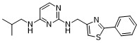
C18H21N5S
|
|---|---|
| 分子量 |
339.461
|
| 精确质量 |
339.152
|
| 元素分析 |
C, 63.69; H, 6.24; N, 20.63; S, 9.44
|
| CAS号 |
1262770-73-9
|
| 相关CAS号 |
KHS101 hydrochloride;1784282-12-7
|
| PubChem CID |
71304818
|
| 外观&性状 |
Solid powder
|
| LogP |
3.775
|
| tPSA |
94.2
|
| 氢键供体(HBD)数目 |
2
|
| 氢键受体(HBA)数目 |
6
|
| 可旋转键数目(RBC) |
7
|
| 重原子数目 |
24
|
| 分子复杂度/Complexity |
361
|
| 定义原子立体中心数目 |
0
|
| SMILES |
CC(C)CN=C1C=CN=C(NCC2=CSC(=N2)C3=CC=CC=C3)N1
|
| InChi Key |
DGRJOOOHPBSAHD-UHFFFAOYSA-N
|
| InChi Code |
InChI=1S/C18H21N5S/c1-13(2)10-20-16-8-9-19-18(23-16)21-11-15-12-24-17(22-15)14-6-4-3-5-7-14/h3-9,12-13H,10-11H2,1-2H3,(H2,19,20,21,23)
|
| 化学名 |
N4-isobutyl-N2-((2-phenylthiazol-4-yl)methyl)pyrimidine-2,4-diamine
|
| 别名 |
KHS-101; KHS 101; N4-isobutyl-N2-((2-phenylthiazol-4-yl)methyl)pyrimidine-2,4-diamine; KHS-101; N4-(2-Methylpropyl)-N2-[(2-phenyl-1,3-thiazol-4-yl)methyl]pyrimidine-2,4-diamine; 4-N-(2-methylpropyl)-2-N-[(2-phenyl-1,3-thiazol-4-yl)methyl]pyrimidine-2,4-diamine; MLS006010727; CHEMBL3186037; KHS101;
|
| HS Tariff Code |
2934.99.9001
|
| 存储方式 |
Powder -20°C 3 years 4°C 2 years In solvent -80°C 6 months -20°C 1 month |
| 运输条件 |
Room temperature (This product is stable at ambient temperature for a few days during ordinary shipping and time spent in Customs)
|
| 溶解度 (体外实验) |
May dissolve in DMSO (in most cases), if not, try other solvents such as H2O, Ethanol, or DMF with a minute amount of products to avoid loss of samples
|
|---|---|
| 溶解度 (体内实验) |
注意: 如下所列的是一些常用的体内动物实验溶解配方,主要用于溶解难溶或不溶于水的产品(水溶度<1 mg/mL)。 建议您先取少量样品进行尝试,如该配方可行,再根据实验需求增加样品量。
注射用配方
注射用配方1: DMSO : Tween 80: Saline = 10 : 5 : 85 (如: 100 μL DMSO → 50 μL Tween 80 → 850 μL Saline)(IP/IV/IM/SC等) *生理盐水/Saline的制备:将0.9g氯化钠/NaCl溶解在100 mL ddH ₂ O中,得到澄清溶液。 注射用配方 2: DMSO : PEG300 :Tween 80 : Saline = 10 : 40 : 5 : 45 (如: 100 μL DMSO → 400 μL PEG300 → 50 μL Tween 80 → 450 μL Saline) 注射用配方 3: DMSO : Corn oil = 10 : 90 (如: 100 μL DMSO → 900 μL Corn oil) 示例: 以注射用配方 3 (DMSO : Corn oil = 10 : 90) 为例说明, 如果要配制 1 mL 2.5 mg/mL的工作液, 您可以取 100 μL 25 mg/mL 澄清的 DMSO 储备液,加到 900 μL Corn oil/玉米油中, 混合均匀。 View More
注射用配方 4: DMSO : 20% SBE-β-CD in Saline = 10 : 90 [如:100 μL DMSO → 900 μL (20% SBE-β-CD in Saline)] 口服配方
口服配方 1: 悬浮于0.5% CMC Na (羧甲基纤维素钠) 口服配方 2: 悬浮于0.5% Carboxymethyl cellulose (羧甲基纤维素) 示例: 以口服配方 1 (悬浮于 0.5% CMC Na)为例说明, 如果要配制 100 mL 2.5 mg/mL 的工作液, 您可以先取0.5g CMC Na并将其溶解于100mL ddH2O中,得到0.5%CMC-Na澄清溶液;然后将250 mg待测化合物加到100 mL前述 0.5%CMC Na溶液中,得到悬浮液。 View More
口服配方 3: 溶解于 PEG400 (聚乙二醇400) 请根据您的实验动物和给药方式选择适当的溶解配方/方案: 1、请先配制澄清的储备液(如:用DMSO配置50 或 100 mg/mL母液(储备液)); 2、取适量母液,按从左到右的顺序依次添加助溶剂,澄清后再加入下一助溶剂。以 下列配方为例说明 (注意此配方只用于说明,并不一定代表此产品 的实际溶解配方): 10% DMSO → 40% PEG300 → 5% Tween-80 → 45% ddH2O (或 saline); 假设最终工作液的体积为 1 mL, 浓度为5 mg/mL: 取 100 μL 50 mg/mL 的澄清 DMSO 储备液加到 400 μL PEG300 中,混合均匀/澄清;向上述体系中加入50 μL Tween-80,混合均匀/澄清;然后继续加入450 μL ddH2O (或 saline)定容至 1 mL; 3、溶剂前显示的百分比是指该溶剂在最终溶液/工作液中的体积所占比例; 4、 如产品在配制过程中出现沉淀/析出,可通过加热(≤50℃)或超声的方式助溶; 5、为保证最佳实验结果,工作液请现配现用! 6、如不确定怎么将母液配置成体内动物实验的工作液,请查看说明书或联系我们; 7、 以上所有助溶剂都可在 Invivochem.cn网站购买。 |
| 制备储备液 | 1 mg | 5 mg | 10 mg | |
| 1 mM | 2.9459 mL | 14.7293 mL | 29.4586 mL | |
| 5 mM | 0.5892 mL | 2.9459 mL | 5.8917 mL | |
| 10 mM | 0.2946 mL | 1.4729 mL | 2.9459 mL |
1、根据实验需要选择合适的溶剂配制储备液 (母液):对于大多数产品,InvivoChem推荐用DMSO配置母液 (比如:5、10、20mM或者10、20、50 mg/mL浓度),个别水溶性高的产品可直接溶于水。产品在DMSO 、水或其他溶剂中的具体溶解度详见上”溶解度 (体外)”部分;
2、如果您找不到您想要的溶解度信息,或者很难将产品溶解在溶液中,请联系我们;
3、建议使用下列计算器进行相关计算(摩尔浓度计算器、稀释计算器、分子量计算器、重组计算器等);
4、母液配好之后,将其分装到常规用量,并储存在-20°C或-80°C,尽量减少反复冻融循环。
计算结果:
工作液浓度: mg/mL;
DMSO母液配制方法: mg 药物溶于 μL DMSO溶液(母液浓度 mg/mL)。如该浓度超过该批次药物DMSO溶解度,请首先与我们联系。
体内配方配制方法:取 μL DMSO母液,加入 μL PEG300,混匀澄清后加入μL Tween 80,混匀澄清后加入 μL ddH2O,混匀澄清。
(1) 请确保溶液澄清之后,再加入下一种溶剂 (助溶剂) 。可利用涡旋、超声或水浴加热等方法助溶;
(2) 一定要按顺序加入溶剂 (助溶剂) 。
 ikB
ikB
 2-PMDQ
 GW590735
 4-PPBP maleate
InvivoChem的所有产品仅用于作科学研究,不面向患者销售
Copyright 2020 InvivoChem LLC | All Rights Reserved 粤ICP备20063088号-1


 463611831
