| 规格 | 价格 | 库存 | 数量 |
|---|---|---|---|
| 1mg |
|
||
| 5mg |
|
||
| 100mg |
|
||
| Other Sizes |
|
| 靶点 |
Secondary metabolite from Aspergillus flavus and Aspergillus parasiticus
|
|---|---|
| 体外研究 (In Vitro) |
黄曲霉毒素(AFs)是一种致肝、致畸、免疫抑制和致癌的真菌代谢物,存在于饲料、坚果、酿酒葡萄、香料和其他谷物作物中。人类通过食用受真菌毒素污染的食物而接触到AFs。本研究旨在确定三留尔法销售的红辣椒粉中AF污染的流行程度。从零售商店、超市、露天集市和养蜂场随机抽取42份样本,检测AFB1、AFB2、AFG1和AFG2毒素的发生和水平。免疫亲和柱预分离后,采用高效液相色谱系统测定AFs。16个样品的AFs含量低于2.5 μg/kg, 13个样品的AFs含量在2.5至10 μg/kg之间,13个样品的AFs含量高于土耳其食品法典和欧盟委员会规定的可容忍限量(10 μg/kg)。还对红辣椒粉加工过程中AF组分的发生情况进行了评价。本研究结果发现,红辣椒粉在接触土壤表面加工产品的排汗和最终干燥过程中AF积累量最高,而在混凝土表面生产的样品中黄曲霉毒素污染没有限值超过[2]。
|
| 体内研究 (In Vivo) |
曲霉和其他真菌孢子的环境发生对人类和动物都是有害的。它们会引起广泛的临床并发症。黄曲霉毒素在农业食品和饲料中的污染是由于黄曲霉和寄生在人类和动物的毒性。曲霉基因组学和黄曲霉毒素管理实践的最新进展令人鼓舞,有助于解决重要曲霉物种[1]带来的挑战。
|
| 酶活实验 |
HPLC分析黄曲霉毒素[2]
采用高效液相色谱(HPLC)和荧光检测器自动进样器对样品中AFB1、AFB2、AFG1和AFG2进行检测和定量。HPLC设备为Shimadzu系统,配备Shimadzu LC-20AD泵、Shimadzu SIL-20 ADHT自动进样器、CTO-20AC柱式烤箱、Shimadzu RF-10AXL荧光检测器(FLD),激发波长为360 nm,发射波长为460 nm。色谱柱为ODS3柱(ODS3 250 mm × 5 μm × 4.6 mm)。流动相为蒸馏水/乙腈(90:10),流速为1 ml/min;注射量为100 μl (AOAC, 999.07)。 |
| 药代性质 (ADME/PK) |
吸收、分布和排泄
给绵羊服用的大部分黄曲霉毒素似乎在体内被破坏,只有8%的剂量能从乳汁、尿液和粪便中回收。 对塞拉利昂64名孕妇的脐带血样本进行分析发现,分别有25%和58%的样本中存在赭曲霉毒素A (OTA) 和黄曲霉毒素。在分娩期间采集的8份母体血液样本中,1份含有OTA,6份检测到黄曲霉毒素。母体血液和脐带血中的霉菌毒素之间没有相关性。本文讨论了这些毒素可能对婴儿出生体重产生的影响。 绵羊单次口服黄曲霉毒素后,约90%的总量会在治疗后48小时内排出体外。6天后,在绵羊的乳汁中检测不到黄曲霉毒素;8天后,在尿液中检测不到黄曲霉毒素;9天后,在粪便中检测不到黄曲霉毒素。在该物种中,单次口服1 mg/kg体重的混合黄曲霉毒素后,仅有8.1%以可识别的形式被回收,其中乳汁中含量为0.1%,尿液中为6.4%,粪便中为1.6%。 黄曲霉毒素以其代谢产物黄曲霉毒素M1的形式在泌乳动物的乳汁中排出。在单次口服黄曲霉毒素的牛中,乳汁和尿液中检测到的总量的85%在给药后的前48小时内被检测到。4天后乳汁中未检测到黄曲霉毒素,6天后尿液和粪便中也未检测到。乳汁中检测到的黄曲霉毒素总量为摄入量的0.39%。……摄入的黄曲霉毒素B1中,乳汁中的排出量不足0.6%。乳汁中黄曲霉毒素的含量与产奶量无关,停止喂食有毒饲料后三到四天内即可从乳汁中清除。 有关黄曲霉毒素(共8种)的更多吸收、分布和排泄(完整)数据,请访问HSDB记录页面。 黄曲霉毒素B1和G1及其代谢物以蛋白质结合物的形式存在于全身血液中。这种结合作用特异性地与血浆白蛋白结合,并由肝脏和肾脏细胞酶促完成。白蛋白-黄曲霉毒素结合物是永久性的,且结合是不可逆的。 三组各四头大白猪母猪在妊娠和哺乳期分别饲喂含有800 ppb纯化黄曲霉毒素B1(第1组)、800 ppb纯化黄曲霉毒素G1(第2组)或400 ppb黄曲霉毒素B1和400 ppb黄曲霉毒素G1(第3组)的日粮。另设一组四头母猪作为对照组,饲喂不含黄曲霉毒素的日粮。在第1组母猪产后5天和25天采集的乳样中检测到了黄曲霉毒素B1和M1,在第2组母猪的乳样中检测到了黄曲霉毒素G1,在第3组母猪的乳样中检测到了全部三种黄曲霉毒素。乳中黄曲霉毒素的浓度比饲料中的浓度低约1000倍,但在产后25天内逐渐升高。 代谢/代谢产物 ……黄曲霉毒素在肝脏中代谢为环氧化物,这些环氧化物半衰期很短,主要作用于肝脏。 目前体内和体外研究表明,不同动物对黄曲霉毒素反应的差异可归因于它们代谢的差异。代谢速率和中间产物的形成是决定黄曲霉毒素B1毒性作用类型的重要因素。根据这些标准,猴子和人类更容易发生急性黄曲霉毒素中毒,但对致癌作用相对具有抵抗力。另一方面,绵羊和老鼠等动物则更容易受到致癌作用的影响。 黄曲霉毒素B1需要通过细胞色素P450依赖性混合功能氧化酶的代谢活化才能转化为活性2,3-环氧化物,后者是最终的致癌物。黄曲霉毒素,例如黄曲霉毒素B1,是具有基因毒性的致癌物,其活性代谢物可与DNA发生反应。在细胞内反应中,与DNA形成的主要加合物是由黄曲霉毒素B1的2位和DNA中鸟嘌呤的N-7位形成的。/B1/ CYP1A2、2B6、3A4、3A5、3A7和GSTM1是介导人体内黄曲霉毒素代谢的酶。这些酶对体内黄曲霉毒素B1代谢的总体贡献不仅取决于它们的亲和力,还取决于它们在人肝脏中的表达水平,其中CYP3A4占主导地位。该酶介导外环氧化物和黄曲霉毒素Q1的形成,而CYP1A2可以生成一些外环氧化物,但也能生成大量的内环氧化物和黄曲霉毒素M1。体外实验证据表明,这两种酶均参与了人体内黄曲霉毒素的代谢,这一结论已通过生物标志物研究得到证实。由CYP1A2和3A4分别产生的黄曲霉毒素M1和Q1存在于接触过黄曲霉毒素的个体的尿液中。 黄曲霉毒素是由一组菌株(主要是曲霉属和青霉属)产生的次级代谢产物。这些真菌毒素是双呋喃香豆素衍生物,根据其在紫外光下发出的蓝色或绿色荧光以及色谱分离结果,可分为四种主要产物:B1、B2、G1 和 G2。黄曲霉毒素 B1 和 B2 在哺乳动物体内代谢后,产生两种代谢物 M1 和 M2,它们是母体化合物的羟基化衍生物…… 在根霉菌中产生黄曲霉毒素 B3……在大鼠中产生黄曲霉毒素 gm1。 静脉注射黄曲霉毒素 B1、黄曲霉毒素 B2 和黄曲霉毒素 G1 后,大鼠迅速代谢为 7 种代谢物,其中 6 种经胆汁排泄。这 3 种毒素均在 2 位和 4 位被羟基化。接受黄曲霉毒素G1处理的大鼠的胆汁中含有葡萄糖醛酸苷。 ……将人肝微粒体与黄曲霉毒素B1或黄曲霉毒素G1孵育……产生了基因毒性代谢物,这些代谢物可诱导鼠伤寒沙门氏菌(TA-1535/psK1002)中umuC基因的表达。基因毒性强度的排序为……黄曲霉毒素B1>黄曲霉毒素G1。用抗p450NF多克隆抗体孵育后,……黄曲霉毒素的微粒体活化被完全抑制,并且肝微粒体制剂中p450NF(硝苯地平氧化酶)的免疫化学测定与……黄曲霉毒素G1和黄曲霉毒素B1的微粒体活化相关。 P450NF 在包含纯化酶和 NADPH 生成系统的重组单加氧酶系统中,将黄曲霉毒素转化为具有遗传毒性的代谢物。…… 黄曲霉毒素在肝脏中通过细胞色素 P-450 依赖性多底物单加氧酶系统代谢为毒性较低的代谢物。黄曲霉毒素代谢的主要反应是羟基化、氧化和去甲基化。(A2973) |
| 毒性/毒理 (Toxicokinetics/TK) |
毒性概述
黄曲霉毒素在紫外线(365纳米)照射下会产生单线态氧。单线态氧反过来会激活黄曲霉毒素,使其成为诱变剂和DNA结合剂。黄曲霉毒素代谢物可以嵌入DNA,并通过其环氧基团烷基化碱基,尤其与N7-鸟嘌呤碱基结合。除了随机诱变DNA外,这种结合还被认为会导致p53基因突变。p53基因在DNA突变时能够阻止细胞周期进程,或发出细胞凋亡信号。 (L1877、A2859、A2972) 相互作用 在巴拉迪兔的饲料中添加低浓度(每种黄曲霉毒素100 ppb [AN])的黄曲霉毒素B1、B2、G1和G2。另一组兔子饲喂相同的受污染饲料,并额外添加0.25%的活性炭(CC)。将这两组兔子与饲喂不含AN饲料的对照组进行比较。在日粮中添加AN降低了采食量、饮水量、体重和存活率。与AN组相比,活性炭组略微提高了采食量、饮水量和生长速度。然而,活性炭组对有机物的消化率的影响大于AN组。AN组和活性炭组的肝脏、肾脏、心脏和肾上腺的相对重量均显著高于对照组。AN组的血红蛋白含量、红细胞压积百分比和血沉均低于对照组。尽管有增加在AN组中,血清钙、无机磷、胆固醇、磷脂和谷丙转氨酶(GPT)水平均升高,而血清氮和谷草转氨酶(GPT)水平降低。活性炭减轻了AN对血清氮、GPT和磷脂的影响。化学分析显示,AN组动物肝脏中的水分、灰分和二氧化硅含量以及肌肉中的水分含量均升高。另一方面,肝脏中的脂肪含量、谷草转氨酶(GOT)和维生素A含量以及肌肉中的灰分含量均降低。在日粮中添加活性炭(CC)可减轻AN对肝脏脂肪、灰分和二氧化硅的影响,但会导致肝脏和肌肉中的水分含量以及肝脏GPT活性升高。活性炭还会导致肝脏中维生素A含量显著降低。黄曲霉毒素处理(AN组和CC组)降低了骨骼中的灰分、二氧化硅和镁含量以及骨体积。活性炭给药增加了骨骼中的钙含量。黄曲霉毒素饲喂(AN组)导致AN残留率较高。肌肉、血清、肝脏、心脏和肾脏中黄曲霉毒素的含量比例分别为 51:24:3:2:1。仅有 1.42% 的饲料中黄曲霉毒素通过粪便排出。活性炭的使用效果良好,因为它能防止黄曲霉毒素在器官中积聚。黄曲霉毒素污染的饲料(AN 组和 CC 组)导致部分器官出现瘫痪、脂肪沉积紊乱、变色和出血。扫描电镜检查显示,AN 或 AN + CC 均未对小肠表面结构造成不良影响。病理检查显示,主要受累器官为肝脏、心脏和脾脏……。病变包括肝圆细胞浸润、小叶结构不规则、局灶性坏死和门静脉周围纤维化。 黄曲霉毒素摄入的更微妙的影响是与各种维生素的协同或拮抗作用。 水貂实验中,水貂分别饲喂含有0、34或102 ppb(μg/kg)黄曲霉毒素的饲料,并分别添加或不添加0.5%的水合硅酸铝钙钠和/或1.0%的活性炭,持续77天。结果显示,饲喂含34 ppb黄曲霉毒素饲料的水貂死亡率为20%,而饲喂含102 ppb黄曲霉毒素饲料的水貂在53天内全部死亡。在含102 ppb黄曲霉毒素的饲料中添加活性炭可降低水貂的死亡率并延长其存活时间,而单独添加或与活性炭联合添加水合硅酸铝钙钠则可预防死亡。对水貂肝脏和肾脏的组织学检查显示,饲喂含102 ppb黄曲霉毒素饲料的水貂肝脏病变极其严重,而饲喂含34 ppb黄曲霉毒素饲料的水貂肝脏病变程度为轻度至中度。在含有102 ppb黄曲霉毒素的饲料中添加水合硅酸铝钙钠和/或活性炭,可减少或基本消除肝脏的组织病理学损伤。在肾脏中未观察到与饲料处理相关的组织病理学改变。 通过对大鼠进行为期18个月的长期喂养实验,确定了氨化法对受黄曲霉毒素污染的花生油饼进行解毒的有效性。加压通入氨气可显著降低油饼中的黄曲霉毒素含量,在2 bar气压下从1000 ppb降至140 ppb,在3 bar气压下降至60 ppb。实验期间未观察到黄曲霉毒素含量回升。未经处理的油饼中肝脏肿瘤的发生率非常高,但经中等压力处理后迅速下降,在3 bar压力下处理后降至零。油饼中残留的黄曲霉毒素含量与观察到的肝脏肿瘤发生率之间存在令人满意的剂量-效应关系。肿瘤。结果表明,氨处理是解决受污染油饼致癌性问题的实用方法。 有关黄曲霉毒素(共11种)的更多相互作用(完整)数据,请访问HSDB记录页面。 非人类毒性值 猴子口服LD50:1750 μg/kg 猴子肌肉注射LD50:2020 mg/kg 大鼠腹腔注射LD50:14,900 μg/kg。 委员会重申了JECFA第四十九次会议的结论,即基于试验动物研究和人类流行病学研究,黄曲霉毒素是已知最强效的致突变和致癌物质之一,并且乙型肝炎病毒(HBV)感染是黄曲霉毒素诱发肝癌的关键因素。在第八十三次会议上,委员会还评估了黄曲霉毒素和伏马菌素的共同暴露。伏马菌素和黄曲霉毒素都是谷物和谷物制品中常见的污染物。黄曲霉毒素是花生和坚果中常见的污染物。在经常食用这些食物的地区,人们很可能同时暴露于这两种霉菌毒素。尽管先前和本次评估中实验室动物的证据表明,伏马菌素和黄曲霉毒素的共同暴露在癌前病变或肝细胞癌的发生发展中具有累加或协同作用,但目前尚无关于此类影响在人类中的数据。委员会的结论是,现有数据很少支持共同暴露是人类疾病的一个促成因素。然而,黄曲霉毒素B1(AFB1)是一种已知具有遗传毒性的化合物,而伏马菌素则有可能诱导再生细胞增殖(尤其是在暴露量高于PMTDI时),两者之间的相互作用仍然令人担忧。这是因为慢性肝病的发病率和在世界上霉菌毒素暴露量高的地区,发育迟缓率很高,并且生物标志物已经证实了同时暴露于霉菌毒素的情况。 |
| 参考文献 | |
| 其他信息 |
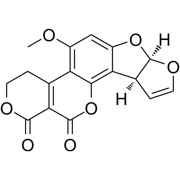
黄曲霉毒素G属于香豆素类化合物。
已有报道称玉米、黄曲霉和寄生曲霉中含有黄曲霉毒素G1,并有相关数据可供参考。 黄曲霉毒素是一类真菌毒素,主要由黄曲霉和寄生曲霉产生,其毒性代谢产物由稠合的香豆素-双(二氢呋喃)环结构组成,具有潜在的急性和慢性毒性。急性暴露于高浓度黄曲霉毒素可导致肝损伤。长期接触黄曲霉毒素会增加患肝癌的风险,这可能是由于黄曲霉毒素代谢物通过DNA嵌入和环氧化物介导的核碱基烷基化作用导致DNA损伤增加,进而可能引发突变。 黄曲霉毒素G是由黄曲霉和寄生曲霉产生的真菌毒素。 黄曲霉毒素G属于二呋喃香豆素内酯类化合物。这类化合物是多环芳香族化合物,其结构中包含一个与二呋喃香豆素骨架上的香豆素部分稠合的δ-戊内酯环。二呋喃香豆内酯是黄曲霉毒素及其相关化合物的一个亚类。 另见:黄曲霉毒素G1(注释已移至)。 作用机制 有研究表明,黄曲霉毒素最可能的作用机制是与DNA相互作用,并抑制负责DNA和RNA合成的聚合酶。 ……黄曲霉毒素8,9-氧化物与鸟嘌呤的N-7位共价结合,导致带有加合物的鸟嘌呤与腺嘌呤而非胞嘧啶配对,从而形成错误的密码子,并将错误的氨基酸插入蛋白质中。此类事件与黄曲霉毒素诱导的ras原癌基因和p53抑癌基因突变有关。 /黄曲霉毒素 8,9-氧化物/ ...在本研究中,异丙肾上腺素(ISO,4x10-9)、AFB(1)(3x10⁻⁶ 和 6x10⁻⁵ M)和 AFG(1)(3x10⁻⁶ 和 6x10⁻⁶ M)使离体豚鼠心房收缩,使该制备物对 ISO 过度敏感。黄曲霉毒素的这些特性值得关注,因为它们可能是文献中描述的某些心脏毒性作用的原因。 在黄曲霉毒素高风险暴露地区的人类肝脏肿瘤和实验动物中,均发现了p53肿瘤抑制基因249位密码子G到T的颠换。 黄曲霉毒素B1、黄曲霉毒素B2和黄曲霉毒素G1抑制大鼠肝细胞核内RNA聚合酶活性和降低RNA含量的能力,与这些化合物的致癌作用以及急性和亚急性毒性作用在性质上相似。黄曲霉毒素G1诱导肝细胞核仁的纤维状和颗粒状部分快速发生宏观分离。 体外人肝研究表明,将肝致癌物黄曲霉毒素B1生物活化为具有遗传毒性的2,3-环氧化物衍生物的主要催化剂是细胞色素P-450NF,这是一种先前已鉴定的蛋白质,它还能催化硝苯地平和其他二氢吡啶类药物、奎尼丁、大环内酯类抗生素、多种类固醇和其他化合物的氧化。……细胞色素P-450NF或其密切相关的蛋白质似乎也是黄曲霉毒素G1和赭曲霉毒素活化的主要催化剂,在这些系统中,赭曲霉毒素的遗传毒性比黄曲霉毒素B1更强。已知多种药物和疾病会影响人肝脏中细胞色素P-450NF的水平和活性,并且可以通过非侵入性检测方法来评估该酶的活性。这些发现为检验以下假设提供了一个测试系统:特定人类疾病状态(肝癌)与黄曲霉毒素摄入量高的人群的氧化代谢水平有关。 黄曲霉毒素 B1、黄曲霉毒素 G1 和黄曲霉毒素 G2 在毒素浓度为 100 umole/3 mL 时抑制了 (14) 碳标记的乳清酸掺入大鼠肝片 RNA 中。相应的抑制率约为90%、40%和20%。黄曲霉毒素B1(20 μmol/3 mL)、黄曲霉毒素G1(150 μmol/3 mL)和黄曲霉毒素G2(230 μmol/3 mL)分别抑制了14C标记的DL-亮氨酸掺入大鼠肝片蛋白质中的32%、35%和38%。 分析了暴露于不同浓度和时间的黄曲霉毒素B1、B2、G1、G2、B2a和M1的大鼠腹腔巨噬细胞的吞噬作用、对白色念珠菌的细胞内杀灭作用以及超氧化物的产生情况,以评估每种霉菌毒素的抑制作用强度。所有极低浓度的黄曲霉毒素均对巨噬细胞功能具有抑制作用。在暴露于黄曲霉毒素B1和M1的巨噬细胞中,吞噬作用、细胞内杀伤作用和自发性超氧化物产生能力均受到显著损害。 在各种有毒的黄曲霉毒素中,黄曲霉毒素B1和G1的生物活性最强,但其他衍生物也具有致癌性。黄曲霉毒素B1需要经细胞色素P450依赖性混合功能氧化酶的代谢活化才能转化为活性2,3-环氧化物,后者是最终的致癌物。黄曲霉毒素,例如黄曲霉毒素B1,是具有基因毒性的致癌物,其活性代谢物可与DNA发生反应。在细胞内反应中,与DNA形成的主要加合物是由黄曲霉毒素B1的2位与DNA中鸟嘌呤的N-7位结合形成的。 |
| 分子式 |
C17H12O7
|
|---|---|
| 分子量 |
328.27298
|
| 精确质量 |
328.058
|
| CAS号 |
1165-39-5
|
| 相关CAS号 |
Aflatoxin G1-13C17;1217444-07-9
|
| PubChem CID |
14421
|
| 外观&性状 |
White to off-white solid powder
|
| 密度 |
1.6±0.1 g/cm3
|
| 沸点 |
612.1±55.0 °C at 760 mmHg
|
| 熔点 |
244-246ºC
|
| 闪点 |
274.1±31.5 °C
|
| 蒸汽压 |
0.0±1.8 mmHg at 25°C
|
| 折射率 |
1.680
|
| LogP |
-0.17
|
| tPSA |
84.2
|
| 氢键供体(HBD)数目 |
0
|
| 氢键受体(HBA)数目 |
7
|
| 可旋转键数目(RBC) |
1
|
| 重原子数目 |
24
|
| 分子复杂度/Complexity |
666
|
| 定义原子立体中心数目 |
0
|
| SMILES |
COC1=C2C3=C(C(=O)OCC3)C(=O)OC2=C4C5C=COC5OC4=C1
|
| InChi Key |
XWIYFDMXXLINPU-UHFFFAOYSA-N
|
| InChi Code |
InChI=1S/C17H12O7/c1-20-9-6-10-12(8-3-5-22-17(8)23-10)14-11(9)7-2-4-21-15(18)13(7)16(19)24-14/h3,5-6,8,17H,2,4H2,1H3
|
| 化学名 |
11-methoxy-6,8,16,20-tetraoxapentacyclo[10.8.0.02,9.03,7.013,18]icosa-1,4,9,11,13(18)-pentaene-17,19-dione
|
| 别名 |
AFLATOXIN G1; Aflatoxin; 1165-39-5; 1402-68-2; AFLATOXINS; Aflatoxin G; Aflatoxin, crude; Aflatoxin G1-d3;
|
| HS Tariff Code |
2934.99.9001
|
| 存储方式 |
Powder -20°C 3 years 4°C 2 years In solvent -80°C 6 months -20°C 1 month |
| 运输条件 |
Room temperature (This product is stable at ambient temperature for a few days during ordinary shipping and time spent in Customs)
|
| 溶解度 (体外实验) |
DMSO : ~10 mg/mL (~30.46 mM)
|
|---|---|
| 溶解度 (体内实验) |
注意: 如下所列的是一些常用的体内动物实验溶解配方,主要用于溶解难溶或不溶于水的产品(水溶度<1 mg/mL)。 建议您先取少量样品进行尝试,如该配方可行,再根据实验需求增加样品量。
注射用配方
注射用配方1: DMSO : Tween 80: Saline = 10 : 5 : 85 (如: 100 μL DMSO → 50 μL Tween 80 → 850 μL Saline)(IP/IV/IM/SC等) *生理盐水/Saline的制备:将0.9g氯化钠/NaCl溶解在100 mL ddH ₂ O中,得到澄清溶液。 注射用配方 2: DMSO : PEG300 :Tween 80 : Saline = 10 : 40 : 5 : 45 (如: 100 μL DMSO → 400 μL PEG300 → 50 μL Tween 80 → 450 μL Saline) 注射用配方 3: DMSO : Corn oil = 10 : 90 (如: 100 μL DMSO → 900 μL Corn oil) 示例: 以注射用配方 3 (DMSO : Corn oil = 10 : 90) 为例说明, 如果要配制 1 mL 2.5 mg/mL的工作液, 您可以取 100 μL 25 mg/mL 澄清的 DMSO 储备液,加到 900 μL Corn oil/玉米油中, 混合均匀。 View More
注射用配方 4: DMSO : 20% SBE-β-CD in Saline = 10 : 90 [如:100 μL DMSO → 900 μL (20% SBE-β-CD in Saline)] 口服配方
口服配方 1: 悬浮于0.5% CMC Na (羧甲基纤维素钠) 口服配方 2: 悬浮于0.5% Carboxymethyl cellulose (羧甲基纤维素) 示例: 以口服配方 1 (悬浮于 0.5% CMC Na)为例说明, 如果要配制 100 mL 2.5 mg/mL 的工作液, 您可以先取0.5g CMC Na并将其溶解于100mL ddH2O中,得到0.5%CMC-Na澄清溶液;然后将250 mg待测化合物加到100 mL前述 0.5%CMC Na溶液中,得到悬浮液。 View More
口服配方 3: 溶解于 PEG400 (聚乙二醇400) 请根据您的实验动物和给药方式选择适当的溶解配方/方案: 1、请先配制澄清的储备液(如:用DMSO配置50 或 100 mg/mL母液(储备液)); 2、取适量母液,按从左到右的顺序依次添加助溶剂,澄清后再加入下一助溶剂。以 下列配方为例说明 (注意此配方只用于说明,并不一定代表此产品 的实际溶解配方): 10% DMSO → 40% PEG300 → 5% Tween-80 → 45% ddH2O (或 saline); 假设最终工作液的体积为 1 mL, 浓度为5 mg/mL: 取 100 μL 50 mg/mL 的澄清 DMSO 储备液加到 400 μL PEG300 中,混合均匀/澄清;向上述体系中加入50 μL Tween-80,混合均匀/澄清;然后继续加入450 μL ddH2O (或 saline)定容至 1 mL; 3、溶剂前显示的百分比是指该溶剂在最终溶液/工作液中的体积所占比例; 4、 如产品在配制过程中出现沉淀/析出,可通过加热(≤50℃)或超声的方式助溶; 5、为保证最佳实验结果,工作液请现配现用! 6、如不确定怎么将母液配置成体内动物实验的工作液,请查看说明书或联系我们; 7、 以上所有助溶剂都可在 Invivochem.cn网站购买。 |
| 制备储备液 | 1 mg | 5 mg | 10 mg | |
| 1 mM | 3.0463 mL | 15.2314 mL | 30.4627 mL | |
| 5 mM | 0.6093 mL | 3.0463 mL | 6.0925 mL | |
| 10 mM | 0.3046 mL | 1.5231 mL | 3.0463 mL |
1、根据实验需要选择合适的溶剂配制储备液 (母液):对于大多数产品,InvivoChem推荐用DMSO配置母液 (比如:5、10、20mM或者10、20、50 mg/mL浓度),个别水溶性高的产品可直接溶于水。产品在DMSO 、水或其他溶剂中的具体溶解度详见上”溶解度 (体外)”部分;
2、如果您找不到您想要的溶解度信息,或者很难将产品溶解在溶液中,请联系我们;
3、建议使用下列计算器进行相关计算(摩尔浓度计算器、稀释计算器、分子量计算器、重组计算器等);
4、母液配好之后,将其分装到常规用量,并储存在-20°C或-80°C,尽量减少反复冻融循环。
计算结果:
工作液浓度: mg/mL;
DMSO母液配制方法: mg 药物溶于 μL DMSO溶液(母液浓度 mg/mL)。如该浓度超过该批次药物DMSO溶解度,请首先与我们联系。
体内配方配制方法:取 μL DMSO母液,加入 μL PEG300,混匀澄清后加入μL Tween 80,混匀澄清后加入 μL ddH2O,混匀澄清。
(1) 请确保溶液澄清之后,再加入下一种溶剂 (助溶剂) 。可利用涡旋、超声或水浴加热等方法助溶;
(2) 一定要按顺序加入溶剂 (助溶剂) 。
 匹伐加宾
匹伐加宾
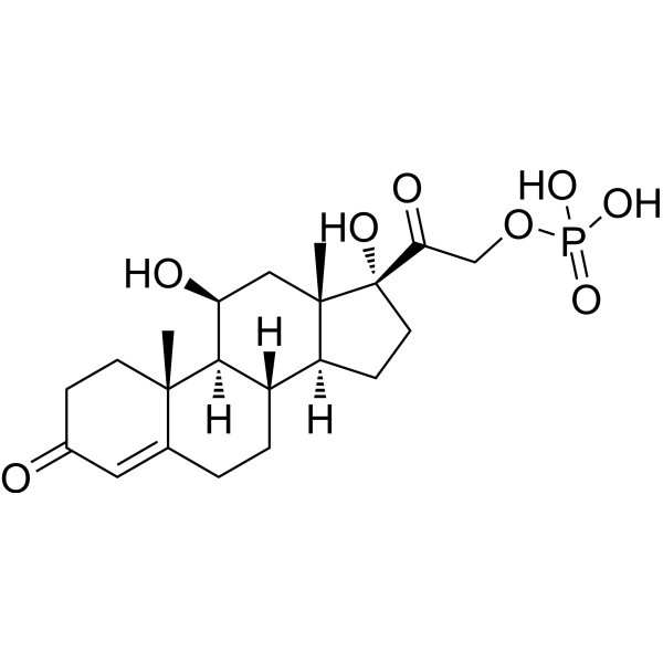 磷酸氢化可的松
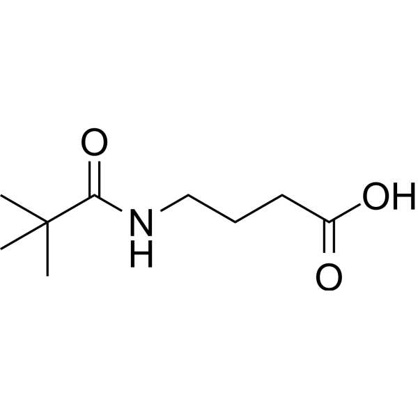
 BAY-784
 Gly-PEG3-endo-BCN
InvivoChem的所有产品仅用于作科学研究,不面向患者销售
Copyright 2020 InvivoChem LLC | All Rights Reserved 粤ICP备20063088号-1


 463611831
