| 规格 | 价格 | 库存 | 数量 |
|---|---|---|---|
| 1mg |
|
||
| 100mg |
|
||
| Other Sizes |
|
| 靶点 |
AMPK
|
|---|---|
| 体外研究 (In Vitro) |
AICA-核糖苷/AICA riboside(5-氨基咪唑-4-甲酰胺-1-β-D-呋喃核糖苷)已被广泛用于细胞中,以激活AMPK(AMP活化蛋白激酶),AMPK是一种参与细胞能量平衡的代谢传感器。在本研究中,我们研究了AICA核糖苷对线粒体氧化的影响;磷酸化。发现AICA核糖苷能剂量依赖性地抑制分离的大鼠肝细胞的寡霉素敏感JO2(耗氧率)。当AICA核糖苷浓度>0.1 mM时,也观察到P(i)(无机磷酸盐)、ATP、AMP和总腺嘌呤核苷酸含量降低。有趣的是,在缺乏α1和α2 AMPK催化亚基的小鼠肝细胞中,与野生型小鼠相比,基础JO2和几种线粒体蛋白的表达显著降低,表明线粒体生物合成受到干扰。然而,AICA核糖苷对JO2的抑制作用在突变小鼠中仍然存在,因此显然不是由AMPK介导的。在透性肝细胞中,这种抑制作用不再明显,表明这可能是由于细胞内Z核苷酸的积累和/或腺嘌呤核苷酸和P(i)的损失。ZMP确实通过对呼吸链复合物I的直接作用抑制了分离的大鼠线粒体的呼吸。此外,在与果糖一起孵育的细胞中,AICA核糖苷对JO2的抑制作用也得到了增强,以耗尽腺嘌呤核苷酸和P(I)。我们得出结论,AICA核糖苷通过AMPK非依赖性机制抑制细胞呼吸,这可能是由于细胞内P(i)耗竭和ZMP积累共同作用的结果。我们的数据还表明,AICA核糖苷的细胞效应不一定是由AMPK激活引起的,对它们的解释应该谨慎。[1]
5-氨基咪唑-4-甲酰胺核糖苷(AICA riboside/AICA-核糖苷;阿卡德新)激活完整细胞中的AMP活化蛋白激酶(AMPK),据报道对哺乳动物中枢神经系统具有保护作用。在大鼠大脑皮质脑切片中,AMPK被代谢应激(缺血>缺氧>多糖血症)和AICA核糖苷(0.1-10mm)激活。平衡核苷转运抑制剂大大减弱了AICA核苷对AMPK的激活。AICA-核糖苷还抑制了大鼠海马CA1区的兴奋性突触传递,这种传递被腺苷A1受体拮抗剂阻止,并被腺苷脱氨酶逆转。然而,AICA-核糖苷既不是腺苷脱氨酶的底物,也不是腺苷受体的激动剂。我们得出结论,代谢应激和AICA核糖核苷都会刺激哺乳动物大脑中的AMPK活性,但AICA核糖苷还有一个额外的作用,即与腺苷竞争核苷转运蛋白的摄取。这导致细胞外腺苷增加,随后腺苷受体激活。AICA核糖苷的神经保护作用可能是通过这种机制介导的,也可能是通过AMPK激活介导的。因此,在将AICA核糖苷的作用归因于AMPK激活时应谨慎,特别是在抑制腺苷再摄取具有生理后果的系统中[2]。 |
| 酶活实验 |
与腺苷脱氨酶作用相关的pH变化[2]
腺苷(10、30和100 µm)和AICA核糖苷(1、3和10 mm)制备为20 生理盐水(0.9%氯化钠;pH 5.72). 将腺苷脱氨酶(0.02单位)一式三份加入其中,用Jenway 3310 pH计测量所得pH变化。在研究AICA-核糖苷对腺苷脱氨酶代谢腺苷能力的影响的实验中,30 µm腺苷,来自5μm的储备溶液 向1、3和10的溶液中加入三份mm mm AICA核糖苷,含有0.02单位的腺苷脱氨酶。 市售AICA核糖苷的纯度/AICA riboside[2] 我们检查了本研究中使用的AICA核糖苷是否有可能被腺苷或可分解为腺苷的腺嘌呤核苷酸污染的迹象。使用了两种独立的HPLC技术,即上述用于组织核苷酸测定的技术,以及第二种技术,其中AICA核糖苷溶液被乙烯化,并用ThermoFenigan FL3000扫描荧光检测器检测成分。用任何一种技术(数据未显示)测量的AICA-核糖苷溶液中都没有可检测的污染物,排除了所述结果可能归因于腺苷或腺嘌呤核苷酸存在的可能性。 |
| 细胞实验 |
完整和透性肝细胞以及分离线粒体线粒体耗氧率的测定[1]
将大鼠或小鼠肝细胞(7-8mg干细胞·ml-1)在37°C的振荡水浴中,在含有2ml Krebs–Ringer碳酸氢盐钙缓冲液[120mM NaCl、4.8mM KCl、1.2mM KH2PO4、1.2mM MgSO4、24mM NaHCO3和1.3mM CaCl2(pH 7.4)]的封闭小瓶中孵育,并补充指定浓度的底物和AICA核糖苷,并与含有O2/CO2的气相平衡(19:1)。在指定时间,将细胞悬浮液再次用O2/CO2饱和1分钟,并立即转移到配备有Clark氧电极的搅拌式氧气计容器中。在连续添加0.5μM寡霉素、150μM DNP(2,4-二硝基苯酚)、0.15μg/ml抗霉素和1 mM TMPD(N,N,N′,N′-四甲基-1,4-苯二胺)加5 mM抗坏血酸之前和之后,在37°C下测量JO2(耗氧率)。 对于线粒体实验,将分离的大鼠线粒体(1 mg蛋白质·ml−1)在37°C下在含有2 ml KCl培养基(如上所述)的血氧计容器中在1 mMAICA核糖苷或指定浓度的ZMP和ZTP存在下孵育。平衡30秒后,在谷氨酸/苹果酸或琥珀酸/苹果酸/鱼藤酮存在的情况下监测线粒体呼吸速率,如上所述用于透性肝细胞。 核苷酸和Pi浓度以及糖异生和酮生的测定[1] 孵育30分钟后,将细胞悬浮液样品在冰冷的25 mM HClO4[5%(w/v)]/25 mM EDTA中淬灭,并离心(13000 g,2分钟)。中和上清液,用高效液相色谱法测定腺嘌呤核苷酸、ZMP和AICA核糖苷的浓度。为了测量细胞内Pi,将0.7ml细胞悬浮液样品通过硅油层离心(13000g,1分钟)至0.25ml HClO4[10%(w/v)]/25mM EDTA中。用比色法测量Pi。对于糖异生和酮生成测量,将细胞悬浮液样品在冰冷的HClO4[5%(w/v)]中淬灭,离心(13000 g,2分钟),中和上清液。如前所述,使用酶法结合NADH的分光光度测定法测量葡萄糖、乙酰乙酸盐和β-羟基丁酸盐的浓度。 大脑皮质切片的治疗[2] 将大脑皮层切片转移到放置在水浴中的孵化室中,将其加热至33-34°C。大约2.5-3之后 h、 将一些切片转移到相同的实验室中,这些实验室含有AICA核糖苷(0.1-10 mm)或AICA核糖糖苷以及摄取抑制剂双嘧达莫和硝基苄硫肌苷的组合(DIPY/NBTI;5 µm和1 μm)。此外,为了诱导代谢应激,切片被转移到没有葡萄糖的腔室中(低血糖;葡萄糖被10 mm蔗糖)或氧气(缺氧;用95%N2/5%CO2鼓泡)或缺乏葡萄糖和氧气(缺血)。将用作时间对照的切片转移到含有标准aCSF的相同腔室中。在适当的时候,切片在液氮中快速冷冻,并储存在-80°C下。 |
| 动物实验 |
切片制备[2]
16-23日龄的Sprague-Dawley大鼠(雌雄不限)采用颈椎脱臼法处死。迅速取出脑组织,置于含11 mM Mg2+的冰冷人工脑脊液(aCSF)中。使用振动切片机(Vibratome)切割400 µm厚的脑皮层切片,切片包含海马和上覆新皮层的矢状切片,具体方法如前所述(Dale等,2000)。将切片置于装有尼龙网的培养箱中,培养箱内盛有持续循环的充氧(95% O2/5% CO2)标准aCSF(1 mM Mg2+),并在室温下孵育至少1小时后方可使用。aCSF溶液的成分为:NaCl 124 mM;KCl 3 mM;CaCl2 2 mM;NaHCO3 26 mM; NaH2PO4 1.25 mM;d-葡萄糖 10 mM;MgSO4 1 mM;pH 7.4,95% O2/5% CO2。 脑皮层切片的处理[2] 将脑皮层切片转移至置于水浴中的培养室,并在33–34°C下加热。约2.5–3小时后,将部分切片转移至相同的实验室,其中分别含有AICA核苷(0.1–10 mM)或AICA核苷加摄取抑制剂双嘧达莫和硝基苄硫代肌苷(DIPY/NBTI;分别为5 µM和1 µM)的组合。此外,为了诱导代谢应激,将脑片转移至无葡萄糖(低血糖;用10 mM蔗糖替代葡萄糖)、无氧(缺氧;通入95% N2/5% CO2混合气体)或同时无葡萄糖和氧气(缺血)的培养室中。用作时间对照的脑片转移至含有标准人工脑脊液(aCSF)的相同培养室中。在适当时间,将脑片速冻于液氮中,并储存于-80°C。 AMPKα1α2LS−/− 敲除小鼠的构建 [1] 为了获得肝脏中两个催化亚基均缺失的小鼠(AMPKα1α2LS−/−),我们首先通过将floxed AMPKα2小鼠与在白蛋白和甲胎蛋白调控元件控制下表达Cre重组酶的AlfpCre转基因小鼠杂交,构建了肝脏特异性AMPKα2敲除小鼠(AMPKα2−/−)。然后,我们通过将肝脏特异性AMPKα2−/−小鼠与AMPKα1−/−小鼠杂交,在AMPKα1−/−背景下构建了肝脏特异性AMPKα1α2LS−/−小鼠。我们使用针对Cre转基因、floxed AMPKα2基因、缺失型AMPKα1基因和野生型AMPKα1基因的特异性引物,对从尾部活检组织中提取的DNA进行PCR,从而对小鼠进行基因分型。 |
| 参考文献 |
[1]. AMP-activated protein kinase-independent inhibition of hepatic mitochondrial oxidative phosphorylation by AICA riboside. Biochem J. 2007 Jun 15;404(3):499-507.
[2]. AICA riboside both activates AMP-activated protein kinase and competes with adenosine for the nucleoside transporter in the CA1 region of the rat hippocampus. J Neurochem. 2004 Mar;88(5):1272-82. |
| 其他信息 |
AICA核糖核苷酸是一种1-(磷酸核糖基)咪唑甲酰胺,属于阿卡地辛,其5'位羟基被转化为单磷酸衍生物。它可用作心血管药物,也是植物、人体、大肠杆菌、酿酒酵母和小鼠的代谢产物。它是一种1-(磷酸核糖基)咪唑甲酰胺和氨基咪唑类化合物,在功能上与阿卡地辛相关。它是5-氨基-1-(5-磷酸-D-核糖基)咪唑-4-甲酰胺(2-)的共轭酸。
5-氨基咪唑-4-甲酰胺核糖核苷酸 (AICAR) 是肌苷酸生成的中间体,也是腺苷酸 (AMP) 的类似物,能够刺激 AMP 依赖性蛋白激酶 (AMPK) 的活性。AICAR 已在临床上用于治疗和预防心肌缺血性损伤。该药物最初于 20 世纪 80 年代用于手术期间维持心脏血流,目前因其可通过改变肌肉的物理组成来提高组织代谢活性,从而成为治疗糖尿病的潜在药物而备受关注。 据报道,AICA 核糖核苷酸存在于拟南芥、人类和其他有相关数据的生物体中。 5-氨基-1-(5-磷酸-D-核糖基)咪唑-4-甲酰胺是酿酒酵母中发现或产生的一种代谢物。 本研究旨在探讨代谢应激和外源激活剂 AICA 核苷(阿卡地辛)对哺乳动物脑内 AMPK 激活的影响。此外,鉴于已有报道称AICA核苷能够保护外周器官和大脑,我们试图在生理背景下,即啮齿动物海马兴奋性突触传递的背景下,确定AICA核苷的作用。 AICA核苷的两个方面,即其作为腺苷调节剂(Mullane 1993)和AMPK(一种参与细胞代谢应激反应的关键细胞内酶)激活剂(Corton等人1995)的(未明确)作用,提示AICA核苷的保护作用可能反映了其对细胞外腺苷和AMPK激活的影响。这些作用中的一种或几种,或它们的组合,可以解释在各种缺血/再灌注损伤(Nawarskas 1999;Matot 和 Jurim 2001)、创伤(Davis 等人 2000)、休克(Ragsdale 和 Proctor 2000)和脓毒症(Melton 等人 1999)的实验模型中,外周器官和组织如何得到保护,以及在人类中,AICA 核苷如何改善冠状动脉旁路移植术后的结果,降低早期心脏死亡和心肌梗死的发生率(Mangano 1997)。然而,很明显,AMPK介导了AICA核苷诱导的糖尿病动物模型中葡萄糖稳态的改善(Halseth等,2002;Song等,2002),因为抗糖尿病药物二甲双胍也能在体内(Zhou等,2001;Fryer等,2002;Hawley等,2002)以及糖尿病患者的骨骼肌中(Musi等,2002)激活AMPK。 同样,在脑部,AICA核苷对沙鼠双侧颈动脉闭塞5分钟引起的神经病理学改变和运动功能障碍具有保护作用(Clough-Helfman和Phillis,1990),并能特异性地抑制小鼠同型半胱氨酸硫内酯诱导的癫痫发作(Marangos等,1990)。虽然它不能诱发戊四唑、咖啡因、苦味素或碘化比库啉引起的癫痫发作(Marangos 等,1990;Zhang 等,1993)。其保护作用归因于腺苷受体的激活、腺苷摄取或代谢的抑制,或清除自由基,所有这些都具有神经保护作用(Lipton,1999;de Mendonça 等,2000)。事实上,低浓度的AICA核苷(20 µM)已被证明能增强由红藻氨酸受体激活诱导的大鼠皮质切片腺苷释放,但对α-氨基-3-羟基-5-甲基-4-异恶唑丙酸(AMPA)或N-甲基-D-天冬氨酸(NMDA)受体无此作用(White 1996)。 然而,不能排除AMPK在脑组织保护中的作用,因为长期(48小时)暴露于200 µM AICA核苷激活AMPK可减少培养的皮质星形胶质细胞中神经酰胺诱导的细胞凋亡(Blazquez等,2001)。此外,研究表明,用极低浓度(10–100 µM)而非较高浓度(0.5–1 mM)的AICA核苷预处理海马神经元培养物1小时,可完全保护神经元免受随后24小时葡萄糖剥夺、氰化物、谷氨酸和β-淀粉样蛋白诱导的毒性损伤(Culmsee等,2001)。这种保护作用与AMPK的激活同时发生,后者可通过磷酸化特异性抗体检测到的α亚基磷酸化水平升高来证实。有趣的是,在竞争性腺苷A1受体拮抗剂DPCPX存在的情况下,仍观察到对葡萄糖剥夺的保护作用;而使用针对催化α1和α2亚基的反义寡核苷酸则可以阻断这种保护作用,这表明AMPK直接参与了该过程(Culmsee等,2001)。显然,AICA核苷通过腺苷A1受体在完整神经组织中发挥作用,能够限制癫痫发作(Dunwiddie 1999)和缺血性脑损伤(de Mendonça et al. 2000),并激活AMPK,从而可能为神经元氧化代谢提供星形胶质细胞酮体(Blazquez et al. 1999)。这些作用赋予AICA核苷多种吸引人的特性,使其成为治疗哺乳动物中枢神经系统急性或慢性功能障碍的潜在药物。 在本研究中,我们发现,在完整脑组织中,缺氧、低血糖和缺血引起的代谢应激可以激活AMPK,且激活程度反映了损伤的严重程度。长时间缺血后AMPK活性明显下降,可能反映了广泛的神经元病变和/或细胞内ATP严重耗竭,即使在富氧/富葡萄糖的人工脑脊液(aCSF)中浸泡30分钟后,ATP水平也几乎没有恢复迹象。ATP水平的降低会削弱上游激酶(AMPKK)磷酸化AMPK的能力,从而导致AMPK活性下降。事实上,在体外灌注的完整大鼠心脏缺血(Marsin等,2000)或单核细胞缺氧或使用ATP合成抑制剂寡霉素处理(Marsin等,2002)期间,也观察到了AMPK活性短暂升高后显著下降的现象。 与非神经元组织类似,AICA核苷通过细胞内ZMP的产生激活AMPK,而不影响ATP/ADP比值。本研究的一项新发现是,AICA核苷转化为ZMP需要通过平衡型腺苷转运蛋白进行转运,这一发现由核苷转运抑制剂DIPY和NBTI对ZMP积累和AMPK活性的抑制作用清晰地证实。此外,AICA核苷转运对神经元活动的功能意义在于导致细胞外腺苷水平升高,而外源性腺苷脱氨酶可以逆转这一过程,但AICA核苷本身可能抑制腺苷脱氨酶的活性,从而进一步增加细胞外腺苷水平。细胞外腺苷的积累能够强烈抑制兴奋性突触传递,这可能是通过抑制突触前腺苷A1受体介导的谷氨酸释放实现的。我们利用两种不同的报告基因检测方法,研究了AICA核苷是否为腺苷受体激动剂。结果表明,AICA核苷在酵母中对A1受体(以及A2A和A2B受体)和在CHO细胞中表达的A3受体均无活性。考虑到我们使用了全部四种腺苷受体和两种检测系统,虽然并非完全不可能,但AICA核苷在两种系统中均无活性,不太可能是由于AMPK激活引发的某种磷酸化事件导致所有四种腺苷受体失活所致。相反,对报告基因检测数据的最简单解释是,AICA 核苷并非腺苷受体的激动剂。 AICA 核苷抑制腺苷再摄取的后果并非仅限于 A1 受体,而是会激活大脑特定区域的主要腺苷受体(例如纹状体中的 A2A 受体)。这可能导致 A1、A2A 和 A3 受体之间的相互作用,进而导致 A1 受体介导的抑制作用脱敏(Dunwiddie 等,1997;Lopes 等,2002)。这些相互作用可能产生有害后果(Ribeiro 等,2002),并可能解释在不同癫痫模型中获得的混合结果。然而,既往研究表明,抑制腺苷摄取可以提高细胞外腺苷水平,并减轻多种中枢神经系统疾病实验模型中的神经病理损伤(de Mendonça et al. 2000)。例如,在新生大鼠幼崽发生缺氧/缺血联合损伤后腹腔注射丙戊托芬,可减少梗死组织体积,并改善纹状体、丘脑、海马和皮层的组织学指标(Gidday et al. 1995),还能增强沙鼠海马的神经保护性缺血预适应现象(Kawahara et al. 1998)。事实上,在最近的一项临床试验中,阿司匹林与DIPY(一种临床常用的抗血小板药物)联合使用可降低卒中复发率,而DIPY已被证实能提高人体血浆腺苷水平,这可能是导致良好预后的一个因素(Picano和Abbracchio,1998)。因此,AICA核苷的神经保护潜能似乎还体现在其提高细胞外腺苷水平的能力上。 总之,我们的研究结果表明,AICA核苷(在激活AMPK的同时)还能通过提高完整神经组织中细胞外腺苷水平发挥作用,这提示我们在解读使用该药物获得的结果时应谨慎。AICA核苷的这两种作用机制中哪一种在体内发挥神经保护作用更为重要,仍有待进一步研究。尽管如此,这两种相互有益的特性的存在,可能提供了一种有价值的协同作用,可用于治疗哺乳动物中枢神经系统疾病。[2] |
| 分子式 |
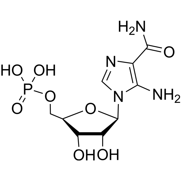
C9H15N4O8P
|
|---|---|
| 分子量 |
338.2112
|
| 精确质量 |
338.063
|
| 元素分析 |
C, 31.96; H, 4.47; N, 16.57; O, 37.84; P, 9.16
|
| CAS号 |
3031-94-5
|
| 相关CAS号 |
3031-94-5; 681006-28-0 (phosphate)
|
| PubChem CID |
65110
|
| 外观&性状 |
White to light brown solid powder
|
| 密度 |
2.3g/cm3
|
| 沸点 |
845.3ºC at 760 mmHg
|
| 熔点 |
198-202ºC dec.
|
| 闪点 |
465ºC
|
| 折射率 |
1.831
|
| LogP |
-3.8
|
| tPSA |
213.19
|
| 氢键供体(HBD)数目 |
6
|
| 氢键受体(HBA)数目 |
10
|
| 可旋转键数目(RBC) |
5
|
| 重原子数目 |
22
|
| 分子复杂度/Complexity |
475
|
| 定义原子立体中心数目 |
4
|
| SMILES |
C1=NC(=C(N1[C@H]2[C@@H]([C@@H]([C@H](O2)COP(=O)(O)O)O)O)N)C(=O)N
|
| InChi Key |
NOTGFIUVDGNKRI-UUOKFMHZSA-N
|
| InChi Code |
InChI=1S/C9H15N4O8P/c10-7-4(8(11)16)12-2-13(7)9-6(15)5(14)3(21-9)1-20-22(17,18)19/h2-3,5-6,9,14-15H,1,10H2,(H2,11,16)(H2,17,18,19)/t3-,5-,6-,9-/m1/s1
|
| 化学名 |
[(2R,3S,4R,5R)-5-(5-amino-4-carbamoylimidazol-1-yl)-3,4-dihydroxyoxolan-2-yl]methyl dihydrogen phosphate
|
| 别名 |
AICA ribonucleotide; 3031-94-5; Z-nucleotide; aminoimidazole carboxamide ribonucleotide; 5'-Phosphoribosyl-5-amino-4-imidazolecarboxamide; 5-amino-4-imidazolecarboxamide ribotide; Acadesine 5'-monophosphate; AICA-ribonucleotide;
|
| HS Tariff Code |
2934.99.9001
|
| 存储方式 |
Powder -20°C 3 years 4°C 2 years In solvent -80°C 6 months -20°C 1 month 注意: 请将本产品存放在密封且受保护的环境中(例如氮气保护),避免吸湿/受潮和光照。 |
| 运输条件 |
Room temperature (This product is stable at ambient temperature for a few days during ordinary shipping and time spent in Customs)
|
| 溶解度 (体外实验) |
May dissolve in DMSO (in most cases), if not, try other solvents such as H2O, Ethanol, or DMF with a minute amount of products to avoid loss of samples
|
|---|---|
| 溶解度 (体内实验) |
注意: 如下所列的是一些常用的体内动物实验溶解配方,主要用于溶解难溶或不溶于水的产品(水溶度<1 mg/mL)。 建议您先取少量样品进行尝试,如该配方可行,再根据实验需求增加样品量。
注射用配方
注射用配方1: DMSO : Tween 80: Saline = 10 : 5 : 85 (如: 100 μL DMSO → 50 μL Tween 80 → 850 μL Saline)(IP/IV/IM/SC等) *生理盐水/Saline的制备:将0.9g氯化钠/NaCl溶解在100 mL ddH ₂ O中,得到澄清溶液。 注射用配方 2: DMSO : PEG300 :Tween 80 : Saline = 10 : 40 : 5 : 45 (如: 100 μL DMSO → 400 μL PEG300 → 50 μL Tween 80 → 450 μL Saline) 注射用配方 3: DMSO : Corn oil = 10 : 90 (如: 100 μL DMSO → 900 μL Corn oil) 示例: 以注射用配方 3 (DMSO : Corn oil = 10 : 90) 为例说明, 如果要配制 1 mL 2.5 mg/mL的工作液, 您可以取 100 μL 25 mg/mL 澄清的 DMSO 储备液,加到 900 μL Corn oil/玉米油中, 混合均匀。 View More
注射用配方 4: DMSO : 20% SBE-β-CD in Saline = 10 : 90 [如:100 μL DMSO → 900 μL (20% SBE-β-CD in Saline)] 口服配方
口服配方 1: 悬浮于0.5% CMC Na (羧甲基纤维素钠) 口服配方 2: 悬浮于0.5% Carboxymethyl cellulose (羧甲基纤维素) 示例: 以口服配方 1 (悬浮于 0.5% CMC Na)为例说明, 如果要配制 100 mL 2.5 mg/mL 的工作液, 您可以先取0.5g CMC Na并将其溶解于100mL ddH2O中,得到0.5%CMC-Na澄清溶液;然后将250 mg待测化合物加到100 mL前述 0.5%CMC Na溶液中,得到悬浮液。 View More
口服配方 3: 溶解于 PEG400 (聚乙二醇400) 请根据您的实验动物和给药方式选择适当的溶解配方/方案: 1、请先配制澄清的储备液(如:用DMSO配置50 或 100 mg/mL母液(储备液)); 2、取适量母液,按从左到右的顺序依次添加助溶剂,澄清后再加入下一助溶剂。以 下列配方为例说明 (注意此配方只用于说明,并不一定代表此产品 的实际溶解配方): 10% DMSO → 40% PEG300 → 5% Tween-80 → 45% ddH2O (或 saline); 假设最终工作液的体积为 1 mL, 浓度为5 mg/mL: 取 100 μL 50 mg/mL 的澄清 DMSO 储备液加到 400 μL PEG300 中,混合均匀/澄清;向上述体系中加入50 μL Tween-80,混合均匀/澄清;然后继续加入450 μL ddH2O (或 saline)定容至 1 mL; 3、溶剂前显示的百分比是指该溶剂在最终溶液/工作液中的体积所占比例; 4、 如产品在配制过程中出现沉淀/析出,可通过加热(≤50℃)或超声的方式助溶; 5、为保证最佳实验结果,工作液请现配现用! 6、如不确定怎么将母液配置成体内动物实验的工作液,请查看说明书或联系我们; 7、 以上所有助溶剂都可在 Invivochem.cn网站购买。 |
| 制备储备液 | 1 mg | 5 mg | 10 mg | |
| 1 mM | 2.9567 mL | 14.7837 mL | 29.5674 mL | |
| 5 mM | 0.5913 mL | 2.9567 mL | 5.9135 mL | |
| 10 mM | 0.2957 mL | 1.4784 mL | 2.9567 mL |
1、根据实验需要选择合适的溶剂配制储备液 (母液):对于大多数产品,InvivoChem推荐用DMSO配置母液 (比如:5、10、20mM或者10、20、50 mg/mL浓度),个别水溶性高的产品可直接溶于水。产品在DMSO 、水或其他溶剂中的具体溶解度详见上”溶解度 (体外)”部分;
2、如果您找不到您想要的溶解度信息,或者很难将产品溶解在溶液中,请联系我们;
3、建议使用下列计算器进行相关计算(摩尔浓度计算器、稀释计算器、分子量计算器、重组计算器等);
4、母液配好之后,将其分装到常规用量,并储存在-20°C或-80°C,尽量减少反复冻融循环。
计算结果:
工作液浓度: mg/mL;
DMSO母液配制方法: mg 药物溶于 μL DMSO溶液(母液浓度 mg/mL)。如该浓度超过该批次药物DMSO溶解度,请首先与我们联系。
体内配方配制方法:取 μL DMSO母液,加入 μL PEG300,混匀澄清后加入μL Tween 80,混匀澄清后加入 μL ddH2O,混匀澄清。
(1) 请确保溶液澄清之后,再加入下一种溶剂 (助溶剂) 。可利用涡旋、超声或水浴加热等方法助溶;
(2) 一定要按顺序加入溶剂 (助溶剂) 。
InvivoChem的所有产品仅用于作科学研究,不面向患者销售
Copyright 2020 InvivoChem LLC | All Rights Reserved 粤ICP备20063088号-1


 463611831
