| 规格 | 价格 | 库存 | 数量 |
|---|---|---|---|
| 50mg |
|
||
| 100mg |
|
||
| Other Sizes |
|
| 靶点 |
Recombinant Fluc protein (Km = 2.06 μM )
|
|---|---|
| 体外研究 (In Vitro) |
用 AkaLumine HCl 处理的 LLC/luc 和 MDA-MB-231/luc 细胞中的信号在较低浓度 (2.5 μM) 时达到峰值,而 D-荧光素和 CycLuc1 在较高剂量下产生更多的生物发光。上升,即使在 250 μM 时也没有达到最大信号 [1]。
|
| 体内研究 (In Vivo) |
AkaLumine 盐酸盐显示来自肺转移的信号显着升高,与 D-荧光素给药相比增强了 8.1 倍。为了评估 AkaLumine HCl 在检测深部组织靶点方面相对于 CycLuc1 的优越性,以 5 mM 剂量(最大剂量)静脉注射 LLC/luc 细胞 15 分钟后,AkaLumine HCl 和 CycLuc1 处理的小鼠的生物发光 比较图像 CycLuc1 的浓度由于其水溶性较低,因此其含量较高。与 CycLuc1 相比,AkaLumine 盐酸盐对播散性肺癌细胞的检测灵敏度提高了 3.3 倍。通过按照 CycLuc1 和 AkaLumine 盐酸盐的顺序腹腔注射 5 mM 底物,并以相反的顺序每 8 小时注射一次,对具有肺转移的相同小鼠进行成像,进一步验证了 AkaLumine 盐酸盐的优越性。成像。与 CycLuc1 相比,AkaLumine 盐酸盐的肺转移信号增加了约四倍 [1]。
可在小鼠体内实现高灵敏度的深部组织成像。将AkaLumine腹腔注射到小鼠体内后,其可与荧光素酶反应,产生的近红外生物发光能够穿透组织,使深部组织如大脑和腹部器官等结构清晰成像,为无创监测活体动物体内生物过程提供了有力工具[1] |
| 酶活实验 |
生物发光发射光谱的测量[1]
使用ATTO AB-1850分光光度计测量d-荧光素、CycLuc1和AkaLumine-HCl的生物发光发射光谱。通过将5 μl基质(100 μM),5 μl QuantiLum重组萤光素酶溶液(1 毫克 ml−1)和5 μl磷酸钾缓冲液(500 mM、pH 8.0)。然后通过注射10 μl ATP Mg(200 μM)加入反应混合物中。生物发光发射光谱在1 nm从400增加到780 nm使用3 最小积分时间。[1] K m值的测量[1] 除底物浓度外,在与生物发光光谱测量相同的条件下测量d-荧光素、CycLuc1和AkaLumine HCl的生物发光强度。基质的最终浓度在0.2到500之间变化 μM和0.1至100 μM。底物的Km和Vmax值由生物发光强度的积分值确定,并使用市售SigmaPlot 9.0软件包中的酶动力学向导通过Lineweaver–Burk图计算。 |
| 细胞实验 |
使用生物组织的生物发光透射测定[1]
用IVIS Spectrum 1测量来自孔的生物发光信号 基质后分钟(最终浓度为2.5 μM,对于d-葡糖苷和AkaLumine HCl,50 CycLuc1的nM)与重组Fluc蛋白(20 μg ml−1)在ATP Mg(最终浓度为20 μM)在黑色96孔板中。将生物组织(4mm厚的牛肉片)放置在孔上,以测量通过组织的生物发光信号,然后通过两层生物组织获取生物发光图像。以下条件用于图像采集:曝光时间=10 s、 装仓=中等:8,视野=12.9×12.9 并且f/stop=1。通过IVIS系统专用的Living Image 4.3软件(PerkinElmer)分析生物发光图像。穿透效率(%)是通过将牛肉覆盖井的信号强度除以相应的未覆盖井的那些信号强度来计算的。[1] 稳定表达luc报告基因的癌症细胞系的分离[1] 小鼠肺癌细胞LLC、人乳腺癌症细胞MDA-MB-231和人癌症细胞PC-3从ATCC获得。LLC/luc和MDA-MB-231/luc细胞在用质粒pEF/luc转染后通过磷酸钙法分离,如先前所述19,20。PC-3/κB-luc也如先前所述被分离21。细胞维持在37 °C的5%FCS-DMEM(Nacalai Tesque,Kyoto,Japan)中添加青霉素(100单位/ml)和链霉素(100 μg ml−1),并通过支原体检查试剂盒定期检查支原体污染。对于每个实验,所有细胞系都被独立地存储并每次从原始库存中回收。[1] 体外BLI[1] 底物在黑色96孔板中与LLC/luc或MDA-MB-231/luc细胞(4×105个细胞/孔)反应。使用IVIS Spectrum 1测量生物发光 分钟后加入基质。以下条件用于图像采集:对总生物发光开放或680±10 用于近红外生物发光的发射滤光片的nm,暴露时间=10 s、 装仓=中等:8,视野=12.9×12.9 cm和f/stop=1。通过IVIS系统专用的Living Image 4.3软件对生物发光图像进行分析。 |
| 动物实验 |
肿瘤模型
对于皮下肿瘤模型,将悬浮于PBS中的LLC/luc(3 × 10⁵个细胞/10 μl)或PC-3/κB-luc(1 × 10⁶个细胞/10 μl)与等体积的Geltrex混合,分别皮下或胫骨内注射到C57B/6白化小鼠(雌性)或重症联合免疫缺陷小鼠(雄性)体内。移植后4天进行实验。对于肺部播散性癌细胞模型,在进行体内生物发光成像(BLI)前15分钟,将悬浮于PBS中的LLC/luc(1 × 10⁵个细胞/100 μl)经尾静脉注射到C57B/6白化小鼠(雄性)体内。对于肺转移模型,将悬浮于PBS中的LLC/luc(5 × 10⁵个细胞/100 μl)经尾静脉注射到C57B/6白化小鼠(雄性)体内。实验在静脉注射后10-20天进行。这些肿瘤模型已建立完善,肿瘤生长稳定。因此,每个实验中6个样本足以评估肿瘤生长情况。 体内生物发光成像 除非另有说明,否则在腹腔注射指定剂量的底物后15分钟,使用IVIS Spectrum采集皮下肿瘤的生物发光图像。由于肺转移瘤的生物发光强度在注射底物后的不同时间达到峰值,因此在腹腔注射底物后,每隔3分钟使用IVIS Spectrum连续采集肺转移瘤的生物发光图像,持续30分钟,并选择采集到的图像中生物发光强度最高的图像进行分析。为了比较使用同一小鼠的不同底物产生的生物发光,分别在注射d-荧光素和CycLucl后4小时和8小时采集AkaLumine-HCl的图像。图像采集条件如下:开放式发射滤光片,曝光时间=60 s,像素合并=中等:8,视野=12.9 × 12.9 cm,光圈=1。对于肺转移模型的三维生物发光成像(BLI),注射了AkaLumine-HCl的小鼠使用三种不同波长(660±10 nm、680±10 nm和700±10 nm)的生物发光滤光片进行BLI成像。图像采集条件如下:曝光时间=60 s,像素合并=中等:8,视野=12.9 × 12.9 cm,光圈=1。生物发光图像使用专为IVIS系统设计的Living Image 4.3软件进行分析。 离体BLI 使用AkaLumine-HCl进行体内BLI成像后,立即处死小鼠并取出肺脏。在以下条件下获得肺部生物发光图像:开放式发射滤光片,曝光时间=30 s,像素合并=中等:8,视野=12.9 × 12.9 cm,光圈=1。使用专为IVIS系统设计的Living Image 4.3软件分析生物发光图像。 首先将AkaLumine溶解于二甲基亚砜(DMSO)中,然后用生理盐水稀释。将配制好的溶液以10 mg/kg的剂量腹腔注射到小鼠体内。注射后不同时间点,使用活体成像系统观察小鼠的生物发光信号,以进行深层组织成像[1] |
| 毒性/毒理 (Toxicokinetics/TK) |
小鼠腹腔注射AkaLumine后未观察到明显的毒性作用。小鼠一般状况无显著变化,也未发现肝肾功能损伤,表明AkaLumine具有良好的生物相容性和低毒性[1]。
|
| 参考文献 | |
| 其他信息 |
AkaLumine 是一种荧光素类似物。传统的荧光素-荧光素酶系统由于其短波长光易被组织吸收,因此在体内信号检测方面存在局限性。AkaLumine 可产生近红外生物发光,具有更好的组织穿透性,从而实现高灵敏度的深层组织成像。它可用于对活体动物的基因表达和生物过程进行无创监测,为生物学研究和临床前研究提供了一种新方法[1]。
|
| 分子式 |
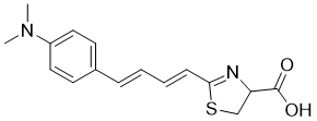
C16H18N2O2S
|
|---|---|
| 分子量 |
302.391322612762
|
| 精确质量 |
302.108
|
| 元素分析 |
C, 63.55; H, 6.00; N, 9.26; O, 10.58; S, 10.60
|
| CAS号 |
1176235-08-7
|
| 相关CAS号 |
1176235-08-7; 2558205-28-8 (HCl)
|
| PubChem CID |
139329217
|
| 外观&性状 |
Typically exists as solid at room temperature
|
| 密度 |
1.2±0.1 g/cm3
|
| 沸点 |
541.4±60.0 °C at 760 mmHg
|
| 闪点 |
281.2±32.9 °C
|
| 蒸汽压 |
0.0±1.5 mmHg at 25°C
|
| 折射率 |
1.598
|
| LogP |
2.06
|
| tPSA |
78.2
|
| 氢键供体(HBD)数目 |
1
|
| 氢键受体(HBA)数目 |
5
|
| 可旋转键数目(RBC) |
5
|
| 重原子数目 |
21
|
| 分子复杂度/Complexity |
449
|
| 定义原子立体中心数目 |
0
|
| SMILES |
S1C(/C=C/C=C/C2C=CC(=CC=2)N(C)C)=NC(C(=O)O)C1
|
| InChi Key |
ULTVSKXCSMDCHR-GGWOSOGESA-N
|
| InChi Code |
InChI=1S/C16H18N2O2S/c1-18(2)13-9-7-12(8-10-13)5-3-4-6-15-17-14(11-21-15)16(19)20/h3-10,14H,11H2,1-2H3,(H,19,20)/b5-3+,6-4+
|
| 化学名 |
2-[(1E,3E)-4-[4-(dimethylamino)phenyl]buta-1,3-dienyl]-4,5-dihydro-1,3-thiazole-4-carboxylic acid
|
| HS Tariff Code |
2934.99.9001
|
| 存储方式 |
Powder -20°C 3 years 4°C 2 years In solvent -80°C 6 months -20°C 1 month |
| 运输条件 |
Room temperature (This product is stable at ambient temperature for a few days during ordinary shipping and time spent in Customs)
|
| 溶解度 (体外实验) |
May dissolve in DMSO (in most cases), if not, try other solvents such as H2O, Ethanol, or DMF with a minute amount of products to avoid loss of samples
|
|---|---|
| 溶解度 (体内实验) |
注意: 如下所列的是一些常用的体内动物实验溶解配方,主要用于溶解难溶或不溶于水的产品(水溶度<1 mg/mL)。 建议您先取少量样品进行尝试,如该配方可行,再根据实验需求增加样品量。
注射用配方
注射用配方1: DMSO : Tween 80: Saline = 10 : 5 : 85 (如: 100 μL DMSO → 50 μL Tween 80 → 850 μL Saline)(IP/IV/IM/SC等) *生理盐水/Saline的制备:将0.9g氯化钠/NaCl溶解在100 mL ddH ₂ O中,得到澄清溶液。 注射用配方 2: DMSO : PEG300 :Tween 80 : Saline = 10 : 40 : 5 : 45 (如: 100 μL DMSO → 400 μL PEG300 → 50 μL Tween 80 → 450 μL Saline) 注射用配方 3: DMSO : Corn oil = 10 : 90 (如: 100 μL DMSO → 900 μL Corn oil) 示例: 以注射用配方 3 (DMSO : Corn oil = 10 : 90) 为例说明, 如果要配制 1 mL 2.5 mg/mL的工作液, 您可以取 100 μL 25 mg/mL 澄清的 DMSO 储备液,加到 900 μL Corn oil/玉米油中, 混合均匀。 View More
注射用配方 4: DMSO : 20% SBE-β-CD in Saline = 10 : 90 [如:100 μL DMSO → 900 μL (20% SBE-β-CD in Saline)] 口服配方
口服配方 1: 悬浮于0.5% CMC Na (羧甲基纤维素钠) 口服配方 2: 悬浮于0.5% Carboxymethyl cellulose (羧甲基纤维素) 示例: 以口服配方 1 (悬浮于 0.5% CMC Na)为例说明, 如果要配制 100 mL 2.5 mg/mL 的工作液, 您可以先取0.5g CMC Na并将其溶解于100mL ddH2O中,得到0.5%CMC-Na澄清溶液;然后将250 mg待测化合物加到100 mL前述 0.5%CMC Na溶液中,得到悬浮液。 View More
口服配方 3: 溶解于 PEG400 (聚乙二醇400) 请根据您的实验动物和给药方式选择适当的溶解配方/方案: 1、请先配制澄清的储备液(如:用DMSO配置50 或 100 mg/mL母液(储备液)); 2、取适量母液,按从左到右的顺序依次添加助溶剂,澄清后再加入下一助溶剂。以 下列配方为例说明 (注意此配方只用于说明,并不一定代表此产品 的实际溶解配方): 10% DMSO → 40% PEG300 → 5% Tween-80 → 45% ddH2O (或 saline); 假设最终工作液的体积为 1 mL, 浓度为5 mg/mL: 取 100 μL 50 mg/mL 的澄清 DMSO 储备液加到 400 μL PEG300 中,混合均匀/澄清;向上述体系中加入50 μL Tween-80,混合均匀/澄清;然后继续加入450 μL ddH2O (或 saline)定容至 1 mL; 3、溶剂前显示的百分比是指该溶剂在最终溶液/工作液中的体积所占比例; 4、 如产品在配制过程中出现沉淀/析出,可通过加热(≤50℃)或超声的方式助溶; 5、为保证最佳实验结果,工作液请现配现用! 6、如不确定怎么将母液配置成体内动物实验的工作液,请查看说明书或联系我们; 7、 以上所有助溶剂都可在 Invivochem.cn网站购买。 |
| 制备储备液 | 1 mg | 5 mg | 10 mg | |
| 1 mM | 3.3070 mL | 16.5349 mL | 33.0699 mL | |
| 5 mM | 0.6614 mL | 3.3070 mL | 6.6140 mL | |
| 10 mM | 0.3307 mL | 1.6535 mL | 3.3070 mL |
1、根据实验需要选择合适的溶剂配制储备液 (母液):对于大多数产品,InvivoChem推荐用DMSO配置母液 (比如:5、10、20mM或者10、20、50 mg/mL浓度),个别水溶性高的产品可直接溶于水。产品在DMSO 、水或其他溶剂中的具体溶解度详见上”溶解度 (体外)”部分;
2、如果您找不到您想要的溶解度信息,或者很难将产品溶解在溶液中,请联系我们;
3、建议使用下列计算器进行相关计算(摩尔浓度计算器、稀释计算器、分子量计算器、重组计算器等);
4、母液配好之后,将其分装到常规用量,并储存在-20°C或-80°C,尽量减少反复冻融循环。
计算结果:
工作液浓度: mg/mL;
DMSO母液配制方法: mg 药物溶于 μL DMSO溶液(母液浓度 mg/mL)。如该浓度超过该批次药物DMSO溶解度,请首先与我们联系。
体内配方配制方法:取 μL DMSO母液,加入 μL PEG300,混匀澄清后加入μL Tween 80,混匀澄清后加入 μL ddH2O,混匀澄清。
(1) 请确保溶液澄清之后,再加入下一种溶剂 (助溶剂) 。可利用涡旋、超声或水浴加热等方法助溶;
(2) 一定要按顺序加入溶剂 (助溶剂) 。
|
|
|
 奈莫雷生盐酸盐
 E55888 HCL
 FITC-Dextran (MW 110000)
FITC-Dextran (MW 2000000)
FITC-Dextran (MW 110000)
FITC-Dextran (MW 2000000)
InvivoChem的所有产品仅用于作科学研究,不面向患者销售
Copyright 2020 InvivoChem LLC | All Rights Reserved 粤ICP备20063088号-1


 463611831
