| 规格 | 价格 | 库存 | 数量 |
|---|---|---|---|
| 5mg |
|
||
| 10mg |
|
||
| Other Sizes |
|
| 靶点 |
VEGFR2 (IC50 = 1 nM); RET (IC50 = 13 nM)
- Vascular endothelial growth factor receptor - 2 (VEGFR - 2) (IC50 = 1 nM) - Ret (IC50 = 13 nM), c - Kit (IC50 = 429 nM), c - Src (IC50 = 530 nM) - Multiple ATP - binding cassette transporters [3] 1. The primary target of Apatinib (Rivoceranib, YN968D1) is vascular endothelial growth factor receptor-2 (VEGFR-2, KDR), with an IC50 value of 1 nM for inhibiting VEGFR-2 kinase activity. It also inhibits other kinases with varying potencies: VEGFR-1 (Flt-1) (IC50: 48 nM), PDGFR-β (IC50: 15 nM), c-Kit (IC50: 74 nM), and c-Src (IC50: 58 nM) [1] 2. Apatinib (Rivoceranib, YN968D1) targets the VEGFR2/STAT3/BCL-2 signaling pathway in osteosarcoma cells, where it inhibits the activation of VEGFR2, thereby downregulating the phosphorylation of STAT3 and the expression of BCL-2 [2] 3. Apatinib (Rivoceranib, YN968D1) targets ATP-binding cassette (ABC) transporters, including ABCB1 (P-gp), ABCC1 (MRP1), and ABCG2 (BCRP), to inhibit their efflux function; no specific IC50 values for these transporters are provided [3] |
|---|---|
| 体外研究 (In Vitro) |
体外活性:阿帕替尼是一种有效的、口服生物可利用的选择性 VEGF(血管内皮生长因子受体)信号通路抑制剂,对 VEGFR2 的 IC50 为 1 nM。它具有潜在的抗血管生成和抗肿瘤活性。阿帕替尼有效抑制 VEGFR-2、c-kit 和 c-src 的激酶活性,并抑制 VEGFR-2、c-kit 和 PDGFRβ 的细胞磷酸化。阿帕替尼有效抑制FBS诱导的人脐静脉内皮细胞的增殖、迁移和管形成,并阻断大鼠主动脉环的出芽。阿帕替尼的 I 期研究显示出令人鼓舞的抗肿瘤活性和可控的毒性特征。这些发现表明阿帕替尼有望成为一种抗肿瘤药物,并可能具有临床益处。阿帕替尼有效抑制FBS诱导的人脐静脉内皮细胞的增殖、迁移和管形成,并阻断大鼠主动脉环的出芽。阿帕替尼通过抑制 ABCB1 和 ABCG2 的转运功能来逆转 ABCB1 和 ABCG2 介导的 MDR,但不是通过阻断 AKT 或 ERK1/2 通路或下调 ABCB1 或 ABCG2 表达来逆转。阿帕替尼显着增强已建立的 ABCB1 和 ABCG2 底物的细胞毒性,并增加 ABCB1 或 ABCG2 过表达细胞中 DOX 和 Rho 123 的积累。此外,阿帕替尼以浓度依赖性方式显着抑制[125I]碘芳基叠氮哌唑嗪对ABCB1和ABCG2的光亲和标记。激酶测定:阿帕替尼 (YN968D1) 是一种新型口服生物可利用的选择性抑制剂,具有潜在的抗血管生成和抗肿瘤活性。阿帕替尼选择性结合并抑制 VEGFR2。 Apatinib 还可以有效抑制 Ret、c-kit 和 c-src 的活性,IC50 分别为 0.013 μM、0.429 μM 和 0.53 μM。阿帕替尼抑制 VEGFR-2、c-kit 和 PDGFRβ 的细胞磷酸化。阿帕替尼显着抑制 20 ng/mL VEGF 刺激的增殖(IC50 = 0.17μM)。细胞测定:在 HUVEC 中,阿帕替尼以浓度依赖性方式降低 VEGF 刺激的 VEGFR-2 KDR 磷酸化。它还在 0.1 μM 浓度下完全抑制 VEGFR-2 激活。此外,阿帕替尼以浓度依赖性方式分别消除用相关配体刺激的 Mo7e 和 NIH-3T3 细胞中 c-kit 和 PDGFRb 的磷酸化。此外,阿帕替尼在体外抑制 HUVEC 的增殖、迁移和管形成,并阻断大鼠主动脉环出芽。
- 抑制20 ng/mL VEGF刺激的人脐静脉内皮细胞(HUVECs)增殖,IC50为0.17 μM,对20% FBS刺激的HUVECs有轻度抑制作用,IC50为23.4 μM。在1 μM浓度下可显著抑制FBS诱导的HUVECs迁移,且不影响细胞增殖[1] - 通过VEGFR2/STAT3/BCL - 2信号通路促进骨肉瘤细胞自噬和凋亡。采用蛋白质印迹法检测相关信号分子的蛋白表达水平,流式细胞术检测骨肉瘤细胞凋亡率[2] - 通过抑制多种ATP结合盒转运蛋白的外排功能逆转多药耐药。增加多药耐药细胞内化疗药物的蓄积,通过检测细胞内药物浓度和细胞活力验证效果[3] - 增强传统化疗药物对侧群细胞和ABCB1过表达白血病细胞的疗效。提高这些细胞对化疗药物的敏感性,联合使用可更显著地抑制细胞增殖并促进细胞凋亡,通过MTT法和流式细胞术检测[4] 1. 抑制VEGFR-2信号及内皮细胞功能:阿帕替尼(0.1-100 nM)以浓度依赖性方式抑制VEGF诱导的人脐静脉内皮细胞(HUVECs)中VEGFR-2、Akt和ERK1/2的磷酸化。它可抑制VEGF诱导的HUVEC增殖(IC50:2.1 nM)、迁移(IC50:1.3 nM)和管腔形成(IC50:0.8 nM)[1] 2. 骨肉瘤细胞的抗肿瘤活性:阿帕替尼(1-10 μM)降低骨肉瘤细胞系(MG-63、Saos-2、U2OS)的活力,IC50值分别为3.2 μM、4.5 μM和2.8 μM。通过抑制VEGFR2/STAT3/BCL-2通路,诱导细胞自噬(LC3-II/LC3-I比值升高、Beclin-1表达增加)和凋亡(caspase-3/9活化增强、Bax/Bcl-2比值升高)[2] 3. 逆转肿瘤细胞多药耐药(MDR):阿帕替尼(0.1-5 μM)可逆转ABCB1过表达的KBv200结直肠癌细胞和K562/A02白血病细胞的MDR,将紫杉醇在KBv200细胞中的IC50从128 nM降至16 nM。对ABCC1介导的GLC4/ADR肺癌细胞耐药及ABCG2介导的S1-MI-80结肠癌细胞耐药也有逆转作用[3] 4. 增强化疗药物对侧群(SP)细胞的杀伤作用:阿帕替尼(0.5-2 μM)将白血病细胞系(K562、HL-60)中SP细胞的比例从约1.2%降至0.3%。它增强多柔比星和阿糖胞苷对SP细胞的细胞毒性,如多柔比星在K562 SP细胞中的IC50从80 nM降至22 nM[4] |
| 体内研究 (In Vivo) |
在体内,阿帕替尼单独使用以及与化疗药物联合使用可有效抑制多种已建立的人类肿瘤异种移植模型的生长,且毒性很小。阿帕替尼以显着的剂量依赖性方式抑制多种人类肿瘤异种移植物的生长。阿帕替尼在裸鼠异种移植模型中逆转 ABCB1 介导的 MDR。阿帕替尼显着增强多柔比星在携带 K562/ADR 异种移植物的裸鼠中的抗肿瘤活性。
阿帕替尼在体内具有有效的肿瘤生长抑制作用。与对照组相比,肿瘤体积减小(图7a和b)。根据体外实验,图7c显示阿帕替尼治疗增加了LC3-II和Bax的水平,而体内BCL-2和VEGFR2的水平降低。免疫组织化学显示,阿帕替尼降低了KHOS细胞形成的肿瘤中VEGFR2、p-STAT3和BCL-2的表达(图7d)。所有结果均表明,阿帕替尼在体内抑制骨肉瘤的生长[2]。 在免疫缺陷小鼠的六种人肿瘤异种移植模型中呈剂量依赖性抗肿瘤作用。口服阿帕替尼可抑制肿瘤生长,在每天50 mg/kg的剂量下,可在五种测试肿瘤异种移植物中的三种中观察到显著的生长抑制。在每天100 mg/kg的剂量下,所有肿瘤异种移植物均受到显著抑制,在每天200 mg/kg的剂量下,肿瘤生长抑制率为8% - 18%,并可在三种异种移植物中观察到完全生长抑制[1] 1. 异种移植模型的抗肿瘤活性:在荷HCT-116人结直肠癌异种移植瘤的裸鼠中,口服阿帕替尼(25 mg/kg/天、50 mg/kg/天)连续21天,与对照组相比,肿瘤生长抑制率分别为45%和72%。在A549肺癌异种移植模型中,50 mg/kg/天的阿帕替尼肿瘤生长抑制率达68%,并使肿瘤内微血管密度(CD31阳性血管)降低55%[1] 2. 骨肉瘤异种移植模型的抗肿瘤效果:在荷MG-63骨肉瘤异种移植瘤的裸鼠中,腹腔注射阿帕替尼(10 mg/kg/天、20 mg/kg/天)连续14天,肿瘤体积分别减少38%和65%。免疫组化分析显示,肿瘤组织中p-STAT3和BCL-2表达降低,自噬标志物LC3-II染色增强[2] 3. 体内MDR模型的耐药逆转:在荷KBv200(ABCB1过表达)异种移植瘤的裸鼠中,阿帕替尼(20 mg/kg/天,口服)与紫杉醇(5 mg/kg/3天,静脉注射)联合治疗的肿瘤生长抑制率为85%,而紫杉醇单药组仅为32%。在K562/A02白血病异种移植模型中也观察到类似结果,阿帕替尼与多柔比星联合的肿瘤抑制率为78%,多柔比星单药组为28%[3] 4. 增强化疗对SP细胞来源异种移植瘤的疗效:在荷K562 SP细胞来源异种移植瘤的小鼠中,口服阿帕替尼(20 mg/kg/天)联合多柔比星(3 mg/kg/3天,静脉注射)的肿瘤生长抑制率为72%,而多柔比星单药组为35%。该联合方案还将肿瘤中SP细胞的比例从1.5%降至0.4%[4] |
| 酶活实验 |
酶联免疫吸附试验。[1]
YN968D1对酪氨酸激酶的抑制活性使用之前描述的ELISA方法测定。VEGFR-2和PDGFR是商业购买的;Her-2、c-kit和c-src是由巴氏杆菌杆状病毒表达载体系统表达并经Ni-NTA旋转柱纯化的活化细胞内蛋白酪氨酸激酶。使用VERSAmax在490nm处测量光密度。抑制活性以IC50表示,IC50是通过Logit方法从三个独立的实验中计算出来的。[1] 阿帕替尼(Apatinib)又名YN968D1,是一种新开发的选择性抑制剂,可口服,可能具有抗血管生成和抗肿瘤特性。阿帕替尼通过特异性结合 VEGFR2 来抑制 VEGFR2。阿帕替尼还对 Ret、c-kit 和 c-src 活性具有有效的抑制作用,IC50 值分别为 0.013 M、0.429 M 和 0.53 M。阿帕替尼可防止 PDGFRβ、c-kit 和 VEGFR-2 在细胞中磷酸化。 atainib 显着抑制 20 ng/mL VEGF 诱导的增殖(IC50 = 0.17μM)。 - 对于VEGFR - 2酪氨酸激酶,进行激酶活性实验。反应体系包含VEGFR - 2、ATP和底物肽,加入不同浓度的阿帕替尼,孵育后通过蛋白质印迹法或ELISA等方法检测底物的磷酸化水平,进而评估阿帕替尼对VEGFR - 2激酶活性的抑制作用,根据抑制率和浓度关系计算IC50值[1] - 对于Ret、c - Kit和c - Src,进行类似的激酶活性实验,使用相应的激酶、ATP和特异性底物,采用相同的检测和计算方法得出其IC50值[1] 1. VEGFR-2激酶活性测定:将重组人VEGFR-2激酶结构域与ATP、生物素化肽底物及不同浓度的阿帕替尼在30°C下孵育60分钟。用EDTA终止反应后,通过链霉亲和素包被的孔板捕获磷酸化肽。使用磷酸化特异性一抗和辣根过氧化物酶偶联二抗进行检测,测定450 nm处的吸光度,计算抑制VEGFR-2的IC50值[1] 2. ABC转运体外排活性测定:检测ABCB1活性时,将KBv200细胞与阿帕替尼(0.1-5 μM)及罗丹明123(ABCB1荧光底物)在37°C下孵育30分钟。洗涤细胞后,通过流式细胞术检测罗丹明123的荧光强度,细胞内荧光增强(外排减少)表明ABCB1功能被抑制。检测ABCC1(以钙黄绿素-AM为底物)和ABCG2(以Hoechst 33342为底物)时采用相同方法[3] |
| 细胞实验 |
将 HUVEC 接种到 96 孔板中。孵育 24 小时后,将测试剂(作为对照的载体)与 20 ng ⁄mL VEGF 或 20% FBS 一起添加到细胞中,并再放置 72 小时。首先用10%三氯乙酸固定细胞,然后用0.4%磺基罗丹明B在37°C染色30分钟。然后用1%乙酸清洗。添加 Tris 溶解复合物后,测量 520 nm 光密度。1]
如前所述,CCK8测定用于评估细胞存活率。40实验前一天,在96孔板上每孔接种5000个细胞。在指定条件下用阿帕替尼孵育细胞。[2] 细胞凋亡分析和细胞周期:对于细胞周期分析,细胞在-20°C下用70%乙醇固定过夜,并用碘化丙啶染色。对于细胞凋亡分析,根据制造商的解释,用Annexin V/FITC试剂盒对细胞进行染色,并在阿帕替尼治疗后通过流式细胞术进行分析。[2] - 将HUVECs接种于培养板中,加入含VEGF或FBS的培养基,再加入不同浓度的阿帕替尼,孵育一定时间后,通过MTT法检测细胞增殖,transwell实验检测细胞迁移[1] - 培养骨肉瘤细胞,加入阿帕替尼,孵育一段时间。利用蛋白质印迹法检测VEGFR2、STAT3、BCL - 2等相关分子的蛋白表达,经Annexin V - FITC/PI双染后流式细胞术检测凋亡[2] - 培养多药耐药细胞,加入阿帕替尼,并与化疗药物共培养。通过荧光检测法检测细胞内化疗药物浓度,MTT法检测细胞活力[3] - 培养侧群细胞和ABCB1过表达白血病细胞,加入阿帕替尼联合化疗药物,MTT法检测细胞增殖,经Annexin V - FITC/PI双染后流式细胞术检测凋亡[4] 1. HUVEC增殖及功能测定:将HUVEC以2×10³个细胞/孔接种到96孔板,加入阿帕替尼(0.01-100 nM)和VEGF(50 ng/mL)。培养72小时后,采用四唑盐法检测细胞活力,计算增殖抑制的IC50。迁移实验中,将HUVEC加入含阿帕替尼的Transwell上室,下室加入VEGF,6小时后计数迁移细胞。管腔形成实验中,将HUVEC接种到基质胶包被的孔板并加入阿帕替尼,18小时后计数管腔分支[1] 2. 骨肉瘤细胞自噬及凋亡测定:MG-63细胞用阿帕替尼(1-10 μM)处理24小时。通过Western blot(检测LC3-II/LC3-I比值和Beclin-1)和免疫荧光(计数LC3斑点)评估自噬;通过流式细胞术(Annexin V-FITC/PI染色)和Western blot(检测活化的caspase-3/9及Bax/Bcl-2比值)检测凋亡[2] 3. 多药耐药逆转细胞实验:KBv200细胞用阿帕替尼(0.1-5 μM)和紫杉醇(0.1-100 nM)处理72小时。采用比色法检测细胞活力,计算逆转倍数(无阿帕替尼时紫杉醇的IC50 / 有阿帕替尼时紫杉醇的IC50)。对GLC4/ADR(ABCC1)和S1-MI-80(ABCG2)细胞,使用相应化疗药物进行相同实验[3] 4. SP细胞分离及细胞毒性测定:K562细胞与阿帕替尼(0.5-2 μM)及Hoechst 33342(5 μg/mL)在37°C下孵育90分钟,通过流式细胞术分离SP细胞。将分离的SP细胞用阿帕替尼和多柔比星处理,48小时后检测细胞活力,评估化疗增效作用[4] |
| 动物实验 |
肿瘤异种移植模型(NCI-H460 人肺癌、HCT 116 人结肠癌或 SGC-7901 人胃癌;BALB/cA 裸鼠)
50、100 和 200 mg/kg 灌胃给药[1] 裸鼠人肿瘤异种移植模型。本研究测试了阿帕替尼 (YN968D1)对皮下接种于 BALB/cA 裸鼠体内的各种人源肿瘤生长的影响。肿瘤生长通过皮下接种细胞诱导。待肿瘤形成并生长至 100–300 mm3 后,将小鼠随机分为实验组。YN968D1 每日灌胃一次,持续指定时间(表 1)。在联合治疗实验中,小鼠分别接受以下给药方案:单独灌胃给予YN968D1;单独静脉注射给予5-氟尿嘧啶(5-FU)、奥沙利铂、多西他赛和阿霉素;或将YN968D1与每种细胞毒性药物联合给药,剂量和给药方案见表2。每隔一天或每三天监测一次肿瘤体积和体重,结果以每组6只(治疗组)或12只(载体对照组)动物的平均值表示。肿瘤体积通过测量最大直径(a)及其垂直距离(b)并根据公式(a × b²)/2计算得出。抑制效果的评价指标为相对肿瘤生长率,计算公式为:T/C (%) = 治疗组肿瘤体积平均增加值/对照组肿瘤体积平均增加值 × 100%。[1] 将2 × 10⁶个KHOS细胞皮下注射到4~6周龄的BALB/c裸鼠右侧腹部。小鼠饲养于SPF级条件下,待肿瘤出现可触及肿块后,随机分为两组,分别给予DMSO或阿帕替尼(Apatinib)50 mg/kg,每日口服,连续30天。每隔一天测量一次肿瘤大小,持续4天。肿瘤体积计算方法为(长×宽²/2)。治疗结束后第13天处死小鼠。制备肿瘤样本用于蛋白质印迹和免疫组化分析。[2] 将阿帕替尼溶解于合适的溶剂中,并口服给药于携带人肿瘤异种移植瘤的免疫缺陷小鼠。剂量分别为每日50 mg/kg、100 mg/kg和200 mg/kg。每日给药一次,定期测量肿瘤体积,并同时监测小鼠体重。[1] 1. 肿瘤异种移植瘤的建立和给药(抗肿瘤疗效):将HCT-116或A549细胞(5×10⁶个细胞/只小鼠)皮下注射到6周龄裸鼠体内。当肿瘤体积达到 100-150 mm³ 时,将小鼠分组(每组 n=6),并进行以下治疗:对照组(口服赋形剂:0.5% 甲基纤维素)、阿帕替尼 25 mg/kg/天(口服)、阿帕替尼 50 mg/kg/天(口服),疗程 21 天。每 3 天测量一次肿瘤体积(体积 = 长×宽²/2),并在疗程结束时处死小鼠,收集肿瘤组织进行微血管密度分析 [1] 2.骨肉瘤异种移植实验:将携带 MG-63 异种移植瘤(肿瘤体积约 100 mm³)的裸鼠分为 3 组:对照组(腹腔注射赋形剂:含 5% DMSO 的生理盐水)、阿帕替尼 10 mg/kg/天(腹腔注射)、阿帕替尼 20 mg/kg/天(腹腔注射),连续 14 天。每 2 天测量一次肿瘤体积,并收集肿瘤组织进行免疫组织化学染色(p-STAT3、BCL-2、LC3-II)[2] 3.异种移植模型中的多药耐药逆转:将携带 KBv200 异种移植瘤的裸鼠分为 4 组:对照组(口服赋形剂 + 静脉注射生理盐水)、紫杉醇单药组(5 mg/kg,静脉注射,每 3 天一次)、阿帕替尼单药组(20 mg/kg/天,口服)和联合用药组(紫杉醇 + 阿帕替尼),疗程 21 天。监测肿瘤生长情况,并在处死时测量肿瘤重量。K562/A02 异种移植瘤采用相同的方案,但使用阿霉素(3 mg/kg,静脉注射,每 3 天一次)[3] 4. SP 细胞异种移植实验:将 K562 SP 细胞(1×10⁶)皮下注射到小鼠体内。当肿瘤体积达到约 80 mm³ 时,小鼠分别接受多柔比星单药治疗(3 mg/kg,静脉注射,每 3 天一次)、阿帕替尼单药治疗(20 mg/kg/天,口服)或二者联合治疗,疗程 18 天。测量肿瘤体积,并在处死小鼠后分离肿瘤细胞,通过流式细胞术分析 SP 细胞比例 [4] |
| 药代性质 (ADME/PK) |
药代动力学评价显示,GNE-3511在体内血浆清除率中等(小鼠、大鼠和食蟹猴)至高(犬),分布容积中等,半衰期短,且脑渗透性足以在神经退行性疾病动物模型中进行研究(表6)。随后,我们在小鼠视神经挤压损伤模型中测试了DLK抑制剂GNE-3511,该模型模拟了青光眼或视神经病变中发生的退行性变。我们之前的研究表明,DLK表达缺失可保护视网膜神经节细胞免受退行性变,并减弱损伤后的下游信号传导。在本研究和其他神经元损伤模型中,c-Jun磷酸化(pc-Jun)在DLK/JNK依赖性途径中被损伤强烈诱导,因此可作为体内DLK抑制的药效学指标。动物在神经挤压伤前30分钟分别口服两种剂量水平的抑制剂GNE-3511或载体对照。损伤后6小时,采用MSD法测定视网膜中pc-Jun的水平。结果显示,抑制剂GNE-3511治疗导致视网膜中pc-Jun的含量呈剂量依赖性降低(图5)。
- 吸收:口服后迅速吸收,约1.7-2.3小时达到血浆峰浓度。 - 分布:广泛分布于组织中[6]。 - 代谢:主要在肝脏代谢,细胞色素P450酶系统(如CYP3A4)参与代谢过程。 - 消除:消除半衰期约为8-9小时,主要经粪便和尿液排泄。 1. 大鼠口服生物利用度:大鼠单次口服阿帕替尼(20 mg/kg)后,血浆峰浓度(Cmax)为1.8 μg/mL,血浆浓度-时间曲线下面积(AUC0-∞)为16.2 μg·h/mL。静脉注射 5 mg/kg 阿帕替尼后,AUC0-∞ 为 4.5 μg·h/mL,口服生物利用度为 89% [1] 2. 小鼠血浆半衰期和组织分布:小鼠单次口服阿帕替尼 (30 mg/kg) 后,血浆消除半衰期 (t1/2) 为 5.8 小时。给药 2 小时后,药物浓度最高的组织是肝脏 (9.2 μg/g) 和肾脏 (4.1 μg/g),其次是肿瘤组织 (2.8 μg/g) 和血浆 (1.1 μg/mL) [1] 3. 人肝微粒体代谢:阿帕替尼在人肝微粒体中通过细胞色素 P450 酶 (CYP3A4、CYP2D6) 代谢。将阿帕替尼(1 μM)与微粒体孵育60分钟,可使母体药物浓度降低75%,主要代谢产物被鉴定为单羟基化衍生物[1] |
| 毒性/毒理 (Toxicokinetics/TK) |
1. 小鼠急性毒性:小鼠单次口服阿帕替尼(剂量高达 300 mg/kg)未见死亡或明显的毒性症状(例如体重减轻、嗜睡、行为异常)。半数致死量 (LD50) 估计 >300 mg/kg [1]
2. 大鼠亚急性毒性:大鼠连续 28 天口服阿帕替尼(10 mg/kg/天、30 mg/kg/天),与对照组相比,体重、血常规(红细胞、白细胞)或血清生化指标(ALT、AST、肌酐)均无显著变化。主要器官(肝脏、肾脏、心脏)的组织病理学检查未发现异常病变[1] 3. 血浆蛋白结合率:采用平衡透析法,阿帕替尼在人血浆中的血浆蛋白结合率为95%,在大鼠血浆中的血浆蛋白结合率为93%[1] 4. 联合治疗无明显毒性:在小鼠中,阿帕替尼(20 mg/kg/天)联合紫杉醇或多柔比星治疗21天,与单独化疗相比,未观察到毒性(例如体重减轻、骨髓抑制)的显著增加[3,4] |
| 参考文献 |
|
| 其他信息 |
Rivoceranib 正在临床试验 NCT02726854(阿帕替尼作为晚期胰腺癌二线治疗)中进行研究。
Rivoceranib 是一种口服生物利用度高的小分子受体酪氨酸激酶抑制剂,具有潜在的抗血管生成和抗肿瘤活性。给药后,Rivoceranib 选择性地结合并抑制血管内皮生长因子受体 2 (VEGF-R2),从而抑制 VEGF 刺激的内皮细胞迁移和增殖,并降低肿瘤微血管密度。此外,该药物还能轻微抑制 c-Kit 和 c-SRC 酪氨酸激酶。 血管生成是细胞发育,尤其是癌症中的重要过程。血管内皮生长因子 (VEGF) 信号通路是血管生成的重要调节因子。目前已开发出多种针对 VEGF 信号转导的疗法,包括 YN968D1,它是一种强效的 VEGF 信号通路抑制剂。本研究在体外和体内探讨了YN968D1(甲磺酸阿帕替尼)的抗肿瘤活性。YN968D1能有效抑制VEGFR-2、c-kit和c-src的激酶活性,并抑制VEGFR-2、c-kit和PDGFRβ的细胞磷酸化。YN968D1能有效抑制FBS诱导的人脐静脉内皮细胞的增殖、迁移和管状结构形成,并阻断大鼠主动脉环的出芽。体内实验表明,YN968D1单药或联合化疗药物均能有效抑制多种已建立的人类肿瘤异种移植模型的生长,且毒性较低。YN968D1的I期临床研究显示出令人鼓舞的抗肿瘤活性和可控的毒性。这些研究结果表明,YN968D1 具有作为抗肿瘤药物的潜力,并可能具有临床应用价值。[1] 过去30年来,骨肉瘤的治愈率并未提高,因此迫切需要寻找新的治疗方法和药物。阿帕替尼是一种高选择性的血管内皮生长因子受体2 (VEGFR2) 酪氨酸激酶抑制剂,在多种肿瘤中均显示出良好的抗肿瘤作用。然而,阿帕替尼在人类骨肉瘤中的抗肿瘤作用尚未见报道。我们研究了阿帕替尼在体外和体内对骨肉瘤的作用。VEGFR2高表达的骨肉瘤患者预后较差。阿帕替尼可以抑制骨肉瘤细胞的生长。除了诱导细胞周期阻滞和凋亡外,阿帕替尼还能诱导自噬。有趣的是,抑制自噬反而增强了阿帕替尼诱导的骨肉瘤细胞凋亡。免疫沉淀实验证实了VEGFR2与信号转导及转录激活因子3 (STAT3) 之间的直接结合。siRNA介导的VEGFR2下调导致KHOS细胞中STAT3活性受到抑制。阿帕替尼在KHOS细胞中抑制VEGFR2和STAT3,且STAT3位于VEGFR2的下游。阿帕替尼可下调STAT3和BCL-2的表达。siRNA介导的STAT3敲低增强了阿帕替尼诱导的自噬和细胞凋亡。BCL-2抑制自噬,其诱导的细胞凋亡也受阿帕替尼抑制。BCL-2的过表达降低了阿帕替尼诱导的细胞凋亡和自噬。阿帕替尼抑制STAT3和BCL-2的表达,并在体内抑制骨肉瘤的生长。综上所述,VEGFR2/STAT3/BCL-2信号通路的失活导致阿帕替尼诱导的骨肉瘤生长抑制。[2] - 阿帕替尼(YN968D1)是一种新型选择性VEGFR-2酪氨酸激酶抑制剂,也是一种具有口服生物利用度的酪氨酸激酶抑制剂。[1] 1. 阿帕替尼(利沃西拉尼,YN968D1)是一种小分子酪氨酸激酶抑制剂,用于治疗实体瘤,对VEGFR-2具有高度选择性,可靶向抑制肿瘤血管生成。临床前研究证实其具有强大的抗肿瘤活性和良好的药代动力学特性(口服生物利用度高,半衰期长)。[1] 2.在骨肉瘤中,阿帕替尼通过抑制血管生成(通过VEGFR-2)和直接诱导肿瘤细胞自噬和凋亡(通过VEGFR2/STAT3/BCL-2通路)发挥双重抗肿瘤作用,为这种侵袭性骨癌提供了一种新的治疗策略[2] 3. 阿帕替尼逆转多药耐药性(MDR)的作用机制在于其能够与ABC转运蛋白的ATP结合位点结合,从而阻止其ATP依赖性药物外排。这使其成为克服ABC转运蛋白过表达癌症化疗耐药性的潜在辅助药物[3] 4. 侧群细胞被认为是具有高MDR潜能的癌症干细胞样细胞。阿帕替尼通过抑制ABC转运蛋白靶向SP细胞,从而增强传统化疗的疗效并降低癌症复发风险[4] |
| 分子式 |
C24H23N5O.CH4O3S
|
|---|---|
| 分子量 |
493.57798
|
| 精确质量 |
397.19
|
| 元素分析 |
C, 72.52; H, 5.83; N, 17.62; O, 4.03
|
| CAS号 |
811803-05-1
|
| 相关CAS号 |
1218779-89-5 (HCl);1218779-75-9 (mesylate);811803-05-1;
|
| PubChem CID |
11315474
|
| 外观&性状 |
Solid powder
|
| 密度 |
1.3±0.1 g/cm3
|
| 沸点 |
578.2±50.0 °C at 760 mmHg
|
| 闪点 |
303.5±30.1 °C
|
| 蒸汽压 |
0.0±1.6 mmHg at 25°C
|
| 折射率 |
1.652
|
| LogP |
4.1
|
| tPSA |
90.7Ų
|
| 氢键供体(HBD)数目 |
2
|
| 氢键受体(HBA)数目 |
5
|
| 可旋转键数目(RBC) |
6
|
| 重原子数目 |
30
|
| 分子复杂度/Complexity |
608
|
| 定义原子立体中心数目 |
0
|
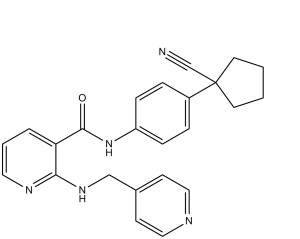
| SMILES |
O=C(NC1=CC=C(C2(C#N)CCCC2)C=C1)C3=C(NCC4=CC=NC=C4)N=CC=C3
|
| InChi Key |
WPEWQEMJFLWMLV-UHFFFAOYSA-N
|
| InChi Code |
InChI=1S/C24H23N5O/c25-17-24(11-1-2-12-24)19-5-7-20(8-6-19)29-23(30)21-4-3-13-27-22(21)28-16-18-9-14-26-15-10-18/h3-10,13-15H,1-2,11-12,16H2,(H,27,28)(H,29,30)
|
| 化学名 |
N-[4-(1-cyanocyclopentyl)phenyl]-2-(pyridin-4-ylmethylamino)pyridine-3-carboxamide
|
| 别名 |
YN968D1; YN-968D1; YN 968D1; Rivoceranib; Apatinib; 811803-05-1; rivoceranib; Apatinib free base; Apatinib (free base); YN968D1; N-(4-(1-Cyanocyclopentyl)phenyl)-2-((pyridin-4-ylmethyl)amino)nicotinamide; N-[4-(1-cyanocyclopentyl)phenyl]-2-(pyridin-4-ylmethylamino)pyridine-3-carboxamide; Apatinib free base
|
| HS Tariff Code |
2934.99.9001
|
| 存储方式 |
Powder -20°C 3 years 4°C 2 years In solvent -80°C 6 months -20°C 1 month |
| 运输条件 |
Room temperature (This product is stable at ambient temperature for a few days during ordinary shipping and time spent in Customs)
|
| 溶解度 (体外实验) |
|
|||
|---|---|---|---|---|
| 溶解度 (体内实验) |
注意: 如下所列的是一些常用的体内动物实验溶解配方,主要用于溶解难溶或不溶于水的产品(水溶度<1 mg/mL)。 建议您先取少量样品进行尝试,如该配方可行,再根据实验需求增加样品量。
注射用配方
注射用配方1: DMSO : Tween 80: Saline = 10 : 5 : 85 (如: 100 μL DMSO → 50 μL Tween 80 → 850 μL Saline)(IP/IV/IM/SC等) *生理盐水/Saline的制备:将0.9g氯化钠/NaCl溶解在100 mL ddH ₂ O中,得到澄清溶液。 注射用配方 2: DMSO : PEG300 :Tween 80 : Saline = 10 : 40 : 5 : 45 (如: 100 μL DMSO → 400 μL PEG300 → 50 μL Tween 80 → 450 μL Saline) 注射用配方 3: DMSO : Corn oil = 10 : 90 (如: 100 μL DMSO → 900 μL Corn oil) 示例: 以注射用配方 3 (DMSO : Corn oil = 10 : 90) 为例说明, 如果要配制 1 mL 2.5 mg/mL的工作液, 您可以取 100 μL 25 mg/mL 澄清的 DMSO 储备液,加到 900 μL Corn oil/玉米油中, 混合均匀。 View More
注射用配方 4: DMSO : 20% SBE-β-CD in Saline = 10 : 90 [如:100 μL DMSO → 900 μL (20% SBE-β-CD in Saline)] 口服配方
口服配方 1: 悬浮于0.5% CMC Na (羧甲基纤维素钠) 口服配方 2: 悬浮于0.5% Carboxymethyl cellulose (羧甲基纤维素) 示例: 以口服配方 1 (悬浮于 0.5% CMC Na)为例说明, 如果要配制 100 mL 2.5 mg/mL 的工作液, 您可以先取0.5g CMC Na并将其溶解于100mL ddH2O中,得到0.5%CMC-Na澄清溶液;然后将250 mg待测化合物加到100 mL前述 0.5%CMC Na溶液中,得到悬浮液。 View More
口服配方 3: 溶解于 PEG400 (聚乙二醇400) 请根据您的实验动物和给药方式选择适当的溶解配方/方案: 1、请先配制澄清的储备液(如:用DMSO配置50 或 100 mg/mL母液(储备液)); 2、取适量母液,按从左到右的顺序依次添加助溶剂,澄清后再加入下一助溶剂。以 下列配方为例说明 (注意此配方只用于说明,并不一定代表此产品 的实际溶解配方): 10% DMSO → 40% PEG300 → 5% Tween-80 → 45% ddH2O (或 saline); 假设最终工作液的体积为 1 mL, 浓度为5 mg/mL: 取 100 μL 50 mg/mL 的澄清 DMSO 储备液加到 400 μL PEG300 中,混合均匀/澄清;向上述体系中加入50 μL Tween-80,混合均匀/澄清;然后继续加入450 μL ddH2O (或 saline)定容至 1 mL; 3、溶剂前显示的百分比是指该溶剂在最终溶液/工作液中的体积所占比例; 4、 如产品在配制过程中出现沉淀/析出,可通过加热(≤50℃)或超声的方式助溶; 5、为保证最佳实验结果,工作液请现配现用! 6、如不确定怎么将母液配置成体内动物实验的工作液,请查看说明书或联系我们; 7、 以上所有助溶剂都可在 Invivochem.cn网站购买。 |
| 制备储备液 | 1 mg | 5 mg | 10 mg | |
| 1 mM | 2.0260 mL | 10.1301 mL | 20.2601 mL | |
| 5 mM | 0.4052 mL | 2.0260 mL | 4.0520 mL | |
| 10 mM | 0.2026 mL | 1.0130 mL | 2.0260 mL |
1、根据实验需要选择合适的溶剂配制储备液 (母液):对于大多数产品,InvivoChem推荐用DMSO配置母液 (比如:5、10、20mM或者10、20、50 mg/mL浓度),个别水溶性高的产品可直接溶于水。产品在DMSO 、水或其他溶剂中的具体溶解度详见上”溶解度 (体外)”部分;
2、如果您找不到您想要的溶解度信息,或者很难将产品溶解在溶液中,请联系我们;
3、建议使用下列计算器进行相关计算(摩尔浓度计算器、稀释计算器、分子量计算器、重组计算器等);
4、母液配好之后,将其分装到常规用量,并储存在-20°C或-80°C,尽量减少反复冻融循环。
计算结果:
工作液浓度: mg/mL;
DMSO母液配制方法: mg 药物溶于 μL DMSO溶液(母液浓度 mg/mL)。如该浓度超过该批次药物DMSO溶解度,请首先与我们联系。
体内配方配制方法:取 μL DMSO母液,加入 μL PEG300,混匀澄清后加入μL Tween 80,混匀澄清后加入 μL ddH2O,混匀澄清。
(1) 请确保溶液澄清之后,再加入下一种溶剂 (助溶剂) 。可利用涡旋、超声或水浴加热等方法助溶;
(2) 一定要按顺序加入溶剂 (助溶剂) 。
| NCT Number | Recruitment | interventions | Conditions | Sponsor/Collaborators | Start Date | Phases |
| NCT03742193 | Active Recruiting |
Drug: Apatinib Drug: GD regimen |
Apatinib Osteosarcoma |
Ruijin Hospital | August 11, 2019 | Phase 2 |
| NCT06081595 | Not yet recruiting | Drug: Fluzoparib Drug: Apatinib |
Relapsed Ovarian Cancer | Jin Li | October 30, 2023 | Phase 2 |
| NCT04824352 | Recruiting | Drug: apatinib | Effect of Drug Toxicity, Drug |
Peking University People's Hospital | April 1, 2021 | Phase 2 |
| NCT05235100 | Recruiting | Drug: Apatinib Mesylate | Trunk Extremity |
Chinese Academy of Medical Sciences |
September 1, 2021 | Phase 2 |
| NCT04863430 | Recruiting | Drug: Apatinib Drug: Oxaliplatin |
Gastric Cancer | Peking University | May 11, 2021 | Phase 2 |
Effects of YN968D1 on various growth factor‐stimulated receptor phosphorylation at the cellular level detected by western blot analysis.Cancer Sci.2011 Jul;102(7):1374-80. |
Inhibition of vascular endothelial growth factor (VEGF)‐stimulated HUVEC proliferation, HUVEC tubule formation, HUVEC migration and microvessel outgrowth from rat aortic ring by YN968D1. |
Antitumor activity of YN968D1 against human tumor xenografts in nude mice.Cancer Sci.2011 Jul;102(7):1374-80. |
 VEGFR2-IN-7
 SYHA1813
 VEGFR-2-IN-38
 BHEP
BHEP
InvivoChem的所有产品仅用于作科学研究,不面向患者销售
Copyright 2020 InvivoChem LLC | All Rights Reserved 粤ICP备20063088号-1


 COA
COA


 463611831
