| 规格 | 价格 | 库存 | 数量 |
|---|---|---|---|
| 50mg |
|
||
| 100mg |
|
||
| 250mg |
|
||
| 500mg |
|
||
| 1g |
|
||
| 2g |
|
||
| Other Sizes |
|

| 靶点 |
EGFR; mTOR
Chrysophanic Acid (Chrysophanol) exerts antiviral activity against herpes simplex virus type 1 (HSV-1) and type 2 (HSV-2) in Vero cells, with an EC50 of 12.5 μg/mL for HSV-1 and 15.8 μg/mL for HSV-2. Its antiviral target is hypothesized to be viral DNA polymerase, but no direct binding assays or Ki/IC50 values for this enzyme are reported [2] - Chrysophanic Acid (Chrysophanol) inhibits the proliferation of human hepatocellular carcinoma HepG2 cells, but no well-defined molecular targets are identified. It may act via indirect regulation of cell cycle and apoptotic pathways, with no IC50/Ki values for specific targets provided [1] |
|---|---|
| 体外研究 (In Vitro) |
体外活性:大黄酸 (Chrysophanol) 是一种 EGFR/mTOR 通路抑制剂。大黄酸 (Chrysophanol) 是一种天然蒽醌,在 EGFR 过表达的 SNU-C5 人结肠癌细胞中具有抗癌活性。大黄酸 (Chrysophanol) 优先阻止 SNU-C5 细胞的增殖,但不会阻止其他 EGFR 表达水平较低的细胞系(HT7、HT29、KM12C、SW480、HCT116 和 SNU-C4)的增殖。 SNU-C5 细胞中的大黄酸 (Chrysophanol) 处理可抑制 EGF 诱导的 EGFR 磷酸化,并抑制下游信号分子的激活,例如 AKT、细胞外信号调节激酶 (ERK) 和哺乳动物雷帕霉素靶标 (mTOR)/核糖体蛋白S6 激酶 (p70S6K)。大黄酸还抑制 2 型和 3 型脊髓灰质炎病毒(小核糖核酸病毒科)的复制以及 BGM(水牛绿猴)肾细胞中脊髓灰质炎病毒诱导的细胞病变效应。细胞测定:将细胞以 5×10³ 个细胞/mL 接种在 96 孔微孔板中,并贴壁 24 小时。将大黄酚(20、50、80 和 120 μM)以不同的浓度(最高 120 μM)添加到培养基中并添加不同的持续时间。处理后,通过细胞计数试剂盒-8 (CCK-8) 评估细胞毒性和/或增殖。简而言之,高水溶性四唑盐 WST-8 产生橙色水溶性产物甲臜。细胞内脱氢酶产生的甲臜染料量与活细胞数量成正比。每孔加入CCK-8(10 μL),37℃孵育3 h,然后通过以下方法评估细胞增殖和细胞毒性:使用酶标仪测量 450 nm 处的吸光度。每个实验条件使用三个重复孔
对HepG2细胞的抗增殖活性(文献[1]): 1. Chrysophanic Acid (Chrysophanol,大黄酚) 呈剂量依赖性抑制HepG2细胞增殖:MTT实验(孵育72小时)显示IC50为45.2 μM;60 μM浓度时,活细胞数量较溶媒对照组(0.1% DMSO)减少68%。 2. 细胞周期分析(PI染色,处理48小时)显示G2/M期阻滞:50 μM Chrysophanic Acid (Chrysophanol,大黄酚) 使G2/M期细胞比例从对照组的14.3%升至32.6%,G1期细胞比例从58.2%降至39.8%。 3. 凋亡诱导(TUNEL染色):50 μM Chrysophanic Acid (Chrysophanol,大黄酚) 处理48小时,凋亡细胞数量较对照组增加2.8倍 [1] - 对HSV的抗病毒活性(文献[2]): 1. Vero细胞空斑减少实验:Chrysophanic Acid (Chrysophanol,大黄酚) 减少HSV-1(KOS株)和HSV-2(333株)空斑形成,EC50分别为12.5 μg/mL(HSV-1)和15.8 μg/mL(HSV-2);25 μg/mL浓度时,对HSV-1和HSV-2空斑形成的抑制率分别为85%和78%。 2. 加药时间实验:该化合物作用于病毒复制的“进入后阶段”——感染后2小时(hpi)加药仍可抑制HSV-1复制70%,感染后6小时加药则抑制率降至25%。 3. 病毒DNA合成抑制(3H-胸苷掺入实验):20 μg/mL Chrysophanic Acid (Chrysophanol,大黄酚) 使HSV-1 DNA合成较感染对照组减少65% [2] |
| 体内研究 (In Vivo) |
大黄酚 (CA) 可改善 C57BL/6 小鼠 HFD 诱导的肥胖。在雄性 C57BL/6J 小鼠中进行大黄酚的体内性能以确定所施用的大黄酚的功效。喂食 HFD 的小鼠体重明显高于喂食标准饮食的小鼠。另一方面,大黄酚组的体重增加明显小于未处理的HFD。 16周后,HFD组的小鼠体重增加了23.92±1.74克,而大黄酚组的小鼠体重增加了16.72±2克
|
| 细胞实验 |
在 96 孔微孔板中,细胞以 5×10³ 细胞/mL 接种,并给予 24 小时贴壁。培养基中添加了不同浓度(最高 120 μM)的大黄酚(20、50、80 和 120 μM),添加时间也不同。 Cell Counting Kit-8 用于测量处理细胞 (CCK-8) 的细胞毒性和/或增殖。简而言之,甲臜是一种橙色水溶性产品,由高水溶性四唑盐 WST-8 生产。活细胞的数量与细胞脱氢酶产生的甲臜染料的量成正比。每孔加入10 μL CCK-8,37℃放置3小时后,用酶标仪测定450 nm处的吸光度,以确定细胞的细胞毒性和增殖情况。对于每个实验条件,使用三个重复孔[1]。
HepG2细胞抗增殖与细胞周期实验(文献[1]): 1. 细胞接种:将HepG2细胞分别接种于96孔板(MTT实验,2×10³个细胞/孔)或6孔板(细胞周期分析,2×10⁵个细胞/孔),用含10%胎牛血清(FBS)的RPMI-1640培养基在37°C(5% CO₂)条件下过夜培养。 2. 药物处理:向细胞中加入系列浓度的Chrysophanic Acid (Chrysophanol,大黄酚)(10–80 μM,溶于DMSO),每个浓度设3个复孔;同时设置溶媒对照组(0.1% DMSO)。 3. MTT实验:孵育72小时后,每孔加入20 μL MTT溶液(5 mg/mL),继续孵育4小时。去除上清液,加入150 μL DMSO溶解甲瓒结晶,测定570 nm处吸光度。细胞活力计算公式为(样品组A570/对照组A570)×100%,使用GraphPad Prism计算IC50。 4. 细胞周期分析:处理48小时后,胰酶消化收集细胞,用冷PBS洗涤2次,4°C下用70%乙醇固定过夜。固定后的细胞用100 μg/mL RNase A在37°C处理30分钟,黑暗中用50 μg/mL碘化丙啶(PI)染色15分钟,通过流式细胞仪(BD FACSCanto)分析 [1] - HSV空斑减少实验(文献[2]): 1. 细胞制备:将Vero细胞接种于6孔板(5×10⁵个细胞/孔),在37°C(5% CO₂)条件下过夜培养形成融合单层。 2. 病毒吸附:将HSV-1(KOS株)或HSV-2(333株)稀释至100空斑形成单位(PFU)/孔,加入细胞单层,37°C孵育1小时以允许病毒吸附。 3. 药物处理:去除未结合的病毒,将系列浓度的Chrysophanic Acid (Chrysophanol,大黄酚)(5–40 μg/mL,溶于DMSO)与1%甲基纤维素(溶于最低必需培养基MEM)混合后覆盖于细胞表面;设置溶媒对照组(0.1% DMSO的1%甲基纤维素溶液)。 4. 空斑计数:37°C孵育72小时后,用4%多聚甲醛固定细胞,0.1%结晶紫染色,计数空斑。空斑减少率计算公式为(对照组空斑数-样品组空斑数)/对照组空斑数×100%,并计算EC50 [2] |
| 动物实验 |
mg/kg 小鼠:实验开始前,使用 4 周龄雄性 C57BL/6J 小鼠进行为期一周的维持期饲养。在无特定病原体 (SPF) 动物房中,小鼠饲养于 12 小时光照/黑暗循环条件下,并可自由饮水和食用实验室饲料。小鼠接受高脂高热量饮食 (HFD) 以诱导肥胖。对照组 (C) 喂食商业标准饲料。HFD 组 (HFD) 小鼠仅喂食 HFD。HFD + CA 组 (CA):在给予 5 mg/kg/天的酪蛋白酚之前,小鼠接受为期四周的 HFD 喂养。在接下来的 16 周内,将小鼠分为三组 (n = 5),分别喂食三种不同的饲料:标准饲料、HFD 和 HFD + 酪蛋白酚。每周测量三次食物摄入量和体重。
|
| 药代性质 (ADME/PK) |
吸收、分布和排泄
本研究对掌叶大黄提取物中的五种蒽醌类化合物(芦荟大黄素、大黄素、大黄酸、金丝桃素和大黄酚)在正常大鼠和血栓性局灶性脑缺血(TFCI)模型大鼠中的口服药代动力学进行了比较研究。血浆样品经固相萃取澄清后,采用经验证的高效液相色谱-荧光联用系统同时测定蒽醌类化合物。结果表明,TFCI模型大鼠体内芦荟大黄素、大黄酸、大黄素和金丝桃素的Cmax、t1/2和AUC0-t值几乎是正常大鼠的两倍,而CL值则显著降低(p < 0.05)。五种蒽醌类药物在大鼠体内的血浆药物浓度-时间数据符合二室开放模型。两组大鼠血浆中这五种蒽醌类药物均表现为吸收迅速、消除缓慢。所得结果有助于评估该药物在临床应用中的疗效和安全性。 民族药理学意义:曲郁清热颗粒(QYQRGs)是治疗血瘀证的常用中药复方制剂。比较正常兔和血瘀证兔服用QYQRG后各成分的药代动力学差异,可提供有价值的信息。本研究的主要目的是比较正常兔和急性血瘀模型兔口服2.0 g/kg体重QYQRG后大黄素和金丝桃素的药代动力学。材料与方法:分别于口服QYQRG后5、10、15、20、30、45、60、75、90、120、240、360和480分钟采集血样。采用高效液相色谱法(HPLC)测定兔血浆中大黄素和金丝桃酚的浓度,并获得主要药代动力学参数。结果:急性血瘀模型兔血浆中大黄素和金丝桃酚的药代动力学参数AUC(0-∞)、T(lag)、Cmax和K21均与正常兔存在显著差异。此外,大黄素的A、β、MRT和T(1/2β)以及金丝桃酚的a和T1/2a在正常兔和急性血瘀模型兔之间也存在显著差异。结论:在急性血瘀兔模型中,大黄素和金丝桃酚的吸收时间加快,吸收量增加,提示大黄素和金丝桃酚可能是QYQRG中的两种有效成分。 研究目的:本研究比较了大黄蒽醌衍生物(AQs)在正常大鼠和四氯化碳(CCl4)诱导的肝损伤大鼠中的组织分布,以探讨其吸收是否存在差异,并探究AQs在病理模型大鼠和正常大鼠组织分布水平上毒性不同的可能原因。材料与方法:将大黄总提取物(基于粗提物量,每日14.49 g/kg体重)灌胃给予正常大鼠和模型大鼠,持续12周。采用液相色谱-串联质谱法(LC-MS)定量分析组织中游离蒽醌类化合物(AQs)的浓度。停药4周后,再次测定组织分布。结果:在肝脏、肾脏和脾脏中均检测到了五种游离AQs——芦荟大黄素、大黄酸、大黄素、金丝桃素和大黄酚,但仅大黄酸、芦荟大黄素和大黄素的浓度达到定量限。正常大鼠组织中大黄酸(p < 0.001)、芦荟大黄素(p < 0.001)和大黄素(p < 0.05)的浓度均高于模型大鼠,肾脏和脾脏组织中大黄酸的浓度顺序为大黄酸>芦荟大黄素>大黄素,肝脏组织中大黄酸>大黄酸>大黄素。停药4周后,组织中未检测到游离AQs。结论:这些结果表明,正常动物体内蒽醌类化合物的组织毒性高于病理模型动物,且大黄的累积毒性较小。该结果与中国传统医学著作《素问》中记载的“幽谷物云”理论相符。 代谢/代谢产物 本研究旨在阐明天然存在的1,8-二羟基蒽醌生物转化过程中涉及的酶,并探讨1,8-二羟基蒽醌的生物转化是否构成一种生物活化途径。我们首先研究了大黄素(1,3,8-三羟基-6-甲基蒽醌)的代谢,大黄素是一种存在于药物制剂中的化合物。在大鼠肝微粒体中,我们观察到了两种大黄素代谢产物——ω-羟基大黄素和2-羟基大黄素的生成。用不同细胞色素P450酶诱导剂预处理的大鼠肝微粒体中ω-羟基大黄素的生成速率没有差异。因此,ω-羟基大黄素的生成似乎是由几种细胞色素P450酶以较低的速率催化的。用3-甲基胆蒽预处理的大鼠肝微粒体中2-羟基大黄素的生成增加,并且被α-萘黄酮、抗大鼠细胞色素P450 1A1/2抗体以及(程度较轻)抗大鼠细胞色素P450 1A1抗体抑制。这些数据表明细胞色素P450 1A2参与了该代谢物的生成。然而,其他细胞色素P450酶似乎也催化了该反应。蒽醌类化合物大黄酚(1,8-二羟基-3-甲基蒽醌)经细胞色素P450依赖性氧化转化为芦荟大黄素(1,8-二羟基-3-羟甲基蒽醌),后者是主要代谢产物。在小鼠淋巴瘤L5178Y细胞的体外微核试验中,比较了母体二羟基蒽醌及其代谢产物的致突变性。与大黄素相比,2-羟基大黄素诱导的微核频率显著更高;与大黄素相比,ω-羟基大黄素诱导的微核频率更低;芦荟大黄素诱导的微核频率显著高于大黄酚。这些数据表明,大黄素和金丝桃酚的细胞色素P450依赖性生物转化可能是这些化合物的生物活化途径。 金丝桃酚是许多传统中药中的主要蒽醌类成分,被认为是一种重要的活性成分,具有抗菌和抗癌等多种药理作用。先前的研究表明,暴露于金丝桃酚会诱导细胞毒性,但其毒性机制尚不清楚。在本代谢研究中,在添加了谷胱甘肽(GSH)的金丝桃酚大鼠和人肝微粒体孵育液中检测到了三种氧化代谢物(M1-M3、芦荟大黄素、7-羟基金丝桃酚和2-羟基金丝桃酚)和五种GSH结合物(M4-M8),除M4和M5外,其他代谢物的生成均依赖于NADPH。 M4 和 M5 直接来源于母体化合物大黄酚,M6 来源于 M2,M7 和 M8 则是由 M4 和 M5 氧化而来。在大鼠暴露于大黄酚后,胆汁中也观察到了代谢物 M5 和 M6;在给予大黄酚的大鼠尿液中检测到了 M1-M3 和一种 NAC 结合物 (M9),尿液代谢物 M9 来源于胆汁中 GSH 结合物 M6 的降解。重组 P450 酶孵育和微粒体抑制实验表明,P450 1A2 是负责大黄酚代谢活化的主要酶,而 P450 2B6 和 P450 3A4 也参与了氧化代谢物的生成。 |
| 毒性/毒理 (Toxicokinetics/TK) |
体外毒性(文献[1]):对于正常人肝细胞(LO2细胞),50 μM 大黄酸(大黄酚)(孵育72小时)可使细胞存活率保持在80%以上(MTT法),表明其固有细胞毒性较低[1]
- 体外毒性(文献[2]):对于Vero细胞,大黄酸(大黄酚)的半数细胞毒性浓度(CC50)为85.3 μg/mL。HSV-1的治疗指数(TI = CC50/EC50)为6.8,HSV-2的治疗指数为5.4[2] - [1]或[2]中未报道大黄酸(大黄酚)的体内毒性数据(例如,LD50、肝毒性、肾毒性、血浆蛋白结合率)[1,2] |
| 参考文献 | |
| 其他信息 |
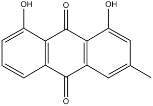
大黄酸呈金黄色片状或棕色粉末状,熔点196℃,微溶于水。淡黄色水溶液遇碱变红,浓硫酸溶液呈红色。(NTP, 1992)
大黄酚是一种三羟基蒽醌,是金盏花素在C-3位被甲基取代的结构。它已从芦荟中分离得到,具有抗病毒和抗炎活性。它可用作抗病毒剂、抗炎剂和植物代谢产物。它在功能上与金星素相关。 据报道,在塔拉霉属(Talaromyces islandicus)、夏枯菌(Ramularia uredinicola)和其他有相关数据的生物体中均发现了金星醇。 另见:弗兰古拉·普尔希亚纳树皮(部分)。 金星酸(金星醇)是一种天然蒽醌衍生物,从大黄属(Rheum)药用植物(例如,大黄,俗称大黄)和其他植物物种中分离得到。它因其抗炎和通便作用,在传统医学中有着悠久的应用历史[1,2] - 抗病毒机制(文献[2]):大黄酸(大黄酚)的抗病毒活性被认为涉及抑制病毒DNA合成,可能是通过干扰HSV DNA聚合酶的活性实现的,但尚未提供直接的实验证据(例如,酶抑制试验)[2] - 抗增殖机制(文献[1]):大黄酸(大黄酚)对HepG2细胞的抗增殖作用与G2/M期细胞周期阻滞和诱导细胞凋亡有关,但本研究并未探讨所涉及的具体信号通路(例如,p53、MAPK)[1] - 临床开发状态:尚未报道大黄酸(大黄酚)的临床开发数据(例如,用于治疗癌症或病毒感染);其生物活性目前仅限于临床前体外研究[1,2] |
| 分子式 |
C15H10O4
|
|
|---|---|---|
| 分子量 |
254.24
|
|
| 精确质量 |
254.057
|
|
| 元素分析 |
C, 70.86; H, 3.96; O, 25.17
|
|
| CAS号 |
481-74-3
|
|
| 相关CAS号 |
|
|
| PubChem CID |
10208
|
|
| 外观&性状 |
Yellow to orange solid powder
|
|
| 密度 |
1.5±0.1 g/cm3
|
|
| 沸点 |
489.5±45.0 °C at 760 mmHg
|
|
| 熔点 |
194-198 °C
|
|
| 闪点 |
263.9±25.2 °C
|
|
| 蒸汽压 |
0.0±1.3 mmHg at 25°C
|
|
| 折射率 |
1.710
|
|
| LogP |
5.03
|
|
| tPSA |
74.6
|
|
| 氢键供体(HBD)数目 |
2
|
|
| 氢键受体(HBA)数目 |
4
|
|
| 可旋转键数目(RBC) |
0
|
|
| 重原子数目 |
19
|
|
| 分子复杂度/Complexity |
405
|
|
| 定义原子立体中心数目 |
0
|
|
| SMILES |
O([H])C1=C([H])C(C([H])([H])[H])=C([H])C2C(C3C([H])=C([H])C([H])=C(C=3C(C=21)=O)O[H])=O
|
|
| InChi Key |
LQGUBLBATBMXHT-UHFFFAOYSA-N
|
|
| InChi Code |
InChI=1S/C15H10O4/c1-7-5-9-13(11(17)6-7)15(19)12-8(14(9)18)3-2-4-10(12)16/h2-6,16-17H,1H3
|
|
| 化学名 |
1,8-dihydroxy-3-methylanthracene-9,10-dione
|
|
| 别名 |
|
|
| HS Tariff Code |
2934.99.9001
|
|
| 存储方式 |
Powder -20°C 3 years 4°C 2 years In solvent -80°C 6 months -20°C 1 month |
|
| 运输条件 |
Room temperature (This product is stable at ambient temperature for a few days during ordinary shipping and time spent in Customs)
|
| 溶解度 (体外实验) |
|
|||
|---|---|---|---|---|
| 溶解度 (体内实验) |
注意: 如下所列的是一些常用的体内动物实验溶解配方,主要用于溶解难溶或不溶于水的产品(水溶度<1 mg/mL)。 建议您先取少量样品进行尝试,如该配方可行,再根据实验需求增加样品量。
注射用配方
注射用配方1: DMSO : Tween 80: Saline = 10 : 5 : 85 (如: 100 μL DMSO → 50 μL Tween 80 → 850 μL Saline)(IP/IV/IM/SC等) *生理盐水/Saline的制备:将0.9g氯化钠/NaCl溶解在100 mL ddH ₂ O中,得到澄清溶液。 注射用配方 2: DMSO : PEG300 :Tween 80 : Saline = 10 : 40 : 5 : 45 (如: 100 μL DMSO → 400 μL PEG300 → 50 μL Tween 80 → 450 μL Saline) 注射用配方 3: DMSO : Corn oil = 10 : 90 (如: 100 μL DMSO → 900 μL Corn oil) 示例: 以注射用配方 3 (DMSO : Corn oil = 10 : 90) 为例说明, 如果要配制 1 mL 2.5 mg/mL的工作液, 您可以取 100 μL 25 mg/mL 澄清的 DMSO 储备液,加到 900 μL Corn oil/玉米油中, 混合均匀。 View More
注射用配方 4: DMSO : 20% SBE-β-CD in Saline = 10 : 90 [如:100 μL DMSO → 900 μL (20% SBE-β-CD in Saline)] 口服配方
口服配方 1: 悬浮于0.5% CMC Na (羧甲基纤维素钠) 口服配方 2: 悬浮于0.5% Carboxymethyl cellulose (羧甲基纤维素) 示例: 以口服配方 1 (悬浮于 0.5% CMC Na)为例说明, 如果要配制 100 mL 2.5 mg/mL 的工作液, 您可以先取0.5g CMC Na并将其溶解于100mL ddH2O中,得到0.5%CMC-Na澄清溶液;然后将250 mg待测化合物加到100 mL前述 0.5%CMC Na溶液中,得到悬浮液。 View More
口服配方 3: 溶解于 PEG400 (聚乙二醇400) 请根据您的实验动物和给药方式选择适当的溶解配方/方案: 1、请先配制澄清的储备液(如:用DMSO配置50 或 100 mg/mL母液(储备液)); 2、取适量母液,按从左到右的顺序依次添加助溶剂,澄清后再加入下一助溶剂。以 下列配方为例说明 (注意此配方只用于说明,并不一定代表此产品 的实际溶解配方): 10% DMSO → 40% PEG300 → 5% Tween-80 → 45% ddH2O (或 saline); 假设最终工作液的体积为 1 mL, 浓度为5 mg/mL: 取 100 μL 50 mg/mL 的澄清 DMSO 储备液加到 400 μL PEG300 中,混合均匀/澄清;向上述体系中加入50 μL Tween-80,混合均匀/澄清;然后继续加入450 μL ddH2O (或 saline)定容至 1 mL; 3、溶剂前显示的百分比是指该溶剂在最终溶液/工作液中的体积所占比例; 4、 如产品在配制过程中出现沉淀/析出,可通过加热(≤50℃)或超声的方式助溶; 5、为保证最佳实验结果,工作液请现配现用! 6、如不确定怎么将母液配置成体内动物实验的工作液,请查看说明书或联系我们; 7、 以上所有助溶剂都可在 Invivochem.cn网站购买。 |
| 制备储备液 | 1 mg | 5 mg | 10 mg | |
| 1 mM | 3.9333 mL | 19.6665 mL | 39.3329 mL | |
| 5 mM | 0.7867 mL | 3.9333 mL | 7.8666 mL | |
| 10 mM | 0.3933 mL | 1.9666 mL | 3.9333 mL |
1、根据实验需要选择合适的溶剂配制储备液 (母液):对于大多数产品,InvivoChem推荐用DMSO配置母液 (比如:5、10、20mM或者10、20、50 mg/mL浓度),个别水溶性高的产品可直接溶于水。产品在DMSO 、水或其他溶剂中的具体溶解度详见上”溶解度 (体外)”部分;
2、如果您找不到您想要的溶解度信息,或者很难将产品溶解在溶液中,请联系我们;
3、建议使用下列计算器进行相关计算(摩尔浓度计算器、稀释计算器、分子量计算器、重组计算器等);
4、母液配好之后,将其分装到常规用量,并储存在-20°C或-80°C,尽量减少反复冻融循环。
计算结果:
工作液浓度: mg/mL;
DMSO母液配制方法: mg 药物溶于 μL DMSO溶液(母液浓度 mg/mL)。如该浓度超过该批次药物DMSO溶解度,请首先与我们联系。
体内配方配制方法:取 μL DMSO母液,加入 μL PEG300,混匀澄清后加入μL Tween 80,混匀澄清后加入 μL ddH2O,混匀澄清。
(1) 请确保溶液澄清之后,再加入下一种溶剂 (助溶剂) 。可利用涡旋、超声或水浴加热等方法助溶;
(2) 一定要按顺序加入溶剂 (助溶剂) 。
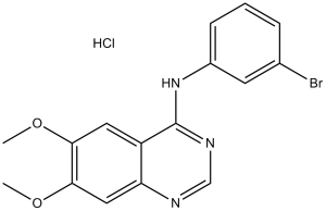 PD153035 HCl (SU5271; ZM252868)
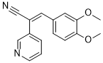 RG 13022
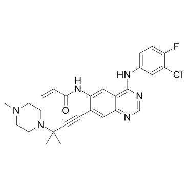 AV-412
 O-去甲基埃罗替尼盐酸盐
InvivoChem的所有产品仅用于作科学研究,不面向患者销售
Copyright 2020 InvivoChem LLC | All Rights Reserved 粤ICP备20063088号-1


 COA
COA
 463611831
