| 规格 | 价格 | 库存 | 数量 |
|---|---|---|---|
| 2mg |
|
||
| 5mg |
|
||
| 10mg |
|
||
| 25mg |
|
||
| 50mg |
|
||
| 100mg |
|
||
| 250mg |
|
||
| 500mg |
|
||
| Other Sizes |
|
| 靶点 |
CDK9 (IC50 = 34 nM); CDK2 (IC50 = 240 nM); CDK1 (IC50 = 250 nM); CDK5 (IC50 = 460 nM); GSK3-β (IC50 = 220 nM); Mk2 (IC50 = 470 nM); Plk1 (IC50 = 980 nM); Chk2 (IC50 = 1100 nM)
PHA-767491 is a dual-specificity inhibitor of Cell Division Cycle 7 (Cdc7)-Dbf4 kinase (DDK) and Cyclin-Dependent Kinase 9 (Cdk9)-Cyclin T1 (P-TEFb) complex, with weak activity against other CDKs or kinases. It targets DNA replication initiation (via DDK) and transcriptional elongation (via Cdk9), exerting synergistic antitumor effects. - For human Cdc7-Dbf4 (DDK) kinase (recombinant, radiometric assay): IC₅₀ = 10 nM [4] - For human Cdk9-Cyclin T1 (P-TEFb) kinase (recombinant, HTRF assay): IC₅₀ = 5 nM [4] - For human Cdk1-Cyclin B1 (recombinant, radiometric assay): IC₅₀ = 450 nM (≈45-fold less potent than DDK) [4] - For human Cdk2-Cyclin E (recombinant, radiometric assay): IC₅₀ = 620 nM [4] - For 20+ other kinases (e.g., Cdk3, Cdk4, EGFR, AKT; panel screening): IC₅₀ > 1000 nM (no significant inhibition) [4] - For DDK-mediated RPA32 phosphorylation in HCT116 cells (cell-based assay): EC₅₀ = 15 nM [4] - For Cdk9-mediated RNA Pol II CTD phosphorylation in HepG2 cells (cell-based assay): EC₅₀ = 8 nM [2] |
|---|---|
| 体外研究 (In Vitro) |
体外活性:PHA-767491 对 Cdk1、Cdk2 和 GSK3-β 的选择性约为 20 倍,对 MK2 和 Cdk5 的选择性约为 50 倍,对 PLK1 和 CHK2 的选择性约为 100 倍。 PHA-767491 可抑制多种人类细胞系的细胞增殖,SF-268 的 IC50 为 0.86 μM,K562 的 IC50 为 5.87 μM,并且与 5-FU 或吉西他滨仅在少数细胞系中起作用。与目前的 DNA 合成抑制剂不同,5 μM 的 PHA-767491 处理会阻止 DNA 复制的启动,但不会阻止复制叉的进展,因为它对 Cdc7 激酶和 Cdc7 依赖性 Ser40 位点的 Mcm2 磷酸化有特异性抑制。 3 μM 的 PHA-767491 处理可显着降低 ABT-737 耐药性 OCI-LY1 和 SU-DHL-4 细胞中上调的 Mcl-1 水平,这可能是由于 Cdk9 的抑制,从而恢复对 ABT-737 敏感。当以 1 μM 浓度应用于静态慢性淋巴细胞白血病 (CLL) 细胞时,通过类似的机制也观察到 PHA-767491 的直接线粒体依赖性促凋亡作用,EC50 为 0.34-0.97 μM。在 CD154 和 IL-4 刺激的 CLL 细胞增殖中,5 μM 的 PHA-767491 处理通过抑制 Cdc7 来消除 DNA 合成,而不是触发细胞死亡。激酶测定:PHA-767491 对 Cdc7 和 Cdk9 的抑制 (IC50) 使用基于强阴离子交换剂(Dowex 1-X8 树脂,甲酸盐形式)的测定来确定。对于每种酶,首先确定 ATP 和特定底物的绝对 Km 值,然后以优化的 ATP/33P-γ-ATP 混合物 (2Km) 和底物 (5Km) 浓度运行每次测定。 Cdc7 激酶测定在含有 50 mM Hepes pH 7.9、15 mM MgCl2、2 mM β-磷酸甘油、0.2 mg/mL BSA、1 mM DTT、3 μM Na3VO4、2Km ATP/33P-γ-ATP 混合物、5Km 的缓冲液中进行Mcm2 (aa 10-294)、37 nM 重组 Cdc7/Dbf4 和浓度不断增加的 PHA-767491,最终体积为 30 μL,并在 25 °C 下孵育 1 小时。使用 50 mM HEPES pH 7.5、10 mM MgCl2、1 mM DTT、3 μM Na3VO4、2Km ATP/33P-γ-ATP 混合物、5Km RNA 聚合酶 CDT 肽和增量溶液中的 50 nM 重组 Cdk9/cyclin T 进行 Cdk9 激酶测定。将 PHA-767491 浓缩至最终体积 30 μL,并在 25 °C 下孵育 1 小时。孵育后,加入 150 μL 树脂/甲酸盐 (pH 3.0) 以停止反应并捕获未反应的 33P-γ-ATP,将其与溶液中的磷酸化底物分离。静置 1 小时后,将 50 μL 上清液转移至 Optiplate 96 孔板中。添加 150 μL Microscint 40 后,在 TopCount 中对放射性进行计数。细胞测定:将细胞(HeLa、MCF7、HCT-116、U2OS、A2780、K562、SF-539、SF-268、Ovcar8、SW480、COLO205、HCT-15、Jurkat、PC3 和 NHDF)暴露于 PHA-767491 24 或 72 小时。将细胞裂解,并使用基于热稳定性萤火虫荧光素酶的测定法测定孔中的 ATP 含量(用作活细胞的测量值)。 caspase-3 和 caspase-7 的激活通过基于荧光素酶的测定(包含特定的发光底物)以处理的样品与未处理的对照之间的比率来测量。 DNA 复制是通过流式细胞术将核苷酸类似物 BrdU 掺入 DNA 来测量的。
1. 对肝癌(HCC)细胞的抗增殖活性及与5-氟尿嘧啶(5-FU)的协同作用:PHA-767491(0.1–10 μM)抑制HepG2和Huh7肝癌细胞活力,GI₅₀分别为0.3 μM(HepG2)和0.4 μM(Huh7)(MTT实验)。与5-FU(0.5 μM)在HepG2细胞中协同作用(组合指数CI = 0.25):细胞活力降低90%,显著高于PHA-767491单药组(40%)或5-FU单药组(35%)。联合处理使活化caspase-3/PARP(western blot)及凋亡细胞(Annexin V-FITC/PI双染:65% vs. 单药组20%)增加 [2] 2. 抑制胶质母细胞瘤(GBM)细胞生长、侵袭及干性:PHA-767491(0.05–5 μM)抑制U87MG和U251 GBM细胞,GI₅₀分别为0.2 μM(U87MG)和0.25 μM(U251)。1 μM时,Transwell侵袭实验显示侵袭能力降低75%(U87MG),球形成实验(CSC标志物)显示球形成能力降低80%。Western blot显示p-Rb(细胞周期)、c-Myc(转录)及CD133(CSC标志物)降低60–70% [3] 3. 抑制DNA复制及诱导细胞周期阻滞:PHA-767491(0.1–2 μM)处理HCT116结肠癌细胞24小时,流式细胞术显示G1/S期阻滞(G1期细胞比例从40%升至70%)。BrdU掺入实验显示1 μM时DNA复制抑制85%。Western blot显示DDK底物(p-RPA32、p-MCM2)及Cdk9底物(p-RNA聚合酶II CTD)减少 [4] 4. 与其他DDK抑制剂的对比:PHA-767491(1 μM)在12种癌细胞系中显示比XL413(选择性DDK抑制剂)更广泛的抗肿瘤活性:8/12种细胞系活力降低>50%,而XL413仅4/12种,差异源于其双重Cdc7/Cdk9抑制作用 [1] |
| 体内研究 (In Vivo) |
PHA-767491 每天两次,持续 5 天,以剂量依赖性方式显着抑制 HL60 异种移植物的生长,剂量为 20 mg/kg 和 30 mg/kg 时,TGI 分别为 50% 和 92%。这也在 A2780、Mx-1 和 HCT-116 异种移植模型以及 DMBA 诱导的乳腺癌中被标记,并且与 Cdc7 抑制以及随后 Cdc7 依赖位点 Ser40 处 Mcm2 磷酸化的降低相关。
PHA-767491对肿瘤模型具有抗肿瘤活性[4] PHA-767491作为一种抗癌药物的潜力首次在裸鼠身上进行了评估,裸鼠携带来自急性髓性白血病(AML) HL60人细胞系的皮下植入肿瘤。在静脉注射20和30 mg kg−1两种剂量水平,每天两次,连续5天后,观察到相对于药物治疗的动物,肿瘤体积呈剂量依赖性减少(图4a)。在治疗结束后的第二天计算,肿瘤生长抑制在低剂量下为50%,在高剂量下为92%,其中8只动物中有5只观察到肿瘤消退的证据。在此条件下,化合物达到微摩尔血浆水平,浓度-时间曲线下面积(AUC)分别为47 μM h−1和71 μM h−1,与基于细胞的活性水平一致。PHA-767491在组织中表现出良好的体积分布(约为体内总含水量的两倍),并能迅速从血浆中清除(在线补充图7)。在这些剂量下,这种化合物似乎具有良好的耐受性,并且没有引起明显的体重减轻;然而,进一步的剂量增加是不能容忍的。在一项毒理学研究中,PHA-767491每天两次,剂量为30 mg kg−1,持续5天,未观察到临床症状或大体病变。从治疗动物身上移植的36个不同器官的组织病理学分析表明,睾丸萎缩,骨髓骨髓中度增生,脾脏淋巴细胞减少,这与报道的Cdc7在睾丸中的高水平表达和Cdc7在高增殖组织中的作用一致。在A2780卵巢癌、Mx-1乳腺腺癌和HCT-116结肠癌异种移植模型中,给药PHA-767491也导致肿瘤生长抑制,治疗5天后,肿瘤生长抑制率约为50%(图4b和在线补充图8)。 然后,我们给患有7,12-二甲基苯(a)蒽(DMBA, 12)诱导的乳腺癌的大鼠注射PHA-767491 10天。在这个实验中,肿瘤生长在治疗期间受到抑制,并在接下来的两周内显著降低(图4c)。为了将抗肿瘤活性与Cdc7抑制作用联系起来,用western blot方法分析了从对照或动物外植的HCT-116肿瘤,并对其进行了5 d周期的PHA-767491处理。在治疗动物的肿瘤中,cdc7依赖位点Ser40的Mcm2磷酸化显著降低(图5a)。肿瘤切片的免疫组织化学(IHC)证实,在处理过的肿瘤活区,大多数细胞的Ser40 Mcm2磷酸化水平较低(图5b),而Ser807/811的Rb磷酸化水平和cyclin a阳性细胞的数量没有减少。PHA-767491处理导致ki67阳性细胞显著增加,原因尚不清楚。 总之,这些结果表明(i) PHA-767491可以在体内抑制Cdc7激酶,(ii) Mcm2磷酸化的丧失是该化合物对活的循环细胞的直接影响,而不是由治疗肿瘤细胞的增殖指数下降或坏死区域的差异引起的-坏死区域是hct -116来源的异种移植肿瘤的特征38。 我们得出结论,PHA-767491在多种临床前癌症模型和至少两种不同物种体内具有抗肿瘤活性。 1. 肝癌异种移植模型疗效(联合5-FU):雌性裸鼠(6–8周龄)皮下注射5×10⁶ HepG2细胞,肿瘤达100–150 mm³后随机分为4组(n=6/组):溶媒组、PHA-767491组(20 mg/kg,腹腔注射,每日1次)、5-FU组(10 mg/kg,腹腔注射,每3天1次)、联合组,处理21天。联合组肿瘤生长抑制率(TGI)达92%,肿瘤重量较溶媒组降低85%,中位生存期从溶媒组的35天延长至62天 [2] 2. 胶质母细胞瘤异种移植模型疗效:携带U87MG异种移植瘤(120–160 mm³)的雄性裸鼠经PHA-767491(15 mg/kg,腹腔注射,每日1次)或溶媒处理28天。TGI达78%,肿瘤重量较溶媒组降低70%。肿瘤免疫组化(IHC)显示Ki-67(增殖标志物,降低60%)、CD133(降低35%)减少,TUNEL阳性细胞(凋亡)增加5倍 [3] 3. 结肠癌细胞异种移植模型抗肿瘤活性:携带HCT116异种移植瘤(100 mm³)的雌性裸鼠经PHA-767491(25 mg/kg,口服灌胃,每日1次)处理21天。TGI达72%,且无显著体重下降。肿瘤western blot显示p-RPA32和p-RNA聚合酶II CTD减少,证实靶点抑制 [4] |
| 酶活实验 |
将 20 ng 纯化的人 DDK 与浓度逐渐升高的 DDK 抑制剂预孵育 5 分钟。接下来,在含有 50 mM Tris-HCl (pH 7.5)、10 mM MgCl2 和 1 mM DTT 的缓冲液中,加入 10 µCi (γ)-32P ATP 和添加 1.5 µM 冷 ATP,并将混合物在 30°C 下孵育 30 分钟。蛋白质在 100°C 的 1X Laemmli 缓冲液中变性后,在 HyBlot CL 胶片上进行放射自显影并进行 SDS-PAGE。 DDK 的自磷酸化是其激酶活性的衡量标准。使用 ImageJ 对 32P 标记带进行定量,并使用 GraphPad 计算 IC50 值。
体外激酶测定。[4] 该化合物对Cdc7和属于我们激酶选择性筛选(KSS)面板的37种其他激酶的效力是通过基于强阴离子交换剂(Dowex 1-X8树脂,甲酸酯形式)的测定或闪烁接近测定来确定的,如前所述25,26。用50 nM重组Cdk9/cyclin T在50 mM HEPES pH 7.5、10 mM MgCl2、1 mM DTT、3 μM Na3VO4、150 μM RNA聚合酶CDT肽和80 μM ATP中测定Cdk9活性。Cdk7实验在相同的缓冲液中进行,使用37 nM纯化激酶,存在200 μM ATP和10 μM髓鞘结合蛋白作为底物。 对于每种酶,首先确定ATP和特定底物的绝对Km值,然后在优化的ATP (2Km)和底物(5Km)浓度下运行每个实验。因为在这些条件下IC50 = 3βKi,这个设置可以直接比较整个KSS面板上PHA-767491的IC50值,以评估其生化选择性。 1. Cdc7-Dbf4(DDK)激酶实验:重组人Cdc7(50 nM)与Dbf4(50 nM)在激酶缓冲液(50 mM Tris-HCl pH 7.5、10 mM MgCl₂、1 mM DTT、0.01% BSA)中预孵育形成DDK复合物。复合物与[γ-³²P]ATP(10 μM)、组蛋白H1(2 μg,底物)及系列浓度PHA-767491(0.001–1000 nM)混合,30°C孵育60分钟,SDS缓冲液终止反应。12% SDS-PAGE分离磷酸化组蛋白H1,放射自显影检测放射性强度,通过剂量-反应曲线计算IC₅₀ [4] 2. Cdk9-Cyclin T1激酶实验(HTRF):重组人Cdk9-Cyclin T1(20 nM)与荧光标记RNA聚合酶II CTD肽(500 nM)、ATP(5 μM)在HTRF缓冲液(25 mM HEPES pH 7.4、10 mM MgCl₂、0.05% Tween-20)中孵育。加入PHA-767491(0.001–1000 nM),37°C孵育45分钟,用抗p-CTD抗体通过TR-FRET(激发485 nm,发射520/620 nm)检测磷酸化肽段,IC₅₀定义为信号抑制50%的浓度 [4] 3. 激酶选择性实验:PHA-767491(1 μM)通过放射或荧光实验筛选24种人激酶(CDK、EGFR、AKT等),活性以相对于溶媒的抑制率量化,选择性定义为对非靶标激酶的IC₅₀高100倍以上 [4] |
| 细胞实验 |
用于测定的96孔板的每个孔中铺有2500个细胞。 24小时后,细胞接受小分子抑制剂处理,然后在37°C下孵育72小时。接下来,细胞进行裂解,并采用 CellTiter-Glo 测定来量化 ATP 含量,作为代谢活跃细胞的标记。利用GraphPad软件确定IC50值。用于测定的六孔板中每孔铺有 100,000 个细胞。一天后将小分子抑制剂应用于细胞,然后培养不同的时间长度。将胰蛋白酶处理的细胞悬浮于 5 毫升磷酸盐缓冲盐水中。将 30 µL 该悬浮液与 30 µL CellTiter-Glo 试剂混合后,在室温下孵育 10 分钟。 EnVision 2104 多标签读板机和 BioTek Synergy Neo 酶标仪用于测量光度。
细胞活力测定[3] 5×10³ U87-MG和U251-MG细胞于处理前24 h接种于96孔板。第二天,用抑制剂(终浓度为10µM)、溶剂对照(水)或不处理细胞。72h后,细胞上加入10µl PrestoBlue细胞活力试剂,测定细胞活力。将溶剂控制的活力设为100%,计算相对细胞活力。实验至少重复了三次。 细胞增殖试验[3] 将U87-MG和U251-MG细胞在添加1% FBS的培养基中保持24 h,然后将1 × 10⁴个U87-MG和U251-MG细胞接种于96孔板中。第二天,用抑制剂(终浓度2.5或10µM)、溶剂对照(水)或不处理细胞。治疗72小时后,按照制造商的说明使用溴脱氧尿苷(BrdU)细胞增殖ELISA试剂盒。将溶剂对照处理的细胞增殖率设为100%,计算细胞的相对增殖率。 1. 抗增殖实验(GI₅₀测定):肝癌(HepG2/Huh7)、胶质母细胞瘤(U87MG/U251)或结肠癌细胞(HCT116)接种于96孔板(1000–2000细胞/孔),过夜孵育(37°C、5% CO₂)。加入PHA-767491(0.01–10 μM)±5-FU(0.5 μM),培养72小时。通过MTT(570 nm吸光度)或CellTiter-Glo(发光法)检测细胞活力,GI₅₀为抑制50%生长的PHA-767491浓度 [1, 2, 3, 4] 2. 凋亡检测(流式细胞术):HepG2细胞经PHA-767491(0.5–2 μM)±5-FU(0.5 μM)处理24小时后收集,用Annexin V-FITC/PI室温避光染色15分钟,流式细胞术分析。凋亡细胞定义为Annexin V阳性(PI阴性/阳性)[2] 3. 胶质母细胞瘤侵袭实验(Transwell):U87MG细胞(5×10⁴细胞/孔)接种于Transwell上室(Matrigel包被),上室含PHA-767491(0.1–1 μM)的无血清培养基,下室含10% FBS培养基。24小时后去除未侵袭细胞,侵袭细胞用4%多聚甲醛固定、结晶紫染色并计数。侵袭率 =(处理组侵袭细胞数/溶媒组)× 100% [3] 4. 靶点/通路蛋白western blot实验:细胞经PHA-767491(0.1–2 μM)处理4–24小时,用含蛋白酶/磷酸酶抑制剂的RIPA缓冲液裂解,30 μg蛋白经10% SDS-PAGE分离后转膜。膜用抗p-RPA32、p-MCM2、p-RNA聚合酶II CTD、p-Rb、c-Myc、活化caspase-3、CD133及β-actin抗体孵育,后续结合HRP二抗,ECL发光显示蛋白条带 [2, 3, 4] |
| 动物实验 |
溶于DMSO,并用生理盐水稀释;50 mg/kg;每日两次静脉或口服给药。雌性SCID小鼠皮下植入HL60细胞,雄性Hsd小鼠,无胸腺nu-nu小鼠皮下植入HCT116细胞、A2780或Mx-1细胞,以及DMBA诱导乳腺癌的雌性Sprague-Dawley大鼠。动物实验。[4]
将HCT-116结肠癌细胞系(购自ATCC)皮下移植到无胸腺小鼠体内。选择携带可触及肿瘤(100-200 mm³)的小鼠,并随机分为对照组和治疗组。随机分组后一天开始治疗。在HL-60研究中,雌性SCID小鼠皮下注射5×10⁶个白血病细胞。当肿瘤大小达到200-250 mm³时开始治疗。 PHA-76749通常以静脉注射给药,剂量为20和30 mg/kg,每日两次,连续五天。每组包含8只动物。实验期间定期使用游标卡尺测量肿瘤大小,并按文献1所述计算肿瘤质量。肿瘤生长抑制率(TGI,%)根据以下公式计算:%TGI = 100 –(治疗组平均肿瘤重量/对照组平均肿瘤重量)* 100。使用7,12-二甲基苯并蒽(DMBA)及其溶剂芝麻油。将20 mg DMBA溶于1.0 ml芝麻油中,单次灌胃给予50日龄雌性Sprague-Dawley大鼠。约50天后,通过触诊检查动物。当至少发现一个直径为 1 cm 的乳腺肿瘤时,将大鼠依次分为两组,每天静脉注射 10 mg/kg/天的 PHA-76749 或其溶剂。每组包含 9 只动物,并在实验期间使用游标卡尺测量 18 个(对照组)或 17 个(PHA-76749 治疗组)原发肿瘤的体积。治疗组在治疗结束后一周内出现 1 例死亡。 毒理学研究。[4] 雄性 Balb Nu/Nu 小鼠连续 5 天,每天两次静脉注射 30 mg/kg 的 PHA-76749。病理学家检查了胃、十二指肠、空肠、回肠、盲肠、结肠、直肠、胸骨/骨髓、关节、胰腺、肝脏、肾脏、心脏、肺、脾脏、颌下腺、舌下腺、腮腺、颌下淋巴结、心脏、骨骼肌、垂体、脑、主动脉、睾丸、附睾、甲状腺、甲状旁腺、食管、气管、坐骨神经、膈肌、前列腺、精囊、脊髓、眼、泪腺和尾部。组织被制成蜡块,切片并用苏木精-伊红染色。检查了May Grunwald-Giemsa染色的骨髓涂片。 1. HepG2 HCC异种移植模型:雌性无胸腺裸鼠(6-8周龄,18-22 g)适应环境7天。将5×10⁶个HepG2细胞(0.2 mL PBS/Matrigel 1:1)皮下注射至小鼠右侧腹部。当肿瘤体积达到100-150 mm³时,将小鼠随机分为4组(每组n=6)。PHA-767491用10% DMSO/90%生理盐水配制,剂量为20 mg/kg,腹腔注射(ip),每日一次(qd),连续21天。5-FU(10 mg/kg,ip)每3天(q3d)给药一次。对照组注射10% DMSO/90%生理盐水。每周测量两次肿瘤体积(V = 长×宽²/2)和小鼠体重。研究结束时,收集肿瘤组织进行蛋白质印迹/免疫组化分析[2] 2. U87MG GBM异种移植模型:将4×10⁶个U87MG细胞(PBS/基质胶1:1)皮下注射到雄性裸鼠体内。当肿瘤体积达到120–160 mm³时,将小鼠分为两组(每组n=6):载体组(10% DMSO/90%生理盐水,腹腔注射)和PHA-767491组(15 mg/kg,腹腔注射,每日一次),持续28天。每日监测小鼠存活情况;濒死小鼠处死。切除肿瘤组织进行重量测量和免疫组化分析(Ki-67、CD133、TUNEL)[3] 3. HCT116结肠癌异种移植模型:将3×10⁶个HCT116细胞皮下注射到雌性裸鼠体内。当肿瘤体积达到100 mm³时,小鼠接受溶于0.5%甲基纤维素的PHA-767491(25 mg/kg,灌胃,每日一次)治疗,持续21天。对照组小鼠接受0.5%甲基纤维素灌胃。每周测量两次肿瘤体积;收集肿瘤组织进行Western blot分析(p-RPA32,p-RNA Pol II CTD)[4] |
| 药代性质 (ADME/PK) |
1. 小鼠口服药代动力学:雄性 CD-1 小鼠(每个时间点 n=3)接受 PHA-767491(25 mg/kg,口服,0.5% 甲基纤维素)。分别于给药后 0.25、0.5、1、2、4、6、8 和 12 小时采集血浆。采用 LC-MS/MS 法测定药物浓度。参数:Cmax = 3.8 μM,Tmax = 1 小时,末端半衰期 (t₁/₂) = 4.5 小时,口服生物利用度 (F) = 38% [4]
2. 静脉注射药代动力学:小鼠接受 PHA-767491(10 mg/kg,静脉注射,10% DMSO/90% 生理盐水)。参数:CL = 12.3 mL/min/kg,Vdss = 0.7 L/kg [4] 3. 血浆蛋白结合:将人血浆 (500 μL) 与 PHA-767491 (0.1–10 μM) 混合,并在 37°C 下透析 4 小时(12–14 kDa 透析膜)。采用 LC-MS/MS 法测定游离药物。结合率 = 96.8% [4] 4. 体外代谢:将 PHA-767491 (1 μM) 与人肝微粒体 (HLM) + NADPH 在 37°C 下孵育。t₁/₂ = 75 分钟,CLint = 22 μL/min/mg 蛋白。主要代谢物:N-去甲基化衍生物 (LC-MS/MS) [4] |
| 毒性/毒理 (Toxicokinetics/TK) |
在一项毒理学研究中,PHA-767491 以 30 mg kg⁻¹ 的剂量每日两次给药,持续 5 天,未观察到任何临床症状或肉眼可见的病变。对从受试动物体内取出的 36 个不同器官进行组织病理学分析,结果显示睾丸萎缩、骨髓中度髓系增生以及脾脏淋巴细胞轻微减少,这与已报道的睾丸中 Cdc7 高表达水平¹⁰ 以及 Cdc7 在高度增殖组织中的作用相符。 [4]
1. 体外毒性:PHA-767491(浓度高达 5 μM)对正常人肝细胞 (L02) 或星形胶质细胞(正常脑细胞)无显著细胞毒性:细胞存活率 > 80%(与溶剂对照组相比,MTT 法)[2, 3] 2. 体内急性毒性:小鼠经 PHA-767491(10–50 mg/kg,腹腔注射,单次给药)处理后未出现死亡。在 50 mg/kg 剂量下,观察到短暂的体重减轻(最大 6%,第 4 天恢复)和轻度嗜睡; H&E染色未发现器官损伤(肝脏、肾脏、脑)[4] 3. 异种移植模型中的亚急性毒性:在HepG2/GBM/HCT116模型中(15–25 mg/kg,腹腔/口服,每日一次,持续21–28天),PHA-767491未引起显著的体重减轻(<5%)或血清标志物异常(ALT/AST/BUN/肌酐)。血常规显示无骨髓抑制(白细胞计数、血小板计数与载体组相比无变化)[2, 3, 4] 4. 联合用药毒性:PHA-767491+5-FU治疗的HepG2异种移植小鼠与单药治疗相比,毒性未增加:未出现严重腹泻、黏膜炎或血液学毒性[2] |
| 参考文献 |
|
| 其他信息 |
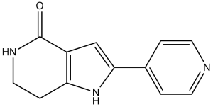
2-吡啶-4-基-1,5,6,7-四氢吡咯并[3,2-c]吡啶-4-酮是一种吡咯并吡啶类化合物。
PHA-767491 是一种 Cdc7/CDK9 抑制剂。 Cdc7-Dbf4 激酶或 DDK(Dbf4 依赖性激酶)通过磷酸化并激活复制性 Mcm2-7 DNA 解旋酶来启动 DNA 复制。DDK 在许多肿瘤细胞中过表达,并且由于 DDK 抑制可导致多种癌细胞类型凋亡,但不会导致正常细胞凋亡,因此 DDK 已成为一种新兴的化疗靶点。PHA-767491 和 XL413 是多种强效 DDK 抑制剂中的两种,它们对纯化的激酶具有低纳摩尔级的 IC50 值。尽管 XL413 对 DDK 具有高度选择性,但其在细胞系中的活性尚未得到充分表征。我们测定了 XL413 对一系列肿瘤细胞系的抗增殖和促凋亡作用,并与活性已得到充分表征的 PHA-767491 进行了比较。两种化合物均为有效的 DDK 生化抑制剂,但令人惊讶的是,它们在不同细胞系中的活性差异很大。与 PHA-767491 不同,XL413 仅对所测试的十种细胞系中的一种表现出显著的抗增殖活性。由于 XL413 未能有效抑制多种细胞系中的 DDK,因此该化合物的生物利用度可能有限。为了寻找其他潜在的 DDK 抑制剂,我们还使用 DDK 热稳定性变化分析 (TSA) 测试了约 400 种已知激酶抑制剂与 DDK 的交叉反应性。我们鉴定出 11 种能够显著稳定 DDK 的化合物。其中一些化合物抑制 DDK 的效力与 PHA-767491 相当,包括 Chk1 和 PKR 激酶抑制剂,但它们的化学骨架与已知的 DDK 抑制剂截然不同。综上所述,这些数据表明,几种已知的激酶抑制剂与DDK存在交叉反应,同时也凸显了设计更多特异性、具有生物活性的DDK抑制剂作为化疗药物的契机。[1] 检查点激酶1 (Chk1) 的激活是肝细胞癌 (HCC) 对5-氟尿嘧啶 (5-FU) 和其他抗代谢类药物产生化疗耐药性的关键因素。在本研究中,我们证实PHA-767491是一种双重抑制剂,可抑制两种细胞周期检查点激酶——细胞分裂周期激酶7 (Cdc7) 和细胞周期蛋白依赖性激酶9 (Cdk9),它与5-FU具有协同抗肿瘤作用,可在体外和体内抑制人HCC细胞。与单独使用各药物相比,PHA-767491 与 5-FU 联合用药表现出更强的细胞毒性,并能显著诱导肝癌细胞凋亡,表现为 caspase 3 活化和聚(ADP-核糖)聚合酶(PARP)片段化显著增加。PHA-767491 可直接拮抗 5-FU 诱导的 Chk1(Cdc7 的底物)磷酸化,并降低抗凋亡蛋白髓系白血病细胞 1(Cdk9 的下游靶点)的表达。在裸鼠肝癌异种移植瘤组织切片中,PHA-767491 的给药也降低了 Chk1 的磷酸化水平,并增加了原位细胞凋亡。我们的研究表明,PHA-767491可通过抑制Chk1磷酸化和下调Mcl1表达(通过抑制Cdc7和Cdk9)来增强5-FU的疗效,因此PHA-767491与5-FU联合用药可能对晚期和耐药性肝细胞癌(HCC)患者有益。[2] 背景:基因组不稳定性是癌细胞的标志性特征,这种细胞现象可能由复制应激引起。可以利用复制应激,并以靶向方式增强其作用来对抗癌细胞。其中一种策略是靶向细胞分裂周期7相关蛋白激酶(CDC7),该蛋白在DNA复制起始调控中起着关键作用。CDC7在多种癌症中均存在过表达,而CDC7小分子抑制剂已被证实具有抗肿瘤作用。本研究旨在探讨CDC7抑制剂作为胶质母细胞瘤治疗新策略的潜力。方法:采用PHA-767491盐酸盐作为CDC7抑制剂。使用两种胶质母细胞瘤细胞系(U87-MG和U251-MG)以及一种对照细胞系(3T3)来表征CDC7抑制剂的作用。分析了CDC7抑制剂对细胞活力、增殖、凋亡、迁移和侵袭的影响。此外,采用实时荧光定量PCR芯片鉴定CDC7抑制剂处理后差异表达的基因。结果:结果表明,CDC7抑制剂可降低胶质母细胞瘤细胞的活力,抑制细胞增殖,并诱导胶质母细胞瘤细胞凋亡。此外,我们还发现CDC7抑制剂可抑制胶质母细胞瘤细胞的迁移和侵袭。为了鉴定 CDC7 抑制的分子靶点,我们使用了实时 PCR 芯片,结果显示多种 mRNA 和 miRNA 的表达失调。结论:综上所述,我们的研究结果表明 CDC7 抑制是治疗胶质母细胞瘤的一种有前景的策略。[3] Cdc7 是一种重要的激酶,它通过激活复制起点来促进 DNA 复制。在此,我们通过生化和细胞实验对强效 Cdc7 抑制剂 PHA-767491 (1) 进行了表征,并测试了其在啮齿动物中的抗肿瘤活性。我们发现该化合物能够阻断 DNA 合成,并影响复制 DNA 解旋酶在 Cdc7 依赖性磷酸化位点的磷酸化。与目前的 DNA 合成抑制剂不同,PHA-767491 能够阻止复制起点的激活,但不会阻碍复制叉的延伸,也不会引发持续的 DNA 损伤反应。 PHA-767491 治疗可导致多种癌细胞类型发生凋亡,并在临床前癌症模型中抑制肿瘤生长。据我们所知,PHA-767491 是首个直接影响 DNA 复制起始而非延伸机制的分子,其活性表明 Cdc7 激酶抑制可能是一种开发抗癌疗法的新策略。[4] 1. 背景:PHA-767491 是首个双重 Cdc7/Cdk9 抑制剂,旨在靶向两个关键的癌症通路:DNA 复制 (DDK) 和转录延伸 (Cdk9)。与选择性 DDK 抑制剂(例如 XL413)不同,其双重活性增强了对高复制/转录速率肿瘤的疗效。[4] 2.作用机制:PHA-767491 与 DDK 和 Cdk9 的 ATP 结合口袋结合。抑制 DDK 可阻断 MCM2 磷酸化和 DNA 复制起始;抑制 Cdk9 可降低 RNA 聚合酶 II CTD 的磷酸化,从而抑制癌基因(c-Myc、细胞周期蛋白)的转录。这种双重作用可诱导癌细胞发生 G1/S 期阻滞和凋亡 [2, 3, 4] 3. 治疗潜力:临床前数据支持 PHA-767491 用于治疗肝细胞癌 (HCC)、胶质母细胞瘤 (GBM)、结肠癌和其他实体瘤,尤其是在与化疗药物(例如 5-氟尿嘧啶)联合使用时。其对癌干细胞的活性(通过降低 CD133 水平)提示其具有预防复发的潜力 [2, 3] 4.局限性:PHA-767491 的口服生物利用度中等 (38%),且对不同癌细胞系的敏感性存在差异(例如,在 p53 突变型非小细胞肺癌细胞系中活性较低 [1])。纳入的文献中未报道临床试验,限制了其在人体中的应用 [1, 4]。 5. 与 XL413 的比较:PHA-767491 由于抑制 Cdk9,其抗肿瘤活性比 XL413(选择性 DDK 抑制剂)更广,但 XL413 对 DDK 的选择性更高(IC₅₀ = 2 nM,而 PHA-767491 为 10 nM)[1]。 |
| 分子式 |
C12H11N3O
|
|
|---|---|---|
| 分子量 |
249.7
|
|
| 精确质量 |
213.09
|
|
| 元素分析 |
C, 67.59; H, 5.20; N, 19.71; O, 7.50
|
|
| CAS号 |
845714-00-3
|
|
| 相关CAS号 |
PHA-767491 hydrochloride;942425-68-5; 845714-00-3; 845538-12-7 (2HCl)
|
|
| PubChem CID |
11715767
|
|
| 外观&性状 |
Off-white to light yellow solid powder
|
|
| 密度 |
1.287
|
|
| 沸点 |
620.6ºC at 760 mmHg
|
|
| 闪点 |
329.1ºC
|
|
| LogP |
2.493
|
|
| tPSA |
57.78
|
|
| 氢键供体(HBD)数目 |
2
|
|
| 氢键受体(HBA)数目 |
2
|
|
| 可旋转键数目(RBC) |
1
|
|
| 重原子数目 |
16
|
|
| 分子复杂度/Complexity |
275
|
|
| 定义原子立体中心数目 |
0
|
|
| SMILES |
O=C1C2=C(NC(C3C=CN=CC=3)=C2)CCN1
|
|
| InChi Key |
DKXHSOUZPMHNIZ-UHFFFAOYSA-N
|
|
| InChi Code |
InChI=1S/C12H11N3O/c16-12-9-7-11(8-1-4-13-5-2-8)15-10(9)3-6-14-12/h1-2,4-5,7,15H,3,6H2,(H,14,16)
|
|
| 化学名 |
2-pyridin-4-yl-1,5,6,7-tetrahydropyrrolo[3,2-c]pyridin-4-one
|
|
| 别名 |
|
|
| HS Tariff Code |
2934.99.9001
|
|
| 存储方式 |
Powder -20°C 3 years 4°C 2 years In solvent -80°C 6 months -20°C 1 month |
|
| 运输条件 |
Room temperature (This product is stable at ambient temperature for a few days during ordinary shipping and time spent in Customs)
|
| 溶解度 (体外实验) |
|
|||
|---|---|---|---|---|
| 溶解度 (体内实验) |
注意: 如下所列的是一些常用的体内动物实验溶解配方,主要用于溶解难溶或不溶于水的产品(水溶度<1 mg/mL)。 建议您先取少量样品进行尝试,如该配方可行,再根据实验需求增加样品量。
注射用配方
注射用配方1: DMSO : Tween 80: Saline = 10 : 5 : 85 (如: 100 μL DMSO → 50 μL Tween 80 → 850 μL Saline)(IP/IV/IM/SC等) *生理盐水/Saline的制备:将0.9g氯化钠/NaCl溶解在100 mL ddH ₂ O中,得到澄清溶液。 注射用配方 2: DMSO : PEG300 :Tween 80 : Saline = 10 : 40 : 5 : 45 (如: 100 μL DMSO → 400 μL PEG300 → 50 μL Tween 80 → 450 μL Saline) 注射用配方 3: DMSO : Corn oil = 10 : 90 (如: 100 μL DMSO → 900 μL Corn oil) 示例: 以注射用配方 3 (DMSO : Corn oil = 10 : 90) 为例说明, 如果要配制 1 mL 2.5 mg/mL的工作液, 您可以取 100 μL 25 mg/mL 澄清的 DMSO 储备液,加到 900 μL Corn oil/玉米油中, 混合均匀。 View More
注射用配方 4: DMSO : 20% SBE-β-CD in Saline = 10 : 90 [如:100 μL DMSO → 900 μL (20% SBE-β-CD in Saline)] 口服配方
口服配方 1: 悬浮于0.5% CMC Na (羧甲基纤维素钠) 口服配方 2: 悬浮于0.5% Carboxymethyl cellulose (羧甲基纤维素) 示例: 以口服配方 1 (悬浮于 0.5% CMC Na)为例说明, 如果要配制 100 mL 2.5 mg/mL 的工作液, 您可以先取0.5g CMC Na并将其溶解于100mL ddH2O中,得到0.5%CMC-Na澄清溶液;然后将250 mg待测化合物加到100 mL前述 0.5%CMC Na溶液中,得到悬浮液。 View More
口服配方 3: 溶解于 PEG400 (聚乙二醇400) 请根据您的实验动物和给药方式选择适当的溶解配方/方案: 1、请先配制澄清的储备液(如:用DMSO配置50 或 100 mg/mL母液(储备液)); 2、取适量母液,按从左到右的顺序依次添加助溶剂,澄清后再加入下一助溶剂。以 下列配方为例说明 (注意此配方只用于说明,并不一定代表此产品 的实际溶解配方): 10% DMSO → 40% PEG300 → 5% Tween-80 → 45% ddH2O (或 saline); 假设最终工作液的体积为 1 mL, 浓度为5 mg/mL: 取 100 μL 50 mg/mL 的澄清 DMSO 储备液加到 400 μL PEG300 中,混合均匀/澄清;向上述体系中加入50 μL Tween-80,混合均匀/澄清;然后继续加入450 μL ddH2O (或 saline)定容至 1 mL; 3、溶剂前显示的百分比是指该溶剂在最终溶液/工作液中的体积所占比例; 4、 如产品在配制过程中出现沉淀/析出,可通过加热(≤50℃)或超声的方式助溶; 5、为保证最佳实验结果,工作液请现配现用! 6、如不确定怎么将母液配置成体内动物实验的工作液,请查看说明书或联系我们; 7、 以上所有助溶剂都可在 Invivochem.cn网站购买。 |
| 制备储备液 | 1 mg | 5 mg | 10 mg | |
| 1 mM | 4.0048 mL | 20.0240 mL | 40.0481 mL | |
| 5 mM | 0.8010 mL | 4.0048 mL | 8.0096 mL | |
| 10 mM | 0.4005 mL | 2.0024 mL | 4.0048 mL |
1、根据实验需要选择合适的溶剂配制储备液 (母液):对于大多数产品,InvivoChem推荐用DMSO配置母液 (比如:5、10、20mM或者10、20、50 mg/mL浓度),个别水溶性高的产品可直接溶于水。产品在DMSO 、水或其他溶剂中的具体溶解度详见上”溶解度 (体外)”部分;
2、如果您找不到您想要的溶解度信息,或者很难将产品溶解在溶液中,请联系我们;
3、建议使用下列计算器进行相关计算(摩尔浓度计算器、稀释计算器、分子量计算器、重组计算器等);
4、母液配好之后,将其分装到常规用量,并储存在-20°C或-80°C,尽量减少反复冻融循环。
计算结果:
工作液浓度: mg/mL;
DMSO母液配制方法: mg 药物溶于 μL DMSO溶液(母液浓度 mg/mL)。如该浓度超过该批次药物DMSO溶解度,请首先与我们联系。
体内配方配制方法:取 μL DMSO母液,加入 μL PEG300,混匀澄清后加入μL Tween 80,混匀澄清后加入 μL ddH2O,混匀澄清。
(1) 请确保溶液澄清之后,再加入下一种溶剂 (助溶剂) 。可利用涡旋、超声或水浴加热等方法助溶;
(2) 一定要按顺序加入溶剂 (助溶剂) 。
|
|---|
|
|
 M1-20
M1-20
 CDK12-Cyclin K Ligand-Linker Conjugates 1
 CGP-74514 dihydrochloride
 (S)-CDK2 degrader 6
InvivoChem的所有产品仅用于作科学研究,不面向患者销售
Copyright 2020 InvivoChem LLC | All Rights Reserved 粤ICP备20063088号-1


 COA
COA


 463611831
