| 规格 | 价格 | |
|---|---|---|
| 100mg | ||
| 500mg | ||
| 1g | ||
| Other Sizes |
| 靶点 |
JAK1 (IC50 = 43 nM); JAK2 (IC50 = 0.2 μM); JAK3 (IC50 = 2.3 μM); Tyk2 (IC50 = 4.7 μM)
|
|---|---|
| 体外研究 (In Vitro) |
在生化实验中,与参与红细胞生成的 JAK-2 相比,Upadacitinib 对 JAK-1 的选择性高出 74 倍,对 JAK-1 的选择性比参与免疫监视的 JAK-3 高 58 倍。 1]。由于 upadacitinib 对 JAK-1 的选择性优于 JAK-2 和 JAK-3,因此整个 RA 患者的获益-风险状况可能会得到改善 [2]。
Upadacitinib具有JAK1选择性,可抑制导致RA病理的细胞因子[3] 为了表征Upadacitini的酶活性,我们评估了利用重组人JAK激酶进行生化测定的效力和选择性。表1总结了数据。Upadacitinib显示出对JAK1(0.045μM)和JAK2(0.109μM)的活性 40 与JAK1相比,JAK3(2.1μM)的选择性是JAK3的100倍,TYK2(4.7μM)是JAK2的100倍。Upadacitinib还显示出对70+激酶的选择性,只有Rock1和Rock2的IC50值低于1μM(附加文件1:表S1)。为了进一步表征JAK1抑制upadactinib的机制,我们评估了不同浓度ATP下JAK1酶的活性。在所有测试浓度下,理论和实验IC50值的接近一致性证实了upadactinib是一种ATP竞争性抑制剂(数据未显示)。 Upadacitinib的JAK家族选择性在细胞检测中得到证实。由于JAK激酶合作性质的复杂性,我们采用了一组工程细胞系来了解upadactinib对每种激酶的细胞效力和选择性。如表1所示,upadactinib> 40 与JAK2(0.593μM)相比,JAK1(0.014μM)具有倍数选择性。Upadacitinib还显示出对JAK3(约130倍)和TYK2(约190倍)的选择性。还评估了upadactinib在生理相关细胞系统中的效力。与Ba/F3细胞数据一致,upadactinib能有效抑制JAK1依赖性细胞因子IL-6、OSM、IL-2和IFNγ,通过抑制STAT磷酸化来衡量。这项活动是~ 60 促红细胞生成素是一种完全依赖JAK2进行信号转导的细胞因子。接下来,我们测量了人全血中IL-6信号传导的抑制情况。upadactinib在CD3+T细胞群中的IC50值为0.207μM,在CD14+单核细胞群中为0.078μM。据报道,托法替尼对人全血中IL-6信号传导的IC50值对CD3+T细胞和单核细胞分别为0.367μM和0.406μM[3]。 |
| 体内研究 (In Vivo) |
在大鼠关节炎模型中,Upadacitinib(0.1-10 mg/kg;口服强饲;每天两次,持续 10 天)已证明有效[3]。ABT-494/Upadacitinib是大鼠AIA炎症和骨丢失的强效抑制剂,与托法替尼相比,在大鼠中以类似的有效剂量避免了相关的基本生理过程,如促红细胞生成素信号传导和外周NK细胞计数。在健康人类受试者中口服ABT-494 14天后,在与大鼠药效学特性一致的预测有效剂量下,ABT-494没有降低网织红细胞或NK细胞计数。
结论ABT-494是一种Jak1选择性抑制剂,在大鼠关节炎模型中显示出疗效。初步证据表明,ABT-494的药效学特性与在啮齿动物模型和健康人体受试者中观察到的一致。总的来说,这些令人鼓舞的观察结果支持在II期随机安慰剂对照试验中在RA患者中进一步测试ABT-494,并表明它可能比现有药物更有可能满足患者的需求。[2] Upadacitinib抑制大鼠佐剂性关节炎的疾病病理[3] 为了了解对炎症和关节炎表型的影响,我们在佐剂诱导的关节炎模型中测试了Upadacitinib,这是一种已建立的RA临床前模型。在第7天出现疾病的最初症状时口服乌帕西替尼,导致爪肿胀的剂量和暴露依赖性减少(图2a)。在疾病诱导后第18天,收获爪子,通过μCT测量骨破坏情况。AIA的正常过程会导致骨体积的显著减少,并且随着upadactinib的给药,骨体积会呈剂量依赖性减少(图2b)。与骨表面受到保护的10mg/kg upadacitnib治疗动物(图2d)相比,经载体处理的动物(图2c)出现了明显的凹陷和骨丢失,显示了破坏的例子。本研究还评估了组织学终点。Upadacitinib给药改善了3和10mg/kg剂量组的滑膜肥大、炎症、软骨损伤和骨侵蚀(数据未显示)。在大鼠胶原诱导性关节炎(CIA)模型中也观察到了类似的结果,这是RA的第二个临床前模型(数据未显示)。托法替尼也在AIA模型中进行了测试,并证明了剂量反应性疗效,尽管与upadactinib相比,暴露反应曲线右移(图2a)。有效浓度定义为达到60%抑制爪肿胀所需的AUC0-12药物浓度(AUC60)。使用AUC60作为参考点的理由是基于与10 mg BID临床剂量的托法替尼相关的AUC暴露量。这为进一步分析建立了一个翻译参考点。计算出乌帕替尼的总有效药物暴露量为83 ng*hr./ml,而托法替尼的暴露量为1205 ng*hr./ml。根据JAK1细胞效力与托法替尼的差异,预计乌帕替尼的体内效力会增加。 与疗效相比,Upadacitinib可避免网织红细胞部署和NK细胞计数减少[3] 我们应用了一种先前描述的方法的变体,以确定Upadacitini和托法替尼对JAK2依赖性Epo受体功能抑制的相对影响。连续两天用PBS或1000IU的Epo静脉注射幼年大鼠,并在第4天测量循环网织红细胞。始终给药upadacinib或tofactinib,并通过流式细胞术定量网织红细胞。我们还试图确定upadactinib和tofacitinib对循环NK细胞计数形式的常见γ链信号传导(JAK1/JAK3)的影响,因为这些细胞依赖IL-15生存。新生大鼠服用乌帕替尼或托法替尼14天,通过流式细胞术定量循环CD3-/CD16+/CD56+NK细胞。将网织红细胞部署、循环NK细胞计数和AIA疗效实验的结果一起绘制,以显示与暴露相关的相对影响(图3)。托法替尼以暴露依赖的方式(AUC60为1230 ng*hr./ml)降低循环NK细胞数量,类似于观察到的抑制爪子肿胀的暴露范围(AUC60 = 1205 ng*hr./ml)。托法替尼以暴露依赖的方式减少网织红细胞部署,达到最大抑制> 40% 在测试的最高浓度下(图3a)。Upadacitinib与AUC60以暴露依赖的方式降低循环NK细胞数量 = 480 ng*hr.ml~ 5 比抑制爪子肿胀所需的药物浓度(AUC60)低一倍 = 83 ng*hr./ml)。网织红细胞的部署也呈剂量反应性减少,并达到最大抑制作用~ 40% (图3b)。在与10 mg BID的托法替尼相关的临床AUC暴露量下,爪子肿胀的减少~ 60% 并且与NK细胞耗竭有明显的重叠(图3a)。使用6 mg BID和12 mg BID临床剂量的乌帕西替尼,爪肿胀的减少> 90%,与NK细胞耗竭明显分离(图3b)。 在AIA模型中,网织红细胞(图4a)和NK细胞(图4b)数据与爪肿胀抑制率进行了重新绘制,以直接比较托法替尼和Upadacitinib的抑制作用与疾病疗效的关系。在较低的有效范围内,托法替尼和乌帕替尼对网织红细胞的相对影响相似,但在较高的有效范围(>60%的爪肿胀)内,差异效应变得更加明显(图4a)。同样,循环NK细胞计数也有明显的差异效应。在AUC60时,托法替尼治疗后循环NK细胞减少了70%,而乌帕替尼治疗导致减少了25%(图4b)。 Upadacitinib在健康志愿者中,相对于IL-6信号传导,保留了常见的γ链信号传导[3] 为了确认Upadacitinib在临床环境中的临床前选择性,在服用1、3、12、24、36或48 mg Upadacitinib或5 mg tofacitinib的健康志愿者的全血中进行了离体细胞因子刺激试验。给药后1小时,抽血并用IL-6或IL-7刺激,以评估upadactinib对这些信号通路的影响。通过流式细胞术评估下游STAT磷酸化(STAT3和STAT5)的抑制作用(图5)。JAK1介导的IL-6诱导的pSTAT3被抑制~ 50% 在3mg剂量的upadactinib下,相当于5mg托法替尼的抑制水平。在36 mg达到最大抑制之前,增加剂量的乌帕西替尼显示了pSTAT3抑制的伴随增加。为了评估体内JAK1/3的效力,使用IL-7驱动的pSTAT5评估了抗常见γ链信号传导的活性。在这种情况下,需要12mg的乌帕替尼才能将pSTAT5抑制到与5mg的托法替尼相同的程度(约70%)。 |
| 酶活实验 |
酶效力和选择性测定[3]
JAK1(aa 845-1142)和JAK3(aa 811-1103)的活性重组人催化结构域在内部制备,并在SF9细胞中表达为谷胱甘肽s转移酶(GST)融合物,并通过谷胱甘肽亲和层析纯化。活性人TYK2(aa880-1185)在室内纯化,含有N端组氨酸标签和C端FLAG标签。通过固定化金属离子亲和层析进行纯化。JAK2的重组激酶结构域购自xxx。使用肽生物素-TYR2(生物素-(Ahx)-AEEYFFLFA酰胺)和生物素-TYR1。在抑制剂和2μM肽的存在下,在100μM ATP下进行反应。对于竞争试验,在不同量的ATP(0.01-1mM)等于或大于激酶的ATP-Km的情况下,测定Upadacitini的JAK1 IC50。使用Cheng-Prusoff方程评估ATP竞争力。ATP竞争性抑制剂在不同ATP浓度下的IC50变化与Cheng-Prusoff方程得出的理论值一致。 |
| 细胞实验 |
Ba/F3细胞效力和选择性测定[3]
TEL-JAK2、TEL-JAK3、TEL-TYK2和BCR-JAK1-Ba/F3工程细胞系购自Advanced Cellular Dynamics。细胞在添加了10%胎牛血清、1×青霉素-链霉素-谷氨酰胺和0.5μg/ml嘌呤霉素的RPMI 1640培养基中生长。 为了测量信号转导子和转录激活子5(pSTAT5)的磷酸化,将细胞洗涤并重新悬浮在密度为2 X 10⁷个细胞/mL的Hank's平衡盐溶液中。将5微升细胞悬浮液加入到含有5μL化合物的384孔、低体积、白壁聚苯乙烯板中(在11点[1:3]滴定系列中)。在进行pSTAT5检测之前,细胞在37°C下与化合物(最终DMSO浓度0.5%)一起孵育30分钟。按照标准制造商的方案,使用SureFire pSTAT5检测试剂盒测量pSTAT5,但在EnVision上检测之前,在添加供体珠后进行过夜温育除外。 细胞因子效力测定[3] 在人红白血病TF-1细胞系中评估了IL-6和肿瘤抑素M(OSM)诱导的STAT3磷酸化。在人UT-7细胞系中评估了促红细胞生成素诱导的STAT5磷酸化。在活化的人T细胞中评估IL-2和IL-15诱导的STAT5磷酸化。根据标准制造商的方案,使用SureFire pSTAT5或pSTAT3检测试剂盒完成磷酸化STAT的检测,但在EnVision上检测之前,在添加供体珠后进行过夜孵育除外。通过流式细胞术评估人PBMC中CD14+单核细胞群中IFNγ诱导的STAT1磷酸化。使用CD14 BV421和STAT1-PE(pY705)。通过流式细胞术评估成人上皮角质形成细胞中IL-4和IL-13诱导的STAT6磷酸化和IL-31诱导的STAT3磷酸化。使用STAT6-PE(pY641)和STAT3-PE(Y705)。 |
| 动物实验 |
动物/疾病模型:雌性Lewis大鼠(佐剂诱导性关节炎大鼠模型)[3]
剂量:0.1、0.3、1、3、10 mg/kg 给药途径:po(灌胃);每日两次,持续10天 实验结果:抑制佐剂诱导性关节炎大鼠的疾病病理。 通过一系列相关的细胞和体内药理学试验,包括骨髓集落形成、佐剂诱导性关节炎(AIA)、促红细胞生成素诱导的网织红细胞部署和NK/NKT细胞抑制,测试了Upadacitinib/ABT-494的疗效和选择性。在健康受试者中,通过口服给药14天,评估了ABT-494在多种互补药效学试验中的效力,并考察了不同剂量下的疗效。[2] 大鼠佐剂诱导关节炎(AIA)模型[3] 通过单次皮内注射0.1 mL结核分枝杆菌乳剂至雌性Lewis大鼠(体重125-150 g)右后足垫(第0天)诱导关节炎。免疫后,大鼠按指示每日两次(BID)灌胃给药,持续10天(第7天-第17天),分别给予赋形剂或研究药物。为评估关节炎的严重程度,每隔一天使用排水量描记法评估爪肿胀情况,直至第17天。第17天,所有大鼠在异氟烷麻醉下经心脏穿刺放血处死。使用μCT扫描左后爪。在包含爪跗骨部分的360 μm垂直切片中测定骨体积和骨密度。 网织红细胞部署试验连续两天,向未经处理的雄性Lewis大鼠静脉注射PBS或1000 IU的促红细胞生成素α。在第4天,使用噻唑橙作为染料,通过流式细胞术测量网织红细胞,方法如前所述[13]。在首次注射促红细胞生成素前30分钟给予Upadacitinib或托法替尼,随后每12小时给药一次,持续3天。 NK细胞分析[3] 连续14天,向Sprague Dawley大鼠口服给予Upadacitinib或托法替尼,剂量如所示。按照制造商说明,使用 BD MultiTest IMK 试剂盒采集血液样本并进行染色。使用 FlowJo 分析软件,通过检测 CD3−/CD16+/CD56+ 细胞群来确定 NK 细胞数量。细胞数/μL 的计算公式为:(细胞群中的事件数/绝对微珠计数区域的事件数)×(每次检测的微珠数/检测体积),其中每次检测的微珠数标注在 BD Trucount 试管标签上。 药代动力学/药效学建模 [3] 直接最大增强模型是确定有效浓度范围和人体有效剂量的最有效预测模型。有效浓度-时间曲线下面积 (AUC) 基于研究最后一天爪肿胀程度与 12 小时内 Upadacitinib 或 tofacitinib 的累积血浆浓度 (AUC0–12) 作图。 临床离体刺激试验 [3] 对于每位受试者,分别在给予 Upadacitinib 或 tofacitinib 后 0、1、6 和 12 小时,通过静脉穿刺采集血液至 2 mL 肝素钠抗凝管中。向血液中加入重组人 IL-6 (400 ng/ml) 或 IL-7 (400 ng/ml),并在 37°C 下孵育 10 分钟。加入表面抗体(CD14-APC、CD3-异硫氰酸荧光素 [FITC]),并在冰上继续孵育 20 分钟。样品裂解后于 37°C 孵育 10 分钟。洗涤后,样品储存于 -70°C。进行细胞内染色时,样品解冻、洗涤后,用 BD Perm 缓冲液 III 重悬,冰上孵育 30 分钟。洗涤后,样品与 pSTAT5-PE 或 pSTAT3-PE 在室温下染色 60 分钟,然后立即使用 FACSCalibur 流式细胞仪进行分析。使用 FlowJo 分析软件计算几何平均值。相关 STAT 磷酸化抑制率的计算公式如下:(1 - (1 小时 pSTAT 诱导值 - 0 小时 pSTAT 基线值) / (0 小时 pSTAT 诱导值 - 0 小时 pSTAT 基线值) * 100)。 |
| 药代性质 (ADME/PK) |
吸收、分布和排泄
在治疗剂量范围内,乌帕替尼的药代动力学特征呈剂量比例关系。口服给药后,达峰时间(Tmax)中位数为2至4小时。每日一次多次给药后,乌帕替尼的稳态血浆浓度可在4天内达到,且蓄积量极低。食物摄入对缓释制剂中乌帕替尼的AUC、Cmax和Cmin无临床相关影响。 单次服用放射性标记的速释制剂后,约53%的总剂量经粪便排出,其中38%为未代谢的母体药物。约43%的总剂量经尿液排出,其中24%为未代谢的母体药物。约 34% 的乌帕替尼总剂量以代谢物的形式排出体外。 对于体重 74 kg 的类风湿性关节炎患者,口服缓释制剂后,乌帕替尼的分布容积估计为 224 L。在一项由健康志愿者参与的药代动力学研究中,服用缓释制剂后,稳态分布容积为 294 L。乌帕替尼在血浆和血液细胞成分之间的分配相似,血浆/血药比为 1.0。 健康志愿者服用缓释制剂后,乌帕替尼的表观口服清除率为 53.7 L/h。 代谢/代谢物 乌帕替尼主要通过 CYP3A4 介导的代谢;然而,乌帕替尼并非 CYP3A4 的敏感底物。它也会少量地被 CYP2D6 代谢。在一项人体放射性标记研究中,约 79% 的血浆总放射性来自母体药物,约 13% 的血浆总放射性来自单氧化后经葡萄糖醛酸化产生的主要代谢物。目前尚无已知的 upadacitinib 活性代谢物。 生物半衰期 服用缓释制剂后,upadacitinib 的平均末端消除半衰期为 8 至 14 小时。在临床试验中,给药后 24 小时内,全身循环中约 90% 的 upadacitinib 被清除。 |
| 毒性/毒理 (Toxicokinetics/TK) |
肝毒性
在upadacitinib治疗类风湿性关节炎患者的上市前临床试验中,肝功能异常较为常见,但通常程度较轻。接受upadacitinib治疗的患者中,ALT升高的比例高达11%,而安慰剂组为7%,但ALT超过正常值上限3倍的患者比例为2%或更低。此外,接受甲氨蝶呤或生物制剂DMARD治疗的患者也出现了类似的ALT升高率。在这些纳入3000多名患者的试验中,未报告临床上明显的肝损伤、严重肝损伤或肝脏相关死亡病例。同样,其他JAK抑制剂,如托法替尼和巴瑞替尼,在治疗期间也常出现轻微的血清转氨酶升高,但未报告临床上明显的肝损伤病例。因此,此类药物被怀疑但尚未证实能够引起肝损伤。 此外,长期使用乌帕替尼和其他 Janus 激酶抑制剂与罕见的乙型肝炎病毒再激活病例相关,这些病例可能病情严重,甚至导致死亡。停用 JAK 抑制剂后,当免疫重建导致机体对病毒复制增强产生免疫反应时,乙型肝炎病毒再激活可能出现临床症状。 可能性评分:D(可能,但罕见,是易感患者出现临床症状性肝损伤(包括乙型肝炎病毒再激活)的原因)。 妊娠和哺乳期用药 ◉ 哺乳期用药概述 目前尚无关于哺乳期使用乌帕替尼的信息。大多数资料建议,服用乌帕替尼的母亲不应哺乳。最好选择其他药物,尤其是在哺乳新生儿或早产儿时。制造商建议在最后一次给药后 6 天内停止母乳喂养。 ◉ 对母乳喂养婴儿的影响 截至修订日期,未找到相关的已发表信息。 ◉ 对泌乳和母乳的影响 截至修订日期,未找到相关的已发表信息。 蛋白结合 乌帕替尼与人血浆蛋白的结合率为 52%。 |
| 参考文献 |
|
| 其他信息 |
药效学
乌帕替尼是一种改善病情抗风湿药 (DMARD),其作用机制是通过抑制 Janus 激酶 (JAK) 发挥作用。JAK 是促炎细胞因子下游细胞信号传导的重要介质。人们认为这些促炎细胞因子在许多自身免疫性炎症疾病(例如类风湿性关节炎)中发挥作用。在临床试验中,乌帕替尼降低了促炎性白细胞介素的活性,短暂地提高了淋巴细胞水平,并使免疫球蛋白水平较基线略有下降。 乌帕替尼是一种口服的 Janus 激酶 (JAK)1 选择性抑制剂,也是一种用于治疗类风湿性关节炎以延缓疾病进展的改善病情抗风湿药 (DMARD)。类风湿性关节炎是一种累及外周关节的慢性自身免疫性炎症疾病。类风湿关节炎的特征是滑膜炎症和增生、自身抗体产生、软骨损伤和骨破坏,并可导致多种并发症。尽管目前有多种治疗药物可用于治疗,但仍有高达40%的患者对包括生物疗法在内的现有疗法无反应。该疾病的病因大多尚不清楚;然而,JAK作为免疫介导疾病的驱动因素已被发现,因此JAK被用作类风湿关节炎的治疗靶点。为了在不显著影响疗效的前提下降低剂量相关毒性(某些泛JAK抑制剂会出现这种情况),研究人员开发了更具选择性的JAK1抑制剂,如upadacitinib和filgotinib。FDA于2019年8月批准了upadacitinib,用于治疗活动性类风湿关节炎、银屑病关节炎、特应性皮炎、溃疡性结肠炎和强直性脊柱炎。 2019年12月,该药物又获得了欧盟委员会和加拿大卫生部的批准。Upadacitinib以商品名RINVOQ上市,用于口服给药。 Upadacitinib是一种Janus激酶抑制剂。其作用机制是作为Janus激酶抑制剂。 Upadacitinib是一种口服选择性Janus相关激酶1 (JAK-1)抑制剂,用于治疗中重度类风湿性关节炎。乌帕替尼治疗期间血清酶升高发生率较低,但尚未发现与临床上明显的急性肝损伤病例相关,尽管它可能对易感患者构成乙型肝炎病毒再激活的风险。 乌帕替尼是一种小分子药物,其临床试验阶段最高为IV期(涵盖所有适应症),于2019年首次获批,用于治疗类风湿性关节炎,并有12项在研适应症。该药物已被美国食品药品监督管理局(FDA)列入黑框警告。 类风湿性关节炎(RA)是一种以滑膜炎症和关节破坏为特征的全身性自身免疫性疾病。生物制剂类疾病修饰抗风湿药(DMARDs)的出现极大地改善了RA的治疗。然而,这些生物制剂需要静脉或皮下注射,部分患者对生物制剂类DMARDs无反应或失去初始疗效。多种细胞因子和细胞表面分子与细胞表面的受体结合,从而激活多种细胞信号通路,包括激酶蛋白的磷酸化。在这些激酶中,非受体酪氨酸激酶家族的 Janus 激酶 (JAK) 在类风湿关节炎 (RA) 的病理过程中起着关键作用。目前已开发出多种 JAK 抑制剂作为 RA 患者的新疗法。这些口服合成 DMARD 可抑制 JAK1、2 和 3。其中一种 JAK 抑制剂托法替尼已在多个国家获批。使用 JAK1/2 抑制剂巴瑞替尼的 III 期临床试验结果显示,其疗效可观且安全性良好。这两种药物对生物制剂和合成 DMARD 疗效不佳的患者均有效。此外,使用特异性 JAK1 抑制剂菲戈替尼和乌帕替尼/ABT-494 的 III 期临床试验目前正在进行中。 JAK抑制剂是类风湿关节炎(RA)的新型疗法,但仍需进一步研究以确定其风险获益比,并筛选最适合接受此类治疗的患者。[1]抗细胞因子疗法已成为治疗类风湿关节炎(RA)疾病症状的主要手段,并可阻止疾病进展。尽管治疗选择众多,但仍有许多RA患者的疾病活动度未能显著降低。近期,临床研究表明JAK激酶阻断可有效控制疾病,并在某些情况下实现缓解。然而,由于剂量限制性耐受性和安全性问题,这些第一代JAK抑制剂未能达到预期效果。ABT-494是一种第二代JAK激酶抑制剂,对JAK1具有高选择性,从而最大限度地减少了JAK2和JAK3抑制相关的潜在副作用。本文描述了临床前和早期临床数据,这些数据表明 ABT-494 有潜力解决类风湿关节炎 (RA) 患者目前一些未被满足的医疗需求。[2] 背景:抗细胞因子疗法,例如阿达木单抗、托珠单抗和小分子 JAK 抑制剂托法替尼,已证实细胞因子及其下游信号通路在类风湿关节炎的发病机制中发挥着重要作用。托法替尼是一种泛 JAK 抑制剂,是首个获批用于治疗 RA 的 JAK 抑制剂,并已被证明能有效控制病情。然而,在 II 期剂量探索研究中,托法替尼存在剂量限制性耐受性和安全性问题,例如贫血。Upadacitinib (ABT-494) 是一种选择性 JAK1 抑制剂,其设计旨在验证以下假设:对 JAK1 的选择性高于其他 JAK 家族成员将带来更佳的获益风险比。 Upadacitinib 选择性靶向 JAK1 依赖性疾病驱动因子,例如 IL-6 和 IFNγ,同时减少对网织红细胞和自然杀伤 (NK) 细胞的影响,这可能是托法替尼耐受性问题的原因之一。 方法:基于结构假设设计了 JAK1 选择性抑制剂 Upadacitinib。通过体外实验(包括生化评估、基因工程细胞系和细胞因子刺激)确定了 JAK 家族选择性。通过 Upadacitinib 和托法替尼在佐剂诱导的大鼠关节炎模型中的疗效、对网织红细胞部署的活性以及对循环 NK 细胞的影响来确定其体内选择性。本研究利用JAK依赖性细胞因子进行体外刺激,在健康志愿者中评估了临床前JAK1选择性的转化。 结果:本文揭示了Upadacitinib对JAK1选择性的结构基础,以及其体外JAK家族选择性谱和后续体内生理效应。在细胞实验中,Upadacitinib对JAK1的选择性比JAK2高约60倍,对JAK3的选择性高100倍以上。虽然Upadacitinib和托法替尼在关节炎大鼠模型中均显示出疗效,但Upadacitinib对JAK1更高的选择性导致其对网织红细胞的激活和NK细胞的清除作用相对于疗效有所降低。从 I 期健康志愿者中获得的离体药效学数据证实了 upadacitinib 在临床环境中的 JAK1 选择性。 结论:本文数据突显了 Upadacitinib 的 JAK1 选择性,并支持其作为治疗 RA 的有效疗法,且具有改善获益风险比的潜力。 |
| 分子式 |
C17H21F3N6O2
|
|---|---|
| 分子量 |
398.382853269577
|
| 精确质量 |
778.325
|
| CAS号 |
2050057-56-0
|
| 相关CAS号 |
1310726-60-3; 1607431-21-9 (tartarte)
|
| PubChem CID |
133053456
|
| 外观&性状 |
White to light yellow solid powder
|
| tPSA |
158
|
| 氢键供体(HBD)数目 |
5
|
| 氢键受体(HBA)数目 |
13
|
| 可旋转键数目(RBC) |
6
|
| 重原子数目 |
55
|
| 分子复杂度/Complexity |
561
|
| 定义原子立体中心数目 |
4
|
| SMILES |
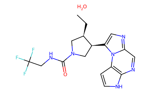
C([C@@H]1CN(C(=O)NCC(F)(F)F)C[C@@H]1C1=CN=C2C=NC3NC=CC=3N12)C.O
|
| InChi Key |
GJMQTRCDSIQEFK-SCDRJROZSA-N
|
| InChi Code |
InChI=1S/2C17H19F3N6O.H2O/c2*1-2-10-7-25(16(27)24-9-17(18,19)20)8-11(10)13-5-22-14-6-23-15-12(26(13)14)3-4-21-15;/h2*3-6,10-11,21H,2,7-9H2,1H3,(H,24,27);1H2/t2*10-,11+;/m11./s1
|
| 化学名 |
(3S,4R)-3-ethyl-4-(1,5,7,10-tetrazatricyclo[7.3.0.02,6]dodeca-2(6),3,7,9,11-pentaen-12-yl)-N-(2,2,2-trifluoroethyl)pyrrolidine-1-carboxamide;hydrate
|
| 别名 |
Upadacitinib hemihydrate; 2050057-56-0; NEW4DV02U5; bis(upadacitinib) hydrate; Upadacitinib hydrate (JAN); UPADACITINIB HYDRATE [JAN]; (3S,4R)-3-Ethyl-4-(3H-imidazo[1,2-a]pyrrolo[2,3-e]pyrazin-8-yl)-N-(2,2,2-trifluoroethyl)pyrrolidine-1-carboxamide hydrate(2:1); 1-Pyrrolidinecarboxamide, 3-ethyl-4-(3H-imidazo(1,2-a)pyrrolo(2,3-E)pyrazin-8-yl)-N-(2,2,2-trifluoroethyl)-, hydrate (2:1), (3S,4R)-;
|
| HS Tariff Code |
2934.99.9001
|
| 存储方式 |
Powder -20°C 3 years 4°C 2 years In solvent -80°C 6 months -20°C 1 month |
| 运输条件 |
Room temperature (This product is stable at ambient temperature for a few days during ordinary shipping and time spent in Customs)
|
| 溶解度 (体外实验) |
May dissolve in DMSO (in most cases), if not, try other solvents such as H2O, Ethanol, or DMF with a minute amount of products to avoid loss of samples
|
|---|---|
| 溶解度 (体内实验) |
注意: 如下所列的是一些常用的体内动物实验溶解配方,主要用于溶解难溶或不溶于水的产品(水溶度<1 mg/mL)。 建议您先取少量样品进行尝试,如该配方可行,再根据实验需求增加样品量。
注射用配方
注射用配方1: DMSO : Tween 80: Saline = 10 : 5 : 85 (如: 100 μL DMSO → 50 μL Tween 80 → 850 μL Saline)(IP/IV/IM/SC等) *生理盐水/Saline的制备:将0.9g氯化钠/NaCl溶解在100 mL ddH ₂ O中,得到澄清溶液。 注射用配方 2: DMSO : PEG300 :Tween 80 : Saline = 10 : 40 : 5 : 45 (如: 100 μL DMSO → 400 μL PEG300 → 50 μL Tween 80 → 450 μL Saline) 注射用配方 3: DMSO : Corn oil = 10 : 90 (如: 100 μL DMSO → 900 μL Corn oil) 示例: 以注射用配方 3 (DMSO : Corn oil = 10 : 90) 为例说明, 如果要配制 1 mL 2.5 mg/mL的工作液, 您可以取 100 μL 25 mg/mL 澄清的 DMSO 储备液,加到 900 μL Corn oil/玉米油中, 混合均匀。 View More
注射用配方 4: DMSO : 20% SBE-β-CD in Saline = 10 : 90 [如:100 μL DMSO → 900 μL (20% SBE-β-CD in Saline)] 口服配方
口服配方 1: 悬浮于0.5% CMC Na (羧甲基纤维素钠) 口服配方 2: 悬浮于0.5% Carboxymethyl cellulose (羧甲基纤维素) 示例: 以口服配方 1 (悬浮于 0.5% CMC Na)为例说明, 如果要配制 100 mL 2.5 mg/mL 的工作液, 您可以先取0.5g CMC Na并将其溶解于100mL ddH2O中,得到0.5%CMC-Na澄清溶液;然后将250 mg待测化合物加到100 mL前述 0.5%CMC Na溶液中,得到悬浮液。 View More
口服配方 3: 溶解于 PEG400 (聚乙二醇400) 请根据您的实验动物和给药方式选择适当的溶解配方/方案: 1、请先配制澄清的储备液(如:用DMSO配置50 或 100 mg/mL母液(储备液)); 2、取适量母液,按从左到右的顺序依次添加助溶剂,澄清后再加入下一助溶剂。以 下列配方为例说明 (注意此配方只用于说明,并不一定代表此产品 的实际溶解配方): 10% DMSO → 40% PEG300 → 5% Tween-80 → 45% ddH2O (或 saline); 假设最终工作液的体积为 1 mL, 浓度为5 mg/mL: 取 100 μL 50 mg/mL 的澄清 DMSO 储备液加到 400 μL PEG300 中,混合均匀/澄清;向上述体系中加入50 μL Tween-80,混合均匀/澄清;然后继续加入450 μL ddH2O (或 saline)定容至 1 mL; 3、溶剂前显示的百分比是指该溶剂在最终溶液/工作液中的体积所占比例; 4、 如产品在配制过程中出现沉淀/析出,可通过加热(≤50℃)或超声的方式助溶; 5、为保证最佳实验结果,工作液请现配现用! 6、如不确定怎么将母液配置成体内动物实验的工作液,请查看说明书或联系我们; 7、 以上所有助溶剂都可在 Invivochem.cn网站购买。 |
| 制备储备液 | 1 mg | 5 mg | 10 mg | |
| 1 mM | 2.5102 mL | 12.5508 mL | 25.1017 mL | |
| 5 mM | 0.5020 mL | 2.5102 mL | 5.0203 mL | |
| 10 mM | 0.2510 mL | 1.2551 mL | 2.5102 mL |
1、根据实验需要选择合适的溶剂配制储备液 (母液):对于大多数产品,InvivoChem推荐用DMSO配置母液 (比如:5、10、20mM或者10、20、50 mg/mL浓度),个别水溶性高的产品可直接溶于水。产品在DMSO 、水或其他溶剂中的具体溶解度详见上”溶解度 (体外)”部分;
2、如果您找不到您想要的溶解度信息,或者很难将产品溶解在溶液中,请联系我们;
3、建议使用下列计算器进行相关计算(摩尔浓度计算器、稀释计算器、分子量计算器、重组计算器等);
4、母液配好之后,将其分装到常规用量,并储存在-20°C或-80°C,尽量减少反复冻融循环。
计算结果:
工作液浓度: mg/mL;
DMSO母液配制方法: mg 药物溶于 μL DMSO溶液(母液浓度 mg/mL)。如该浓度超过该批次药物DMSO溶解度,请首先与我们联系。
体内配方配制方法:取 μL DMSO母液,加入 μL PEG300,混匀澄清后加入μL Tween 80,混匀澄清后加入 μL ddH2O,混匀澄清。
(1) 请确保溶液澄清之后,再加入下一种溶剂 (助溶剂) 。可利用涡旋、超声或水浴加热等方法助溶;
(2) 一定要按顺序加入溶剂 (助溶剂) 。
A Study to Assess Change in Disease Activity and Adverse Events of Oral Upadacitinib in Adult and Adolescent Participants With Moderate to Severe Hidradenitis Suppurativa Who Have Failed Anti-TNF Therapy
CTID: NCT05889182
Phase: Phase 3 Status: Recruiting
Date: 2024-11-25
 LysoPI(16:0/0:0)
 16:0 Lyso PI ammonium salt
 15S-曲伏前列腺素
 5,6-反式-拉坦前列腺素
InvivoChem的所有产品仅用于作科学研究,不面向患者销售
Copyright 2020 InvivoChem LLC | All Rights Reserved 粤ICP备20063088号-1


 463611831
