| 规格 | 价格 | 库存 | 数量 |
|---|---|---|---|
| 50mg |
|
||
| 100mg |
|
||
| 250mg |
|
||
| 1g |
|
||
| Other Sizes |
|

| 靶点 |
Topoisomerase
|
|---|---|
| 体外研究 (In Vitro) |
Exatecan作为高效TOP1抑制剂的生化和结构基础[1]
DXd是通过2-羟基乙酰基(COCH2OH)基团对F环上的NH2进行化学修饰而从艾替康衍生而来的(补充图S1A)。在一组癌症细胞系中,Exatecan的体外细胞毒性平均是DXd的10至20倍,具有亚纳米级IC50(补充图S1B;补充表S1)。 基于细胞的TOP1抑制试验和结构建模提供了艾替康更高细胞毒性效力的机制。通过改进的DNA加合物回收快速方法(RADAR)测定法检测艾替康与DXd和SN-38相比的DNA捕获TOP1(TOP1ccs)水平(21)。Exatecan是最有效的药物,其诱导TOP1ccs的浓度低于DXd和SN-38(图1A;补充图S1C)。捕获的DNA TOP1通常从DNA中去除并降解。为了测量TOP1降解率,细胞用艾替康或DXd/SN-38处理2小时,然后在没有药物的情况下生长30分钟,以逆转TOP1cs和TOP1降解。Exatecan以剂量依赖的方式诱导TOP1降解,比DXd和SN-38更有效(图1B)。 Exatecan对MDR机制的敏感性低于DXd和SN38[1] 在Caco-2细胞中测定了艾考替康、DXd和SN-38的MDR转运蛋白ABCG2和P-gp介导的外排(补充图S1E)。DXd显示出比没有或有ABCG2抑制剂新生霉素、P-gp抑制剂维拉帕米或双抑制剂GF120918的艾替康高一个数量级的外排率(图1D)。有趣的是,SN-38的流出率高于艾替康,但低于DXd。艾替康和DXd细胞内积累的免疫荧光(IF)检测表明,在ABCG2高表达细胞系NCI-H460中,DXd的积累低于艾替康(图1E;补充图S1F-1J)。 与MDR底物实验结果一致,DXd和艾替康之间的细胞毒性效力差异与内源性ABCG2/P-gp表达(mRNA和蛋白质;补充图S2A-S2C)相关。一般来说,在ABCG2或P-gp表达较高的细胞中,DXd/exatecan的IC50比值较高(图1F;补充图S2D和S2E)。ABCG2或P-gp的抑制提高了DXd/SN-38的细胞毒性,但对艾替康的影响要小得多(图1G)。即使DXd/SN-38抑制剂的IC50有所提高,艾替康仍然更有效(补充图S2F和S2G)。总的来说,exatecan对MDR基因的敏感性低于DXd/SN38,这可能在癌症细胞中赋予更高的细胞毒性以及更普遍的耐药性机制。 |
| 体内研究 (In Vivo) |
使用DNA拓扑异构酶I抑制剂DXd/SN-38的抗体-细菌偶联物(ADC)已经改变了癌症的治疗,但需要更有效的ADC来克服耐药性。我们设计了一种ADC类,使用一种新型的自焚T部分,用于艾替康的无痕结合和释放,艾替康是一种更有效的拓扑异构酶I抑制剂,对多药耐药性(MDR)的敏感性较低。以增强的治疗指数、更高的稳定性和改善的肿瘤内药效学反应为特征,靶向HER2、HER3和TROP2的抗体-T部分艾考替康偶联物克服了低靶表达、大和MDR+肿瘤中等效DXd/SN-38 ADC的内在或治疗耐药性。T部分脱羧酶ADC在患者来源的异种移植物和类器官模型中显示出持久的抗肿瘤活性,这些模型代表了未满足的临床需求,包括EGFR ex19del/T790M/C797S三突变肺癌和BRAF/KRAS-TP53双突变癌症,并显示出与PARP/ATR抑制剂和抗PD-1治疗的协同作用。T部分艾考替康ADC类在非人灵长类动物中的高耐受性支持其将反应患者群体和肿瘤类型扩展到当前ADC之外的潜力。
意义:结合新型自焚部分和拓扑异构酶I抑制剂exatecan作为有效载荷的ADC在低靶表达和耐DXd/SN-38 ADC的MDR+肿瘤中显示出深度和持久的反应,而不会增加毒性。这种新型ADC有可能使现有选择之外的更多患者受益。见Gupta等人的相关评论,第817页。这篇文章在《in This Issue》特刊第799[1]页中突出显示。
|
| 细胞实验 |
MTX-1000/DS-8201a在血浆中的体外稳定性[1]
在小鼠、大鼠、猴子和人血浆中评估了37°C下10 mg/mL浓度下MTX-1000/DS-8201a在21天内艾替康/DXd的释放速率。 ELISA[1] 对于结合试验,免疫板在包被缓冲液中用2.5 mg/mL His标记的HER2-ECD蛋白包被,并在4°C下保持过夜。洗涤后,将板封闭,并将每种连续稀释的物质加入孔中。在37°C下孵育1.5小时后,清洗平板,并在37°C下用HRP偶联的抗人IgG二抗孵育1小时。洗涤后,加入TMB溶液,用微孔板读数器测量每个孔中的A450。为了检测磷酸化Akt(pAkt),将SK-BR-3细胞在96孔板中预孵育4天,然后与每种物质孵育24小时。孵育后,根据制造商的说明,裂解细胞,使用PathScan Phospho-Akt1(Ser473)夹心ELISA试剂盒和PathScan total-Akt1夹心ELISA试剂袋检测细胞间pAkt和总Akt。通过将处理过的归一化pAkt值除以未处理的归一化pAk值来计算每个样品孔的相对pAkt。 免疫印迹[1] 用每种物质处理KPL-4细胞。24、48或72小时后,收获细胞并用含有Halt蛋白酶和磷酸酶抑制剂混合物的M-PER裂解缓冲液裂解。通过SDS-PAGE对样品进行装载和分离,并将其印迹到聚偏二氟乙烯膜上。用抗磷酸化Chk1(Ser345;133D3)兔单克隆抗体、抗Chk1(2G1D5)小鼠单克隆抗体、反切割PARP(Asp214)抗体、抗β-肌动蛋白(8H10D10)小鼠mAb、抗磷酸化组蛋白H2A将膜封闭并探测过夜。X(Ser139)抗体和抗组蛋白H2A。4°C下的X抗体(Abcam)。然后,用SNAP皮内洗涤膜并用荧光标记的二抗孵育10分钟。使用Odyssey成像系统检测荧光信号。 |
| 动物实验 |
细胞系和PDX研究[1]
所有荷瘤模型均建立于购自上海市计划生育研究所的雌性BALB/c裸鼠中。所有体内研究均按照多药联合生物技术有限公司机构动物护理和使用委员会的当地指南进行,并经该委员会批准。简而言之,将4至6周龄的小鼠饲养于无菌笼中,并维持在所需的无病原体条件下。将细胞悬液或肿瘤组织块皮下接种于雌性裸鼠体内。当肿瘤生长至适当体积后,根据肿瘤体积将荷瘤小鼠随机分为治疗组和对照组,并开始给药。抗体和抗体药物偶联物(ADC)通过静脉注射给药。小分子药物通过口服给药。肿瘤体积定义为1/2 × 长 × 宽²,每周测量两次(3-4天)。 TGI (%) 的计算方法如下:TGI (%) = [1 – (评估日治疗组肿瘤体积均值)/(评估日对照组肿瘤体积均值)] × 100。当小鼠达到终点(肿瘤体积超过 3,000 mm³、体重下降超过 10% 或出现表明出于伦理原因应实施安乐死的临床症状)时,使用二氧化碳气体对小鼠实施安乐死。 同源模型 [1] 雌性 BALB/c 小鼠,体重约 18 至 20 g,饲养于特定病原体清除级动物房内,每笼 5 只,笼子独立通风,并经高压灭菌。适应环境 9 天后,在小鼠右侧腹部皮下接种 CT26-hHER2 细胞(3 × 10⁵ 个细胞/0.1 mL/只),以诱导肿瘤发生。接种后10天,将36只肿瘤体积在100至200 mm³(平均肿瘤体积178 mm³)的小鼠纳入疗效研究,并随机分为6个治疗组。治疗于随机分组当日(定义为D0)开始。MTX-1000、DS-8201a(10 mg/kg)和抗PD-1抗体(5 mg/kg)均以10 mL/kg的体积静脉注射给小鼠。作为对照,以与药物相同的体积注射ABS缓冲液(10 mmol/L醋酸盐缓冲液,5%山梨醇,pH 5.5)。在第0天和第7天分别给予MTX-1000和DS-8201a。在第0天、第3天、第7天和第10天分别给予抗PD-1抗体。为了进行T细胞、树突状细胞和肿瘤细胞的流式细胞术分析,当肿瘤平均体积达到约250至400 mm³(肿瘤接种后8天)时,小鼠接受载体或DS-8201a(10 mg/kg,单次,静脉注射;第0天)治疗。小鼠在第8天用二氧化碳窒息法处死,并将肿瘤切成小块,使用gentleMACS Octo Dissociator(带加热器)和肿瘤解离试剂盒进行解离。将所得单细胞用小鼠 BD Fc 阻断剂封闭,并用针对小鼠 CD3、CD4、CD8、CD11c、CD45、CD86、颗粒酶 B、MHC I 类、MHC II 类、PD-L1 和人 HER2 的抗体进行染色。 组织多重免疫组化 [1] 根据制造商的方案,使用 Opal 多重免疫组化试剂盒对福尔马林固定、石蜡包埋的肿瘤组织切片进行 DAPI、CD45、CD4 和 CD8 染色。使用 Vectra 3.0 仪器进行切片扫描。所有图像均使用 HALO™ 图像分析平台进行分析。分析整个切片,并排除大面积坏死区域和间质区域。单独计数单阳性细胞。图像经光谱解混后,评估染色强度和形态,基于组织标记物进行组织分割,基于核和膜标记物进行细胞分割,并进行表型评分。 MTX-1000/DS-8201a 在大鼠/小鼠中的药代动力学[1] 采用经验证的配体结合试验测定血浆中抗体药物偶联物 (ADC) 和总抗体的浓度;定量下限为 0.02 μg/mL。简而言之,将免疫板用 1 μg/mL 人 HER2 蛋白(含 His 标签)包被于包被缓冲液中,并在 4°C 下过夜。洗涤后,封闭免疫板,并将各系列稀释的样品加入孔中。在37°C孵育1至2小时后,洗涤微孔板,然后分别加入HRP标记的抗人IgG Fc二抗(用于总抗体测定)或生物素标记的抗依沙替康/DXd抗体(用于ADC测定)。在37°C反应1至2小时后,直接加入TMB溶液,或先加入链霉亲和素蛋白和HRP,在37°C孵育40至60分钟后再加入TMB溶液。使用微孔板读数仪测量每个孔的A450值。采用经验证的LC/MS-MS方法测定血浆中依沙替康/DXd的浓度,定量下限为0.05 ng/mL。将MTX-1000/DS-8201a以4.0 mg/kg的剂量静脉注射给大鼠。给药后21天内测定血浆中ADC、总抗体和DXd的浓度。 |
| 药代性质 (ADME/PK) |
部分偶联将依沙替康转化为更优的抗体药物偶联物:理化和药理学特性、稳定性及毒性[1]
T 部分设计:PABC 间隔基的亲水性修饰以实现无痕偶联[1] 为了实现依沙替康的无痕偶联和释放,我们使用了二肽 VA 连接子和一种修饰的自裂解间隔基对氨基苄基 (pAB),称为 T 部分(图 2A;补充图 S4A 中的 S1)。选择 VA 是因为与其他肽连接子 VC 或 Gly–Gly–Phe–Gly (GGFG)(补充图 S4A 中的 S0;参考文献 29–30)相比,它能生成亲水性更强、DAR 更高的抗体药物偶联物。使用未修饰的肽连接子VA/VC/GGFG直接偶联HER2靶向药物Tras和依沙替康会导致高度聚集(补充图S4B)。将T部分与PEG基团整合(T900;补充图S4A中的S3)可显著降低聚集程度至可接受的2%。然而,用相同的PEG修饰MC(称为M部分,M900;补充图S4A中的S2)会导致>50%的高度聚集(补充图S4B),这凸显了谨慎选择修饰位置的重要性。考虑到基于PEG的生物药物可能引起致命的不良反应(31),我们决定使用聚肌氨酸(pSAR),因为它具有更高的溶解度和生物相容性(补充图S4C;参考文献32)。用10个单元的聚肌氨酸(pSAR10)修饰的pAB(T1000)可得到均一的抗体药物偶联物(ADC),其聚集程度可忽略不计(补充图S4B)。值得注意的是,中间体结构T800通过对pAB进行m-甲基氨基甲基修饰来连接pSAR10,可得到均一的、高药物抗体比(DAR)且无聚集的ADC。相比之下,将相同的m-甲基氨基甲基基团连接到VA连接子上(VK-pABC,M800,CLogP = 4.98)会导致依沙替康的聚集率达到10%,即使T800(CLogP = 4.73)和M800被认为化学性质相同,这支持了选择pAB作为最佳修饰位点。T部分ADC的偶联产率>90%。在T1000中用GGFG连接子替换VA连接子也得到了均一的ADC,且未观察到聚集现象(T1001;补充图S4B)。 Tras–T1000–依沙替康(MTX-1000)展现出更优异的理化性质[1] 为了比较Tras–T1000–依沙替康(MTX-1000)与DS-8201a的理化参数,我们使用了市售的DS-8201a或内部生产的Tras–GGFG–DXd,二者的理化性质(补充图S5A和S5B)、体内效力(补充图S5C–S5E)以及小鼠药代动力学(PK)(补充图S5F)均高度匹配。与 Tras–GGFG–DXd 类似,依沙替康与 HER2 抗体的连接并不影响 HER2 抗体与靶点的结合(通过流式细胞术或 ELISA 检测)(补充图 S6A 和 S6B)。使用 T800 或 T900 的 Tras–依沙替康偶联物也比 Tras–GGFG–DXd 具有更强的亲水性(补充图 S6C 和 S6D)。DAR8 和 DAR4 ADC 均可轻松制备(补充图 S6E)。Tras–T 部分–依沙替康 ADC(Tras–T800–依沙替康/MTX-800 和 Tras–T1000–依沙替康/MTX-1000,均为 DAR8)的稳定性与 Tras–GGFG–DXd 相似或更优。通过在 37°C 下长时间孵育以及反复冻融循环后形成的聚集体来测定其稳定性(补充图 S6F)。T 部分-依沙替康抗体药物偶联物 (ADC) 的热稳定性和光稳定性也优于 DS-8201a(补充图 S6G 和 S6H)。值得注意的是,VA 和 pSAR ADC (T1000) 的稳定性优于其他连接子 (GGFG) 和修饰 (PEG;T900 或 T1001;补充图 S6C、S6D、S6F 和 S6G),且聚集程度更低,这凸显了连接子和偶联化学选择对 ADC 理化性质的影响。 |
| 毒性/毒理 (Toxicokinetics/TK) |
依沙替康甲磺酸盐在大鼠中耐受性良好[1]
我们对大鼠(每组6只动物)进行了为期4周(第1、8、15、22和29天)的间歇性静脉注射依沙替康甲磺酸盐毒性研究,并设置了4周的恢复期(6只动物中的3只)。在剂量高达10 mg/kg(以依沙替康游离碱计算)时,大鼠对依沙替康甲磺酸盐耐受性良好。当剂量达到30 mg/kg时,依沙替康甲磺酸盐导致动物体重下降、发病率增加(食物摄入量减少、临床症状异常)和/或死亡(发生在给药后5或6天)。在 3 mg/kg 和 10 mg/kg 组中观察到剂量依赖性的短暂体重增加抑制(补充图 S3A)、血细胞计数下降(补充图 S3B–S3E)和关键血清酶水平升高(补充图 S3F 和 S3G),但在 4 周恢复期结束时基本可逆(补充表 S2),表明依沙替康与 DXd 或 SN-38 具有相似的耐受性。 |
| 参考文献 |
|
| 其他信息 |
在非人灵长类动物研究中,T 基团-依沙替康抗体偶联物(ADC)在不增加 HNSTD 毒性的情况下,仍保持了较高的疗效,其疗效水平与其他 TOP1 抑制剂 ADC 相当或更高。值得注意的是,T 基团-依沙替康 ADC 给药后的副作用通常更明显地出现在胃肠道(腹泻)和血液系统(网织红细胞减少)中,但这些症状均可完全逆转。T 基团-依沙替康 ADC 的血液系统和消化器官毒性与依沙替康单药在人体试验中的毒性相似。然而,与依沙替康相比,T 基团-依沙替康 ADC 在胃肠道方面表现出更佳的安全性,且骨髓毒性极低,这可能反映了该 ADC 设计具有更优异的理化特性。与游离有效载荷相比,T 基团-依沙替康 ADC 具有更高的稳定性、更长的半衰期、更低的游离有效载荷释放以及更低的骨髓毒性,这些因素可能共同促成了其良好的毒性特征。在临床试验中,接受基于 DXd 的 ADC 治疗的患者中,间质性肺病的发生率略高于 10%。尽管我们未在接受 T 基团-依沙替康 ADC 治疗的动物肺部观察到任何结构损伤或炎症迹象,但长期治疗是否会导致额外的肺毒性仍不确定。然而,可以设想,T 基团-依沙替康 ADC 可能具有与基于 DXd 的 ADC 不同的靶点非依赖性组织分布,从而导致不同的症状表现。
对于对 TOP1 抑制剂反应较差或耐药的肿瘤,可能需要具有不同作用机制的有效载荷类别。T 基团是一种模块化结构,与多种连接子化学兼容,适用于构建具有双有效载荷的 ADC。综上所述,T 部分-依沙替康 ADC 有潜力满足当前 ADC 无法满足的患者需求,而 T 部分的通用性和可扩展性可以促进引入具有完全不同作用机制的有效载荷,以应对药物耐药性的持续挑战。[1] |
| 分子式 |
C13H13NO5
|
|---|---|
| 分子量 |
263.25
|
| 精确质量 |
263.079
|
| CAS号 |
102978-40-5
|
| 相关CAS号 |
Exatecan Intermediate 1;110351-94-5;(R)-Exatecan Intermediate 1;110351-91-2
|
| PubChem CID |
359849
|
| 外观&性状 |
White to light yellow solid powder
|
| LogP |
0.089
|
| tPSA |
85.6
|
| 氢键供体(HBD)数目 |
1
|
| 氢键受体(HBA)数目 |
5
|
| 可旋转键数目(RBC) |
1
|
| 重原子数目 |
19
|
| 分子复杂度/Complexity |
574
|
| 定义原子立体中心数目 |
0
|
| SMILES |
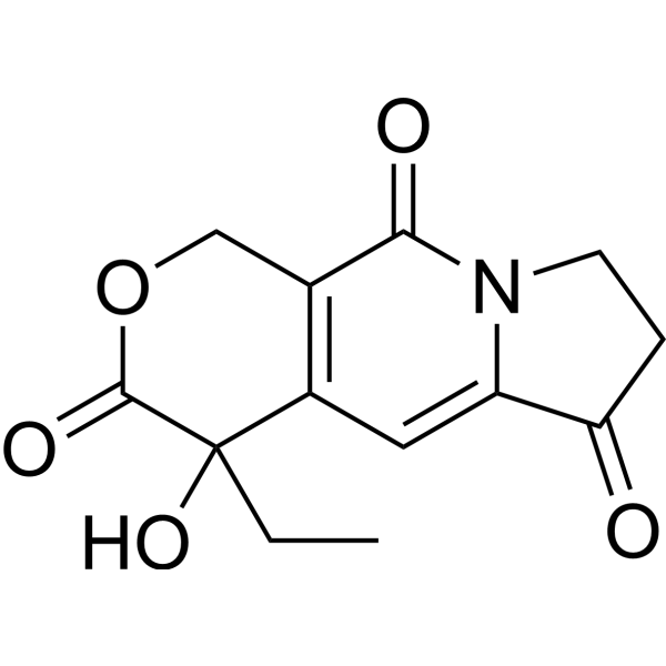
CCC1(C2=C(COC1=O)C(=O)N3CCC(=O)C3=C2)O
|
| InChi Key |
IGKWOGMVAOYVSJ-UHFFFAOYSA-N
|
| InChi Code |
InChI=1S/C13H13NO5/c1-2-13(18)8-5-9-10(15)3-4-14(9)11(16)7(8)6-19-12(13)17/h5,18H,2-4,6H2,1H3
|
| 化学名 |
4-ethyl-4-hydroxy-7,8-dihydro-1H-pyrano[3,4-f]indolizine-3,6,10-trione
|
| HS Tariff Code |
2934.99.9001
|
| 存储方式 |
Powder -20°C 3 years 4°C 2 years In solvent -80°C 6 months -20°C 1 month |
| 运输条件 |
Room temperature (This product is stable at ambient temperature for a few days during ordinary shipping and time spent in Customs)
|
| 溶解度 (体外实验) |
May dissolve in DMSO (in most cases), if not, try other solvents such as H2O, Ethanol, or DMF with a minute amount of products to avoid loss of samples
|
|---|---|
| 溶解度 (体内实验) |
注意: 如下所列的是一些常用的体内动物实验溶解配方,主要用于溶解难溶或不溶于水的产品(水溶度<1 mg/mL)。 建议您先取少量样品进行尝试,如该配方可行,再根据实验需求增加样品量。
注射用配方
注射用配方1: DMSO : Tween 80: Saline = 10 : 5 : 85 (如: 100 μL DMSO → 50 μL Tween 80 → 850 μL Saline)(IP/IV/IM/SC等) *生理盐水/Saline的制备:将0.9g氯化钠/NaCl溶解在100 mL ddH ₂ O中,得到澄清溶液。 注射用配方 2: DMSO : PEG300 :Tween 80 : Saline = 10 : 40 : 5 : 45 (如: 100 μL DMSO → 400 μL PEG300 → 50 μL Tween 80 → 450 μL Saline) 注射用配方 3: DMSO : Corn oil = 10 : 90 (如: 100 μL DMSO → 900 μL Corn oil) 示例: 以注射用配方 3 (DMSO : Corn oil = 10 : 90) 为例说明, 如果要配制 1 mL 2.5 mg/mL的工作液, 您可以取 100 μL 25 mg/mL 澄清的 DMSO 储备液,加到 900 μL Corn oil/玉米油中, 混合均匀。 View More
注射用配方 4: DMSO : 20% SBE-β-CD in Saline = 10 : 90 [如:100 μL DMSO → 900 μL (20% SBE-β-CD in Saline)] 口服配方
口服配方 1: 悬浮于0.5% CMC Na (羧甲基纤维素钠) 口服配方 2: 悬浮于0.5% Carboxymethyl cellulose (羧甲基纤维素) 示例: 以口服配方 1 (悬浮于 0.5% CMC Na)为例说明, 如果要配制 100 mL 2.5 mg/mL 的工作液, 您可以先取0.5g CMC Na并将其溶解于100mL ddH2O中,得到0.5%CMC-Na澄清溶液;然后将250 mg待测化合物加到100 mL前述 0.5%CMC Na溶液中,得到悬浮液。 View More
口服配方 3: 溶解于 PEG400 (聚乙二醇400) 请根据您的实验动物和给药方式选择适当的溶解配方/方案: 1、请先配制澄清的储备液(如:用DMSO配置50 或 100 mg/mL母液(储备液)); 2、取适量母液,按从左到右的顺序依次添加助溶剂,澄清后再加入下一助溶剂。以 下列配方为例说明 (注意此配方只用于说明,并不一定代表此产品 的实际溶解配方): 10% DMSO → 40% PEG300 → 5% Tween-80 → 45% ddH2O (或 saline); 假设最终工作液的体积为 1 mL, 浓度为5 mg/mL: 取 100 μL 50 mg/mL 的澄清 DMSO 储备液加到 400 μL PEG300 中,混合均匀/澄清;向上述体系中加入50 μL Tween-80,混合均匀/澄清;然后继续加入450 μL ddH2O (或 saline)定容至 1 mL; 3、溶剂前显示的百分比是指该溶剂在最终溶液/工作液中的体积所占比例; 4、 如产品在配制过程中出现沉淀/析出,可通过加热(≤50℃)或超声的方式助溶; 5、为保证最佳实验结果,工作液请现配现用! 6、如不确定怎么将母液配置成体内动物实验的工作液,请查看说明书或联系我们; 7、 以上所有助溶剂都可在 Invivochem.cn网站购买。 |
| 制备储备液 | 1 mg | 5 mg | 10 mg | |
| 1 mM | 3.7987 mL | 18.9934 mL | 37.9867 mL | |
| 5 mM | 0.7597 mL | 3.7987 mL | 7.5973 mL | |
| 10 mM | 0.3799 mL | 1.8993 mL | 3.7987 mL |
1、根据实验需要选择合适的溶剂配制储备液 (母液):对于大多数产品,InvivoChem推荐用DMSO配置母液 (比如:5、10、20mM或者10、20、50 mg/mL浓度),个别水溶性高的产品可直接溶于水。产品在DMSO 、水或其他溶剂中的具体溶解度详见上”溶解度 (体外)”部分;
2、如果您找不到您想要的溶解度信息,或者很难将产品溶解在溶液中,请联系我们;
3、建议使用下列计算器进行相关计算(摩尔浓度计算器、稀释计算器、分子量计算器、重组计算器等);
4、母液配好之后,将其分装到常规用量,并储存在-20°C或-80°C,尽量减少反复冻融循环。
计算结果:
工作液浓度: mg/mL;
DMSO母液配制方法: mg 药物溶于 μL DMSO溶液(母液浓度 mg/mL)。如该浓度超过该批次药物DMSO溶解度,请首先与我们联系。
体内配方配制方法:取 μL DMSO母液,加入 μL PEG300,混匀澄清后加入μL Tween 80,混匀澄清后加入 μL ddH2O,混匀澄清。
(1) 请确保溶液澄清之后,再加入下一种溶剂 (助溶剂) 。可利用涡旋、超声或水浴加热等方法助溶;
(2) 一定要按顺序加入溶剂 (助溶剂) 。
InvivoChem的所有产品仅用于作科学研究,不面向患者销售
Copyright 2020 InvivoChem LLC | All Rights Reserved 粤ICP备20063088号-1


 463611831
