| 规格 | 价格 | 库存 | 数量 |
|---|---|---|---|
| 1mg |
|
||
| 5mg |
|
||
| 10mg |
|
||
| 25mg |
|
||
| 50mg |
|
||
| 100mg |
|
||
| 250mg |
|
||
| Other Sizes |
|
| 靶点 |
ACVR1 (IC50 = 5 nM); BMPR1A (IC50 = 30 nM); ALK2 (IC50 = 5 nM), ALK3 (IC50 = 30 nM)
LDN-193189 2HCl targets bone morphogenetic protein (BMP) type I receptors (ALK1/2/3/6), with an IC50 of ~5 nM for inhibiting BMP4-induced phosphorylation of Smad1/5/8 in PASMCs; it also inhibits TGF-β signaling with an IC50 ≥ 1 μM and targets ActRIIA (type II activin receptor) [1] LDN-193189 2HCl inhibits myostatin/GDF8 signaling by targeting BMP type I receptors (ALK1/2/3/6) and ActRIIA; it represses GDF8-induced Smad2/3 signaling with an IC50 of 0.05 μM for inhibiting GDF8-induced (CAGA)₁₂-luciferase activity and 0.05 μM for inhibiting BMP2-induced BRE-luciferase activity [2] LDN-193189 2HCl targets BMP type I receptors in breast cancer cells, interfering with the BMP pathway regulated by the ZNF217 oncogene [3] |
|---|---|
| 体外研究 (In Vitro) |
LDN-193189 的 IC50 值分别为 5 nM 和 30 nM,可有效抑制 BMP I 型受体 ALK2 和 ALK3 的转录活性[1]。 LDN-193189 的 IC50 值低于 500 nM,对激活素和 TGF-β I 型受体 ALK4、ALK5 和 ALK7 的影响可以忽略不计[1]。 ActRIIA 与 LDN-193189 结合,Kd 值为 14 nM[2]。 LDN-193189(0.5 μM;30 分钟)靶向抑制 GDF8 触发的肌源性转录因子和 Smad2/3 信号传导[2]。有效抑制 GDF8 诱导的 Smad3/4 报告基因活性的是 LDN-193189(0.05、0.5 和 5 μM)[2]。 GDF8 处理的成肌细胞看到 LDN-193189 (0–5 μM) 恢复了其肌生成[2]。
LDN-193189 2HCl在PASMCs中强效抑制BMP4诱导的Smad1/5/8磷酸化(IC50约5 nM),对TGF-β诱导的信号通路抑制作用较弱(IC50≥1 μM)[1] LDN-193189 2HCl在COS细胞中以浓度依赖的方式降低组成型活性ALK2突变体(ALK2^R206H和ALK2^Q207D)的转录活性,抑制Id1启动子活性[1] LDN-193189 2HCl(100 nM)可阻断BMP4诱导的C2C12细胞成骨分化,且不影响细胞活力[1] LDN-193189 2HCl(0.5 μM)在未分化和分化的原代人成肌细胞及C2C12前成肌细胞中,抑制GDF8诱导的Smad2/3和p38磷酸化[2] LDN-193189 2HCl(0.05 μM)在C2C12细胞中抑制GDF8诱导的(CAGA)₁₂-荧光素酶活性和BMP2诱导的BRE-荧光素酶活性;在0.5 μM浓度下抑制TGF-β诱导的(CAGA)₁₂-荧光素酶活性[2] LDN-193189 2HCl可挽救GDF8处理的成肌细胞的成肌分化,上调成肌转录因子(MyoD、肌细胞生成素),并增加C2C12细胞和原代人成肌细胞中MHC阳性肌管的形成[2] LDN-193189 2HCl(0.5 μM)在体外促进C2C12细胞中肌管网络的收缩活性,可通过延时微分干涉相差显微镜检测到这一效应[2] |
| 体内研究 (In Vivo) |
LDN-193189(腹腔注射;3 mg/kg;每天;35 天)可能会影响乳腺癌细胞与其周围骨骼的相互作用[3]。 LDN-193189(腹腔注射;3 mg/kg;单剂量)可减少功能损伤和异位骨化 [1]。
BMP I型受体激酶的选择性抑制剂LDN-193189(参考文献6)抑制由腺病毒特异性Cre诱导的表达caALK2的组织中BMP信号效应器SMAD1、SMAD5和SMAD8的激活(Ad.Cre)。这种治疗减少了异位骨化和功能损害。与Ad.Cre对caALK2的局部诱导(这会引起炎症)相反,caALK2在出生后的整体表达(在不使用Ad.Cre的情况下诱导,因此没有炎症)不会导致异位骨化。然而,如果在这种情况下,用对照腺病毒提供炎症刺激,则会诱导异位骨形成。与LDN-193189一样,皮质类固醇抑制Ad.Cre-injected突变小鼠的骨化,表明caALK2表达和炎症环境都是该模型异位骨化发展所必需的。这些结果支持ALK2激酶活性失调在FOP发病机制中的作用,并表明小分子抑制BMP I型受体活性可能有助于治疗FOP和与过量BMP信号相关的异位骨化综合征。[1] 在本研究中,研究人员旨在调查LDN-193189化合物(BMP I型受体的强效抑制剂)对体内转移发展的影响。将ZNF217-revLuc细胞注射到裸鼠(n=16)的左心室中,而对照组小鼠(n=13)接种对照pcDNA6-revLuc电池。每组小鼠用LDN-193189治疗或不治疗35天。我们发现,对小鼠进行全身LDN-193189治疗,通过增加转移的数量和大小,显著促进了转移的发展。在注射pcDNA6-revLuc的小鼠中,LDN-193189也影响了转移出现的动力学。总之,这些数据表明,在体内,LDN-193189可能影响癌症细胞与骨环境之间的相互作用,有利于多发转移的出现和发展。因此,我们的报告强调了骨转移治疗中药物选择和治疗策略的重要性。[3] LDN-193189 2HCl处理可减少Ad.Cre注射的条件性caALK2表达小鼠的异位骨化和关节融合,保留关节间隙,并减轻踝关节被动活动度损伤(P < 0.001)[1] LDN-193189 2HCl降低Ad.Cre注射的caALK2突变小鼠后肢肌肉中核p-Smad1/5/8和Runx2的表达,减少碱性磷酸酶染色(成骨细胞活性和软骨内骨形成的标志物)[1] LDN-193189 2HCl处理经心内注射ZNF217-revLuc或pcDNA6-revLuc乳腺癌细胞的裸鼠,可显著促进转移灶的发展,增加转移灶的数量和大小,并改变转移出现的动力学[3] LDN-193189 2HCl增加ZNF217-revLuc和pcDNA6-revLuc注射小鼠的总转移负荷(通过生物发光检测,P < 0.05)[3] |
| 酶活实验 |
Id1和纤溶酶原激活物抑制剂-1启动子萤光素酶报告基因测定[1]
我们使用Fugene6,用0.3μg Id1启动子荧光素酶报告子构建体(BRE-Luc30,由P.ten Dijke提供)和0.6μg表达组成型活性形式的BMP I型受体(caALK2、caALK3或caALK631,由K.Miyazono提供)的质粒在六孔板中瞬时转染生长至50%汇合的小鼠PASMC。为了评估激活素和TGF-βI型受体的功能,我们用0.3μg PAI1(纤溶酶原激活物抑制剂-1)启动子萤光素酶报告子构建体(CAGA-Luc32,由P.ten Dijke提供)与0.6μg表达I型受体组成型活性形式(caALK4、caALK5和caALK733)的质粒联合瞬时转染PASMC。对于两个报告质粒,我们使用0.2μg的pRL-TK雷尼拉萤光素酶来控制转染效率。我们在转染后1小时开始用LDN-193189(2 nM–32μM)或载体孵育PASMC。我们采集了细胞提取物,并用双荧光素酶测定试剂盒通过萤火虫与雷尼拉萤光素酶活性的比率来量化相对启动子活性。
采用等温滴定量热法(ITC)检测LDN-193189 2HCl与ActRIIA的结合亲和力,将抑制剂滴定至纯化的ActRIIA蛋白溶液中,记录结合过程中的热变化以计算解离常数(Kd),记录了背索吗啡与ActRIIA的Kd为58 nM [2] 在PASMCs中开展激酶活性实验,评估LDN-193189 2HCl对BMP4诱导的Smad1/5/8磷酸化的抑制作用;向细胞中加入BMP4(10 ng ml⁻¹)和不同浓度的LDN-193189 2HCl,随后通过定量免疫印迹分析细胞裂解物,确定抑制Smad1/5/8磷酸化的IC50 [1] |
| 细胞实验 |
Smad1、Smad5和Smad8磷酸化的免疫印迹分析[1]
我们在SDS裂解缓冲液(62.5mM Tris-HCl(pH 6.8)、2%SDS、10%甘油、50mM二硫苏糖醇和0.01%溴酚蓝)中机械匀浆细胞提取物,通过SDS-PAGE分离蛋白质,用磷酸化Smad1、Smad5和Smad8特异性多克隆抗体、磷酸化Smad2或兔Smad1或Smad2特异性单克隆抗体免疫印迹,并用ECL-Plus观察免疫反应蛋白。
将原代人成肌细胞和C2C12前成肌细胞血清饥饿后,用LDN-193189 2HCl(0.5 μM)预处理30分钟,再用GDF8(8 nM)刺激45分钟;对细胞裂解物进行免疫印迹,检测磷酸化Smad2/3、p38及总Smad/p38蛋白,并对条带强度进行密度定量[2] 将Smad3/4响应的(CAGA)₁₂-荧光素酶或Smad1/5响应的BRE-荧光素酶报告载体与海肾荧光素酶载体共转染C2C12细胞;血清饥饿后,用LDN-193189 2HCl(0.05–0.5 μM)处理细胞,并加入GDF8(20 nM)、BMP2(10 nM)或TGF-β(100 pM)刺激6小时,随后检测荧光素酶活性并以海肾荧光素酶活性校正[2] 将C2C12细胞和原代人成肌细胞培养至汇合后,换用含LDN-193189 2HCl(0.5 μM)的分化培养基(加或不加GDF8 8 nM);分化3天后,对细胞进行骨骼肌MHC(肌球蛋白重链)和DAPI免疫染色,通过数字图像分析MHC阳性区域和细胞核数量,定量成肌分化程度[2] 将表达条件性caALK2^Q207D转基因的PASMCs感染Ad.Cre或Ad.GFP,并预处理LDN-193189 2HCl(100 nM);通过免疫印迹分析细胞裂解物中的磷酸化Smad1/5/8和总Smad1,评估BMP信号通路的抑制效果[1] |
| 动物实验 |
溶于 DMSO,然后用水稀释;3 mg/kg;腹腔注射
在 P7 时将 Ad.Cre 注射到条件性 caALK2 转基因小鼠和野生型小鼠中。条件性表达、组成型活性 ALK2 转基因小鼠[1] 先前已描述了在 C57BL/6 背景下表达编码组成型活性 ALK2Q207D 的基因的单个条件性表达等位基因 (CAG-Z-EGFP-caALK2) 的小鼠的构建。我们从杰克逊实验室获得了CAGGS-CreER小鼠,该小鼠在巨细胞病毒即刻早期增强子和鸡β-肌动蛋白启动子/增强子20的控制下普遍表达他莫昔芬诱导型Cre重组酶。 如前所述(Bellanger等人,2017),将pcDNA6-revLuc或ZNF217-revLuc细胞(2.5 × 10⁵)分别注射到n = 18或n = 20只6周龄无胸腺NMRI裸鼠的左心室中。细胞植入通过体内生物发光成像立即进行监测。只有生物发光信号扩散至全身的小鼠才被认为植入成功(分别为13/18和16/20,见补充图1A),并被纳入以下实验组:pcDNA6-revLuc组(n = 5)、pcDNA6-revLuc + LDN-193189组(n = 8)、ZNF217-revLuc组(n = 8)和ZNF217-revLuc + LDN-193189组(n = 8)。随后,从第0天到第35天,pcDNA6-revLuc小鼠或ZNF217-revLuc小鼠每天接受腹腔注射LDN-193189(3 mg/kg体重,溶于蒸馏水)或载体(蒸馏水)。 LDN-193189 的实验方案基于之前的体内研究(Yu 等,2008;Lee 等,2011;Balboni 等,2013)。LDN-193189 处理的小鼠未出现体重下降,表明该抑制剂没有严重的毒副作用。生物发光成像每周进行一次,方法如前所述(Bellanger 等,2017)。p 值 < 0.05 被认为具有统计学意义。[3] 条件性表达 caALK2 的小鼠(P7)左侧腓肠肌和比目鱼肌肌内注射 Ad.Cre;从注射 Ad.Cre 开始,给予 LDN-193189 2HCl,并在 P13、P15、P30 和 P60 时通过放射成像、μCT 和组织学评估小鼠的异位骨化情况 [1] 将 ZNF217-revLuc (n=16) 或 pcDNA6-revLuc (n=13) 乳腺癌细胞经心脏注射到裸鼠体内;将LDN-193189 2HCl全身给药35天,并在注射后35-42天通过生物发光成像和微型CT监测转移灶的发展情况[3] 在注射了Ad.Cre病毒的caALK2突变小鼠中,分别用LDN-193189 2HCl或载体治疗,通过测量P15和P30时踝关节和胫骨被动背屈形成的最小角度,评估踝关节的被动活动范围[1] |
| 毒性/毒理 (Toxicokinetics/TK) |
LDN-193189 2HCl治疗未在条件性表达caALK2的小鼠中诱发骨折、骨质疏松或骨骼异常[1]
LDN-193189 2HCl(100 nM)不影响体外培养的C2C12细胞的活力[1] |
| 参考文献 |
|
| 其他信息 |
进行性骨化性纤维发育不良症 (FOP) 是一种先天性疾病,其特征是出生后软组织进行性广泛骨化,目前尚无有效的治疗方法。受影响的个体携带 ACVR1 基因的保守突变,这些突变被认为会导致骨形态发生蛋白 (BMP) I 型受体——激活素受体样激酶 2 (ALK2) 的组成型激活。本文研究表明,在小鼠肌肉内表达编码组成型激活 ALK2 (caALK2) 的诱导型转基因(该转基因由氨基酸 207 位谷氨酰胺替换为天冬氨酸引起)会导致异位软骨内成骨、关节融合和功能障碍,从而模拟人类 FOP 的关键表型特征。选择性BMP I型受体激酶抑制剂LDN-193189(参考文献6)可抑制腺病毒Cre(Ad.Cre)诱导表达caALK2的组织中BMP信号通路效应分子SMAD1、SMAD5和SMAD8的激活。该治疗可减少异位骨化和功能障碍。与Ad.Cre诱导的局部caALK2表达(伴有炎症)不同,全身性出生后caALK2表达(无需使用Ad.Cre,因此不伴有炎症)不会导致异位骨化。然而,如果在此情况下使用对照腺病毒提供炎症刺激,则会诱导异位骨形成。与 LDN-193189 类似,皮质类固醇可抑制 Ad.Cre 注射突变小鼠的骨化,提示 caALK2 表达和炎症环境均是该模型中异位骨化发生的必要条件。这些结果支持 ALK2 激酶活性失调在进行性骨化性肌炎 (FOP) 发病机制中的作用,并提示小分子抑制剂 BMP I 型受体活性可能有助于治疗 FOP 和与 BMP 信号过度激活相关的异位骨化综合征。[1]
GDF8,又称肌肉生长抑制素,是 TGF-β 超家族分泌型多肽生长因子成员。GDF8 是体内外肌生成的强效负调控因子。我们发现,小分子 ATP 竞争性抑制剂 dorsomorphin 和 LDN-193189 可抑制 GDF8 信号通路。这些化合物此前已被证实能通过与I型BMP受体ALK1/2/3/6结合而有效抑制BMP信号通路。我们解析了II型受体ActRIIA与dorsomorphin的晶体结构,并证实dorsomorphin或LDN-193189靶向GDF8可诱导Smad2/3信号通路并抑制肌源性转录因子。因此,这两种抑制剂均能挽救经GDF8处理的成肌细胞的肌生成。定量活细胞显微镜观察显示,dorsomorphin或LDN-193189处理可促进体外肌管网络的收缩活性。因此,我们认为这些抑制剂是促进功能性肌生成的合适工具。[2] 目前抗癌疗法对骨转移性乳腺癌基本无效。骨形态发生蛋白(BMP)通路是一个极具吸引力的治疗靶点,因为它参与骨代谢、癌细胞形成及其向远处器官(如骨骼)的转移。我们之前报道过,在乳腺癌细胞中,ZNF217 癌基因驱动 BMP 通路激活,增加骨转移生长速度,并加速小鼠严重溶骨性病变的发生。 [3] LDN-193189 2HCl是一种选择性ATP竞争性BMP I型受体激酶抑制剂,是dorsomorphin的衍生物。[1] LDN-193189 2HCl通过阻断失调的ALK2激酶活性,抑制进行性骨化性纤维发育不良(FOP)小鼠模型中的异位骨化,其疗效与该模型中的皮质类固醇(地塞米松)治疗相当。[1] LDN-193189 2HCl通过抑制肌肉生长抑制素/GDF8信号通路促进功能性成肌细胞分化,使其成为体外增强肌生成的潜在工具。[2] LDN-193189 2HCl通过改变癌细胞与骨骼之间的相互作用,增强小鼠乳腺癌转移。微环境,突显了BMP通路抑制在不同病理背景下的双重作用[3] |
| 分子式 |
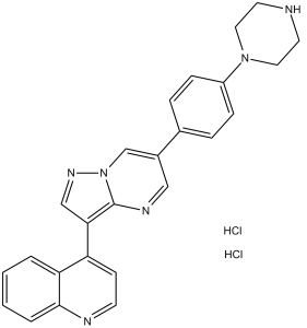
C25H24CL2N6
|
|
|---|---|---|
| 分子量 |
479.4
|
|
| 精确质量 |
478.143
|
|
| 元素分析 |
C, 62.63; H, 5.05; Cl, 14.79; N, 17.53
|
|
| CAS号 |
1435934-00-1
|
|
| 相关CAS号 |
LDN193189;1062368-24-4;LDN193189 Tetrahydrochloride;2310134-98-4
|
|
| PubChem CID |
91900717
|
|
| 外观&性状 |
Typically exists as orange to red solids at room temperature
|
|
| tPSA |
58.4Ų
|
|
| 氢键供体(HBD)数目 |
3
|
|
| 氢键受体(HBA)数目 |
5
|
|
| 可旋转键数目(RBC) |
3
|
|
| 重原子数目 |
33
|
|
| 分子复杂度/Complexity |
587
|
|
| 定义原子立体中心数目 |
0
|
|
| SMILES |
N1=C2C(C=CC=C2)=C(C2=C3N(N=C2)C=C(C2=CC=C(N4CCNCC4)C=C2)C=N3)C=C1.[H]Cl.[H]Cl
|
|
| InChi Key |
CMQXLLAILGGLRV-UHFFFAOYSA-N
|
|
| InChi Code |
InChI=1S/C25H22N6.2ClH/c1-2-4-24-22(3-1)21(9-10-27-24)23-16-29-31-17-19(15-28-25(23)31)18-5-7-20(8-6-18)30-13-11-26-12-14-30;;/h1-10,15-17,26H,11-14H2;2*1H
|
|
| 化学名 |
4-[6-(4-piperazin-1-ylphenyl)pyrazolo[1,5-a]pyrimidin-3-yl]quinoline;dihydrochloride
|
|
| 别名 |
|
|
| HS Tariff Code |
2934.99.9001
|
|
| 存储方式 |
Powder -20°C 3 years 4°C 2 years In solvent -80°C 6 months -20°C 1 month 注意: 请将本产品存放在密封且受保护的环境中,避免吸湿/受潮。 |
|
| 运输条件 |
Room temperature (This product is stable at ambient temperature for a few days during ordinary shipping and time spent in Customs)
|
| 溶解度 (体外实验) |
|
|||
|---|---|---|---|---|
| 溶解度 (体内实验) |
注意: 如下所列的是一些常用的体内动物实验溶解配方,主要用于溶解难溶或不溶于水的产品(水溶度<1 mg/mL)。 建议您先取少量样品进行尝试,如该配方可行,再根据实验需求增加样品量。
注射用配方
注射用配方1: DMSO : Tween 80: Saline = 10 : 5 : 85 (如: 100 μL DMSO → 50 μL Tween 80 → 850 μL Saline)(IP/IV/IM/SC等) *生理盐水/Saline的制备:将0.9g氯化钠/NaCl溶解在100 mL ddH ₂ O中,得到澄清溶液。 注射用配方 2: DMSO : PEG300 :Tween 80 : Saline = 10 : 40 : 5 : 45 (如: 100 μL DMSO → 400 μL PEG300 → 50 μL Tween 80 → 450 μL Saline) 注射用配方 3: DMSO : Corn oil = 10 : 90 (如: 100 μL DMSO → 900 μL Corn oil) 示例: 以注射用配方 3 (DMSO : Corn oil = 10 : 90) 为例说明, 如果要配制 1 mL 2.5 mg/mL的工作液, 您可以取 100 μL 25 mg/mL 澄清的 DMSO 储备液,加到 900 μL Corn oil/玉米油中, 混合均匀。 View More
注射用配方 4: DMSO : 20% SBE-β-CD in Saline = 10 : 90 [如:100 μL DMSO → 900 μL (20% SBE-β-CD in Saline)] 口服配方
口服配方 1: 悬浮于0.5% CMC Na (羧甲基纤维素钠) 口服配方 2: 悬浮于0.5% Carboxymethyl cellulose (羧甲基纤维素) 示例: 以口服配方 1 (悬浮于 0.5% CMC Na)为例说明, 如果要配制 100 mL 2.5 mg/mL 的工作液, 您可以先取0.5g CMC Na并将其溶解于100mL ddH2O中,得到0.5%CMC-Na澄清溶液;然后将250 mg待测化合物加到100 mL前述 0.5%CMC Na溶液中,得到悬浮液。 View More
口服配方 3: 溶解于 PEG400 (聚乙二醇400) 请根据您的实验动物和给药方式选择适当的溶解配方/方案: 1、请先配制澄清的储备液(如:用DMSO配置50 或 100 mg/mL母液(储备液)); 2、取适量母液,按从左到右的顺序依次添加助溶剂,澄清后再加入下一助溶剂。以 下列配方为例说明 (注意此配方只用于说明,并不一定代表此产品 的实际溶解配方): 10% DMSO → 40% PEG300 → 5% Tween-80 → 45% ddH2O (或 saline); 假设最终工作液的体积为 1 mL, 浓度为5 mg/mL: 取 100 μL 50 mg/mL 的澄清 DMSO 储备液加到 400 μL PEG300 中,混合均匀/澄清;向上述体系中加入50 μL Tween-80,混合均匀/澄清;然后继续加入450 μL ddH2O (或 saline)定容至 1 mL; 3、溶剂前显示的百分比是指该溶剂在最终溶液/工作液中的体积所占比例; 4、 如产品在配制过程中出现沉淀/析出,可通过加热(≤50℃)或超声的方式助溶; 5、为保证最佳实验结果,工作液请现配现用! 6、如不确定怎么将母液配置成体内动物实验的工作液,请查看说明书或联系我们; 7、 以上所有助溶剂都可在 Invivochem.cn网站购买。 |
| 制备储备液 | 1 mg | 5 mg | 10 mg | |
| 1 mM | 2.0859 mL | 10.4297 mL | 20.8594 mL | |
| 5 mM | 0.4172 mL | 2.0859 mL | 4.1719 mL | |
| 10 mM | 0.2086 mL | 1.0430 mL | 2.0859 mL |
1、根据实验需要选择合适的溶剂配制储备液 (母液):对于大多数产品,InvivoChem推荐用DMSO配置母液 (比如:5、10、20mM或者10、20、50 mg/mL浓度),个别水溶性高的产品可直接溶于水。产品在DMSO 、水或其他溶剂中的具体溶解度详见上”溶解度 (体外)”部分;
2、如果您找不到您想要的溶解度信息,或者很难将产品溶解在溶液中,请联系我们;
3、建议使用下列计算器进行相关计算(摩尔浓度计算器、稀释计算器、分子量计算器、重组计算器等);
4、母液配好之后,将其分装到常规用量,并储存在-20°C或-80°C,尽量减少反复冻融循环。
计算结果:
工作液浓度: mg/mL;
DMSO母液配制方法: mg 药物溶于 μL DMSO溶液(母液浓度 mg/mL)。如该浓度超过该批次药物DMSO溶解度,请首先与我们联系。
体内配方配制方法:取 μL DMSO母液,加入 μL PEG300,混匀澄清后加入μL Tween 80,混匀澄清后加入 μL ddH2O,混匀澄清。
(1) 请确保溶液澄清之后,再加入下一种溶剂 (助溶剂) 。可利用涡旋、超声或水浴加热等方法助溶;
(2) 一定要按顺序加入溶剂 (助溶剂) 。
Inhibitor binding to ActRII.J Biol Chem.2015 Feb 6;290(6):3390-404.
Dorsomorphin and LDN-193189 inhibit GDF8-induced signaling pathways in undifferentiated and in differentiated primary human myoblasts and in C2C12 premyoblasts.J Biol Chem.2015 Feb 6;290(6):3390-404. |
|---|
Ligand-specific effects of kinase inhibitors on Smad2/3 and Smad1/5 phosphorylation.
Dorsomorphin treatment facilitates myotube formation.J Biol Chem.2015 Feb 6;290(6):3390-404. |
Dorsomorphin and LDN-193189 efficiently inhibit GDF8 induced Smad3/4 reporter gene activity.J Biol Chem.2015 Feb 6;290(6):3390-404. |
Dorsomorphin and LDN-193189 counteract GDF8-induced repression of myogenic differentiation.J Biol Chem.2015 Feb 6;290(6):3390-404. |
|---|
Dorsomorphin and LDN-193189 promote the formation of a contractile myotube network.J Biol Chem.2015 Feb 6;290(6):3390-404. |
 TGF-β/Smad-IN-2
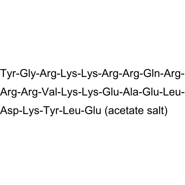 TAT-QFNP12 acetate
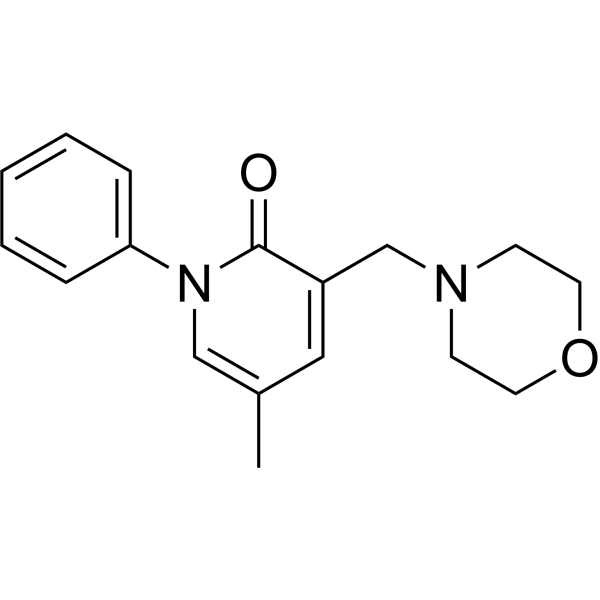
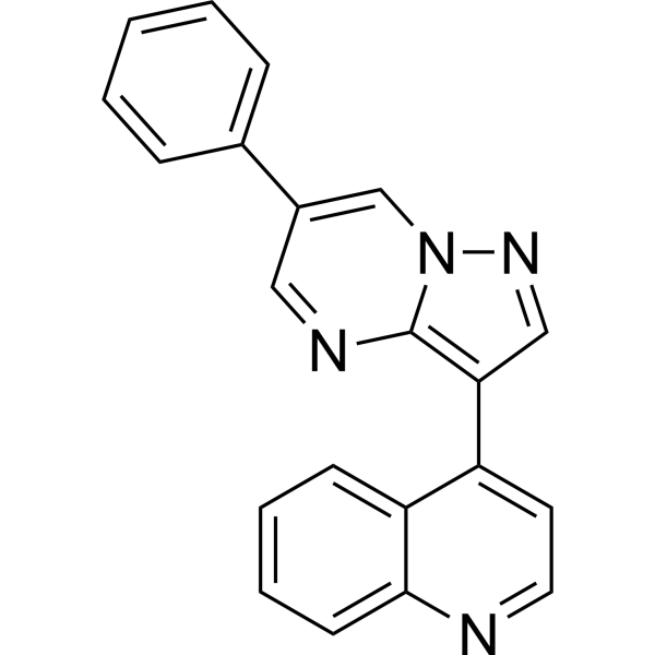 Depiperazine-DM3189
 CMF9
CMF9
InvivoChem的所有产品仅用于作科学研究,不面向患者销售
Copyright 2020 InvivoChem LLC | All Rights Reserved 粤ICP备20063088号-1


 COA
COA


 463611831
