| 规格 | 价格 | |
|---|---|---|
| 500mg | ||
| 1g | ||
| Other Sizes |
| 靶点 |
PPARβ/δ (IC50 = 22.9 μM)
|
|---|---|
| 体外研究 (In Vitro) |
BS-181 diHClide(0–40 μM;72 小时)可抑制癌细胞的生长,同时还可抑制乳腺癌细胞系(IC50 值:15.1 μM–20 μM)和结直肠癌细胞系(IC50 值)的生长。 :11.5 μM–20 μM)。 15.3 μM,对肺癌、骨肉瘤、前列腺癌和肝癌细胞系的IC50值分别为11.5 μM至37.3 μM [1]。 RNA 聚合酶 II 的 C 末端结构域 (CTD) 丝氨酸 5 (P-Ser5) 的磷酸化可被 BS-181 二盐酸盐(0-50 μM;4 小时)抑制。虽然它对其他 CDK 或细胞周期蛋白没有影响,但它会下调 CDK4 和细胞周期蛋白 D1 的表达 [1]。在低浓度下,当暴露于 BS-181 二盐酸盐(0-50 μM;24 小时)时,处于 G1 期的细胞数量增加,而处于 S 和 G2/M 期的细胞数量减少。然而,较高浓度会导致细胞积聚在亚 G1 期,这表明细胞凋亡 [1]。
BS-181的合成和体外激酶抑制[1] BS-181由二氯吡唑并[1,5-a]嘧啶2合成,通过使用苄胺依次选择性取代C-7氯化物,Boc保护,在Buchwald-Hartwig反应条件下使用二-Boc-1,6-己二胺钯催化取代C-5氯化物,并在酸性甲醇中脱保护(补充图S1)。通过将越来越多的BS-181与纯化的重组CDK7/CycH/MAT1复合物孵育来测量CDK7活性的抑制,然后使用萤光素酶测定法(PkLight,Cambrex,UK)测量反应中剩余的游离ATP,萤光素酶活性因此提供了CDK7活性抑制的衡量标准。BS-181抑制CDK7活性,IC50=21 nM(表1;补充数据图S2),而用罗斯科维汀达到的IC50为510 nM,与之前关于罗斯科维廷抑制CDK7的报告一致(13)。BS-181对CDK1/cycB、CDK4/cycD1、CDK5/p35NCK、CDK6/cycD1和CDK9/cycT的抑制IC50值远高于1μM,对CDK2/cycE的抑制IC50=880 nM,比CDK7的IC50高约40倍。 来自许多不同类别的70种蛋白激酶被BS-181测试了抑制作用。使用高浓度(10μM)的BS-181观察到对几种激酶活性的一些抑制(补充表5)。对表现出最大抑制作用的激酶CDK2/cycA、CK1和DYRK1A进行IC50测量,结果显示IC50分别为730 nM、7.36μM和2.3μM。这些数据证实BS-181是CDK7活性的高选择性抑制剂。 BS-181促进细胞周期阻滞并抑制癌症细胞生长[1] 为了评估BS-181的抗增殖活性,一组代表一系列肿瘤类型的细胞系,包括乳腺癌、肺癌、前列腺癌和结直肠癌癌症,用浓度增加的BS-181治疗72小时。使用磺基罗丹明B测定法测定增殖表明,所有受试细胞系的生长都受到抑制,IC50值范围为11.5至37μM(表2)。对BS-181观察到的生长抑制与对Roscovitine观察到的相似,其IC50值在8至33.5μM的范围内。 BS-181或Roscovitine治疗4小时后,CDKs和细胞周期蛋白的免疫印迹显示CDK4和细胞周期素D1下调(图2C),其他CDKs和周期蛋白的水平不受影响。此外,BS-181降低了抗凋亡蛋白XIAP和Bcl-xL的水平,而Bcl-2的水平没有变化。 用低浓度BS-181处理24小时后,G1期细胞数量增加,同时S期和G2/M期细胞数量减少(图3A;图S3)。然而,在较高浓度下,细胞在亚G1期积聚,表明细胞凋亡。BS-181处理24小时后,细胞的Annexin V染色证实了这一点,其中30%和83%的细胞分别用25μM和50μM BS-181对AnnexinⅤ染色呈阳性(图3B)。roscovitine未观察到明显的细胞凋亡。 BS-181-抑制癌症细胞增殖、迁移和侵袭[2] 为了评估BS-181在GC中的抗增殖能力,将包括MKN28、SGC-7901、AGS和BGC823在内的几种不同细胞系用越来越高浓度的BS-181处理48小时。CCK-8试验表明,BS-181抑制GC细胞生长,抑制浓度(IC50)范围为17至22μM。对于正常胃上皮RGM-1细胞系,IC50为6.5μM(表1)。此外,我们还研究了BS-181对细胞迁移和侵袭能力的影响。正如预期的那样,BS-181以剂量依赖的方式显著抑制细胞迁移和侵袭能力(分别为P<0.05;图1)。 BS-181诱导癌症细胞凋亡和细胞周期阻滞[2] 使用流式细胞术测定细胞凋亡。与对照组相比,在BS-181处理的BGC823细胞中观察到凋亡细胞的显著增加(分别为P<0.05;图2A)。我们的研究结果还表明,BS-181以剂量和时间依赖的方式诱导细胞凋亡。此外,与对照组相比,经BS-181处理的细胞中caspase-3和Bax的表达显著增加,而Bcl-2水平降低(分别为P<0.05)(图2B)。这些结果表明,BS-181可以诱导GC细胞凋亡。此外,CDK7活性的抑制导致关键抗凋亡蛋白XIAP和细胞周期调节因子细胞周期蛋白D1的显著减少(P<0.05)(图2C)。因此,BS-181可能通过下调BGC823细胞中XIAP和细胞周期蛋白的表达来调节细胞凋亡和细胞周期进程。在本研究中,通过流式细胞术分析了细胞周期分布(图3)。BS-181治疗显示G0/G1期细胞增加,同时S期和G2/M期细胞数量减少。这些结果表明,BS-181诱导细胞周期阻滞在G0/G1期,并延迟了细胞周期的进展。 BS-181抑制癌症细胞CDK7活性[2] BS-181是一种特异性CDK7抑制剂。在这项研究中,我们还证实了BS-181是CDK7的特异性抑制剂。如表2所示,BS-181抑制CDK7活性,IC50=0.019μM,而用罗斯科维汀达到的IC50为0.48μM。此外,BS-181对其他CDKs的抑制IC50值>1μM,远高于CDK7。此外,免疫印迹显示,BS-181抑制了RNA聚合酶II CTD在成熟的CDK7磷酸化位点丝氨酸5(P-Ser5)的磷酸化。 |
| 体内研究 (In Vivo) |
用 BS-181 二盐酸盐(10 mg/kg、20 mg/kg;单剂量)治疗的小鼠对药物反应良好,体重没有显着变化 [1]。 BS-181 二盐酸盐以剂量依赖性方式抑制肿瘤生长(腹腔注射;5 mg/kg 或 10 mg/kg,每日两次;每日总剂量 10 mg/kg 或 20 mg/kg;14 天)。与对照组相比,每天 10 mg/kg 和 20 mg/kg/天的肿瘤生长速度分别减慢了 25% 和 50% [1]。
BS-181 体内稳定,小鼠腹腔注射 10 mg/kg 后血浆消除半衰期为 405 分钟。 BS-181 以剂量依赖性方式抑制裸鼠模型中 MCF-7 异种移植物的生长,10 mg/kg/天和 20 mg/kg 治疗 2 周后,肿瘤生长减少 25% 和 50% /天,分别无明显毒性。
体内肿瘤生长抑制[1] 腹腔注射(i.p)的BS-181的最大耐受单剂量为30mg/kg,其中10mg/kg和20mg/kg耐受良好(数据未显示)。因此,对于异种移植物肿瘤生长抑制研究,在14天内,每天两次向动物腹腔注射5mg/kg或10mg/kg,以给予10mg/kg或20mg/kg的总日剂量。与对照组相比,BS-181的10mg/kg/天和20mg/kg/天剂量分别以剂量依赖的方式抑制了肿瘤生长,肿瘤生长减少了25%和50%(图4A)。根据对动物体重没有明显不良影响的判断,在这些剂量下没有明显的毒性(图4B)。 BS-181-抑制体内肿瘤生长并提高存活率[2] 如前所述,7腹腔注射BS-181的最大耐受单剂量为30mg/kg/d,BALB/c-nu小鼠对10mg/kg/d或20mg/kg/d的剂量耐受良好。在这项研究中,小鼠接受BS-181腹腔注射,每天两次,每次5mg/kg/d或10mg/kg/d,在2周内给予10mg/kg或20mg/kg的日剂量。此外,另一组15只大鼠接受了为期14天的roscovitine(20mg/kg/d)注射。我们观察到,与对照组相比,BS-181以剂量依赖的方式显著抑制了肿瘤生长(分别为P<0.05)(图5A)。然而,在14天的观察期间,各组小鼠的体重没有显著差异(图5B)。这表明,每日剂量为10mg/kg或20mg/kg时没有明显的毒性。此外,所有动物都被再饲养30天进行存活观察。如图5C所示,在对照组中,每10只小鼠中有8只(80%)死亡,在roscovitine组中有5只(50%)死亡,而在BS-181治疗组中,有6只(60%,10mg/kg/d)和3只(30%,20mg/kg/d)死亡。BS-181治疗组和未治疗组大鼠的存活率总体差异显著(分别为P<0.05)。 |
| 酶活实验 |
将纯化的重组 CDK7/CycH/MAT1 复合物与浓度不断增加的 BS-181 一起孵育,以测量受抑制的 CDK7 活性的量。然后使用荧光素酶测定来测量反应中剩余的游离 ATP 的量,并且荧光素酶活性提供了用于 IC50 计算的 CDK7 活性抑制的量度。
体外激酶测定[1] 纯化的重组CDK2/cycE(0050-0055-1)、CDK4/cycD1(0142-0143-1),CDK5/p35NCK(0356-0355-1)和CDK7/CycH/MAT1(0366-0360-4)和CDK9/CycT(0371-0345-1)购自Proqinase GmbH。激酶测定根据制造商的方案进行,使用购自Proqinase GmbH的底物肽,如下所述。根据制造商的方案,使用萤光素酶测定来确定激酶反应结束时剩余的ATP,这提供了激酶活性的测量。 体外激酶测定[2] 如前所述,进行了7次激酶测定,以评估BS-181对体外CDK活性的抑制作用。使用购自ProQinase GmbH的底物肽进行激酶测定。根据制造商的方案,使用萤光素酶测定法 测定激酶反应结束时剩余的ATP。 |
| 细胞实验 |
细胞增殖实验[1]
细胞类型:乳腺癌 细胞类型: MCF-7、MDA-MB-231、T47D、ZR-75- 1等结直肠癌 细胞类型: COLO-205、HCT-116、HCT-116 (p53-/-) 肺癌 细胞类型: > A549、NCI-460 骨肉瘤 细胞类型: U2OS、SaOS2 前列腺癌 细胞类型: PC3、LNCaP 测试浓度: 0-40 μM 孵育时间:72小时 实验结果:一组具有抗增殖作用的细胞活动系统,包括乳腺癌、肺癌、前列腺癌和结直肠癌。 蛋白质印迹分析[1] 细胞类型: 乳腺癌 细胞类型: MCF-7 细胞 测试浓度: 0 μM; 25μM; 50 μM 孵育时间:4小时 实验结果:抑制CDK7底物的磷酸化。 细胞凋亡分析 [1] 细胞类型: 乳腺癌 细胞类型: MCF-7 细胞 已测试浓度:0μM; 25μM; 50 μM 孵育持续时间: 24 小时 (hrs (小时)) 实验结果: 导致细胞 G1 期停滞和凋亡。 细胞生长试验[1] 所有细胞均购自ATCC,并在添加了10%胎牛血清(FCS)的DMEM中常规培养。如所述,使用磺基罗丹明B(SRB)测定法评估细胞生长。 流式细胞术[1] 将MCF-7细胞(4×10⁵)接种在含有10%FCS的DMEM中的6孔板中,并使其粘附24小时,然后加入化合物或DMSO并孵育24小时。将细胞胰蛋白酶化,以1100 rpm离心5分钟,然后重新悬浮在5 ml冰冷的PBS中,如上所述离心,轻轻重新悬浮在2 ml冰冷的70%乙醇中,并在4°C下孵育一小时。用5ml冰冷的PBS洗涤细胞两次,然后重新悬浮在100μl PBS中,PBS中含有100μg/ml RNase和1ml 50μg/ml碘化丙啶。在4°C的黑暗中孵育过夜,用70μm纱布过滤到FACS管中以去除细胞团块后,使用Beckman Coulter Elite ESP上的RXP细胞组学软件采集染色细胞,并使用Flow Jo v7.2.5分析数据。对于碘化丙啶和膜联蛋白V的双重标记,将细胞胰蛋白酶化,用培养基收集,以10 rpm离心5分钟,用5 ml含有2%(w/V)BSA的冰冷PBS洗涤两次。根据制造商的说明,使用膜联蛋白V-FITC凋亡检测试剂盒I用膜联蛋白V-FITC标记细胞。使用Beckman Coulter Elite ESP上的RXP细胞组学软件在1小时内采集标记细胞,并使用Flow Jo v7.2.5分析数据。对三个独立实验进行了统计分析,使用非配对学生t检验来确定p值。 免疫印迹[1] 将1×10⁶个细胞铺在10cm板上24小时后用化合物处理。4小时后,通过加入500μl预热至100°C的热裂解缓冲液(4%SDS(w/v)、20%甘油(v/v)、0.1%溴酚蓝(w/v。 细胞周期和凋亡分析[2] 将细胞接种在含有10%胎牛血清的Dulbecco改良Eagle培养基中的平板上。在0μM、1μM、10μM和20μM下用BS-181孵育12小时、24小时、48小时和72小时后,收获细胞,用冷PBS冲洗,用70%冰冷乙醇固定30分钟,然后在流式细胞术前用碘化丙啶(PI)孵育30分钟。采用Annexin V-FITC/PI双染色法通过流式细胞术测定细胞凋亡。膜联蛋白V阳性和PI阴性细胞被鉴定为凋亡细胞。使用CellQuest软件测定凋亡率。 细胞活力测定[2] 根据供应商的介绍,使用细胞计数试剂盒检测细胞活力。简而言之,在有或没有BS-181的情况下,以每孔104个细胞的速度接种BGC823细胞48小时。然后,在每个孔中在450nm处检测吸光度(在650nm处检测参考)。 |
| 动物实验 |
动物/疾病模型: 7周龄雌性nu/nu-BALB/c无胸腺裸鼠,MCF-7细胞[1]
剂量: 5 mg/kg或10 mg/kg;10 mg/kg或20 mg/kg 给药途径: 腹腔注射;每日两次或每日一次;持续14天 实验结果: 显著抑制肿瘤生长。 将5×10⁶个BGC823细胞(0.1 mL)皮下注射到小鼠侧腹部。每周测量两次肿瘤大小,并使用以下公式计算肿瘤体积:肿瘤大小 = (长度 × 宽度2)/2。最终,选取30只肿瘤体积在100至200 mm³之间的实验小鼠,并随机分为三组。如前所述,BS-181的配制条件为:10%二甲基亚砜/50 mM HCl/5% Tween 20/85%生理盐水。连续14天,每天两次腹腔注射BS-181,剂量分别为10 mg/kg/d(BS-181组)或20 mg/kg/d(Roscovitine组)。对照组小鼠注射等体积的溶剂。在14天的治疗期间,每日测量肿瘤体积和动物体重。为了观察动物的存活情况,所有实验小鼠还需额外饲养30天。在小鼠中,腹腔注射 BS-181,剂量为 5 mg/kg 或 10 mg/kg,每日两次。人肿瘤异种移植模型[1]:使用 7 周龄雌性 nu/nu-BALB/c 无胸腺裸鼠。在接种细胞前,皮下植入 0.72 mg 17β-雌二醇 60 天缓释片。将 5×10⁶ 个 MCF-7 细胞以不超过 0.1 ml 的体积皮下注射到动物侧腹。每周测量两次肿瘤体积,并使用公式 1/2 [长度 (mm)] × [宽度 (mm)]² 计算肿瘤体积。将动物随机分组,当肿瘤体积达到 100–200 mm³ 时,将动物分配到不同的治疗组,并开始接受试验药物或载体对照治疗。动物每天两次腹腔注射化合物,共持续14天。化合物配制于10% DMSO/50mM HCl/5% Tween 20/85%生理盐水溶剂中。对照组小鼠注射溶剂。化合物按体重精确给药,注射体积不超过0.2ml。14天治疗结束后,处死小鼠。在整个14天治疗期间,每天测量动物体重,隔天测量肿瘤体积。 动物制备和人肿瘤异种移植模型[2] 人肿瘤异种移植模型的建立方法如前所述7。总共将5×10⁶个BGC823细胞(0.1 mL)皮下注射到小鼠侧腹部。每周测量两次肿瘤大小,并使用以下公式计算肿瘤体积:肿瘤大小 = (长度[mm] × 宽度2 [mm])/2。最后,选取30只小鼠(肿瘤体积100–200 mm³)并随机分为三组。如前所述,将BS-181配制于10%二甲基亚砜/50 mM HCl/5% Tween 20/85%生理盐水中。小鼠每日两次腹腔注射BS-181,剂量分别为BS-181[10 mg/kg/d或20 mg/kg/d]或roscovitine[20 mg/kg/d],共14天。对照组小鼠注射溶剂。在14天的治疗期间,每日测量动物体重和肿瘤体积。此外,所有小鼠继续饲养30天进行生存观察。小鼠每日两次腹腔注射BS-181,剂量为5 mg/kg或10 mg/kg。 |
| 药代性质 (ADME/PK) |
体内药代动力学研究和肿瘤生长抑制[1]
腹腔注射(ip)BS-181的最大耐受单次剂量为30 mg/kg,10 mg/kg和20 mg/kg耐受性良好(数据未显示)。因此,在异种移植瘤生长抑制研究中,动物每天腹腔注射两次,每次5 mg/kg或10 mg/kg,每日总剂量分别为10 mg/kg或20 mg/kg,持续14天。肿瘤生长呈剂量依赖性抑制,与对照组相比,BS-181 10 mg/kg/天和20 mg/kg/天剂量组的肿瘤生长分别减少了25%和50%(图4A)。在这些剂量下,未观察到明显的毒性,动物体重未出现显著不良反应(图 4B)。 静脉注射 (iv) 和腹腔注射 (ip) 10 mg/kg BS-181 均显示快速清除(补充数据图 S4)。腹腔注射和静脉注射的终末半衰期分别为 405 分钟和 343 分钟,15 分钟时测得的血浆浓度分别为 1950 (SEM = 203) 和 2530 (SEM = 269) ng/mL,腹腔注射 BS-181 的生物利用度为 37%(补充信息表 3 和表 4)。 药代动力学研究表明,腹腔注射或静脉注射 BS-181 均可快速清除。腹腔注射时,BS-181 的最大血药浓度为 1317 ng/mL。此外,生物利用度仅为37%,表明需要进一步优化BS-181的结构以提高其稳定性和生物利用度。目前,本文所述研究表明,需要进行持续静脉输注或重复给药以进行进一步的体内评估。尽管血浆浓度较低(低于体外生长抑制的IC50值),但观察到的疗效可能至少部分归因于腹腔注射后产生的活性更高的代谢物。阐明可能的代谢物的结构及其活性将是未来研究的主题。[1] |
| 参考文献 |
|
| 其他信息 |
细胞周期的正常进程需要细胞周期蛋白依赖性激酶 CDK1、CDK2、CDK4 和 CDK6 的顺序作用。几乎所有癌症都存在 CDK 活性直接或间接失调,这促使人们开发 CDK 抑制剂作为抗癌药物。CDK 激活激酶 (CAK) 通过介导 CDK1、CDK2、CDK4 和 CDK6 的激活磷酸化,在细胞周期调控中发挥着关键作用。因此,作为 TFIIH 基础转录因子的一部分,CDK7 也参与转录调控,是开发抗癌药物的理想靶点。我们利用 CDK7 结构的计算机建模设计了潜在的高效 CDK7 抑制剂。本文中,我们发现一种吡唑并[1,5-a]嘧啶衍生物 BS-181 能以 21 nmol/L 的 IC50 值抑制 CAK 活性。对其他 CDK 以及另外 69 种激酶的测试表明,BS-181 仅在浓度低于 1 μmol/L 时抑制 CDK2,其对 CDK2 的抑制效力(IC50 为 880 nmol/L)比对 CDK7 的抑制效力低 35 倍。在 MCF-7 细胞中,BS-181 抑制 CDK7 底物的磷酸化,促进细胞周期阻滞和凋亡,从而抑制癌细胞系的生长,并在体内显示出抗肿瘤作用。该药物在体内稳定,小鼠腹腔注射 10 mg/kg 后,血浆消除半衰期为 405 分钟。相同剂量的药物可抑制裸鼠体内 MCF-7 人源异种移植瘤的生长。因此,BS-181 是首个具有抗癌潜力的高效选择性 CDK7 抑制剂。 [1]
总之,我们通过计算机辅助药物设计发现了迄今为止最有效的 CDK7 选择性抑制剂。BS-181 具有纳摩尔级的酶活性,并且在低微摩尔浓度下即可抑制所有测试的细胞系。在给定的给药途径下(生物利用度为 37%),该药物在人肿瘤异种移植模型中显示出体内活性。BS-181 值得进一步进行临床前和临床评估,作为潜在的癌症治疗药物。[1] 细胞周期蛋白依赖性激酶 (CDK) 家族成员已被认为是极具吸引力的癌症治疗靶点。在本研究中,我们旨在研究选择性 CDK7 抑制剂 BS-181 在胃癌 (GC) 细胞系中的抗癌作用。将人胃癌细胞 (BGC823) 在不同浓度的 BS-181 存在或不存在的情况下培养 24-72 小时。 BS-181显著降低了胃癌细胞中CDK7的活性,并下调了细胞周期蛋白D1和XIAP的表达。BS-181处理诱导了细胞周期阻滞和凋亡。在BS-181处理的细胞中,Bax和caspase-3的表达显著升高,而Bcl-2的表达降低。此外,BS-181对CDK7的抑制作用降低了胃癌细胞的增殖、迁移和侵袭能力。这些结果表明,选择性CDK7抑制剂BS-181在BGC823细胞中具有抗癌活性,提示CDK7可能成为胃癌治疗的新靶点。总之,我们的研究表明,选择性CDK7抑制剂BS-181在体外和体内均能抑制细胞生长,诱导G1期阻滞和凋亡,并抑制胃癌细胞的迁移和侵袭。因此,BS-181 是一种强效且选择性的 CDK7 抑制剂,具有作为抗胃癌药物的潜力。[2] |
| 分子式 |
C22H34CL2N6
|
|---|---|
| 分子量 |
453.4516
|
| 精确质量 |
452.222
|
| 元素分析 |
C, 58.27; H, 7.56; Cl, 15.64; N, 18.53
|
| CAS号 |
1883548-83-1
|
| 相关CAS号 |
BS-181 hydrochloride;1397219-81-6;BS-181;1092443-52-1
|
| PubChem CID |
91826108
|
| 外观&性状 |
Typically exists as solid at room temperature
|
| tPSA |
80.3
|
| 氢键供体(HBD)数目 |
5
|
| 氢键受体(HBA)数目 |
5
|
| 可旋转键数目(RBC) |
11
|
| 重原子数目 |
30
|
| 分子复杂度/Complexity |
425
|
| 定义原子立体中心数目 |
0
|
| SMILES |
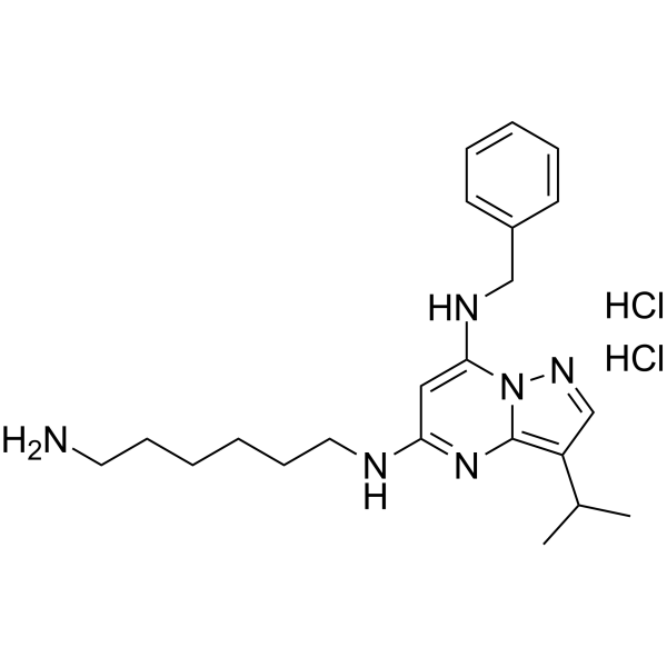
Cl[H].Cl[H].N12C(=C([H])C(=NC1=C(C([H])=N2)C([H])(C([H])([H])[H])C([H])([H])[H])N([H])C([H])([H])C([H])([H])C([H])([H])C([H])([H])C([H])([H])C([H])([H])N([H])[H])N([H])C([H])([H])C1C([H])=C([H])C([H])=C([H])C=1[H]
|
| InChi Key |
XYXAMTBYYTXHSO-UHFFFAOYSA-N
|
| InChi Code |
InChI=1S/C22H32N6.2ClH/c1-17(2)19-16-26-28-21(25-15-18-10-6-5-7-11-18)14-20(27-22(19)28)24-13-9-4-3-8-12-23;;/h5-7,10-11,14,16-17,25H,3-4,8-9,12-13,15,23H2,1-2H3,(H,24,27);2*1H
|
| 化学名 |
5-N-(6-aminohexyl)-7-N-benzyl-3-propan-2-ylpyrazolo[1,5-a]pyrimidine-5,7-diamine;dihydrochloride
|
| 别名 |
BS 181 dihydrochloride; BS-181 (dihydrochloride); 1883548-83-1; 5-N-(6-aminohexyl)-7-N-benzyl-3-propan-2-ylpyrazolo[1,5-a]pyrimidine-5,7-diamine;dihydrochloride; BS 181 2HCl; IAD54883; AKOS025293513;
|
| HS Tariff Code |
2934.99.9001
|
| 存储方式 |
Powder -20°C 3 years 4°C 2 years In solvent -80°C 6 months -20°C 1 month |
| 运输条件 |
Room temperature (This product is stable at ambient temperature for a few days during ordinary shipping and time spent in Customs)
|
| 溶解度 (体外实验) |
May dissolve in DMSO (in most cases), if not, try other solvents such as H2O, Ethanol, or DMF with a minute amount of products to avoid loss of samples
|
|---|---|
| 溶解度 (体内实验) |
注意: 如下所列的是一些常用的体内动物实验溶解配方,主要用于溶解难溶或不溶于水的产品(水溶度<1 mg/mL)。 建议您先取少量样品进行尝试,如该配方可行,再根据实验需求增加样品量。
注射用配方
注射用配方1: DMSO : Tween 80: Saline = 10 : 5 : 85 (如: 100 μL DMSO → 50 μL Tween 80 → 850 μL Saline)(IP/IV/IM/SC等) *生理盐水/Saline的制备:将0.9g氯化钠/NaCl溶解在100 mL ddH ₂ O中,得到澄清溶液。 注射用配方 2: DMSO : PEG300 :Tween 80 : Saline = 10 : 40 : 5 : 45 (如: 100 μL DMSO → 400 μL PEG300 → 50 μL Tween 80 → 450 μL Saline) 注射用配方 3: DMSO : Corn oil = 10 : 90 (如: 100 μL DMSO → 900 μL Corn oil) 示例: 以注射用配方 3 (DMSO : Corn oil = 10 : 90) 为例说明, 如果要配制 1 mL 2.5 mg/mL的工作液, 您可以取 100 μL 25 mg/mL 澄清的 DMSO 储备液,加到 900 μL Corn oil/玉米油中, 混合均匀。 View More
注射用配方 4: DMSO : 20% SBE-β-CD in Saline = 10 : 90 [如:100 μL DMSO → 900 μL (20% SBE-β-CD in Saline)] 口服配方
口服配方 1: 悬浮于0.5% CMC Na (羧甲基纤维素钠) 口服配方 2: 悬浮于0.5% Carboxymethyl cellulose (羧甲基纤维素) 示例: 以口服配方 1 (悬浮于 0.5% CMC Na)为例说明, 如果要配制 100 mL 2.5 mg/mL 的工作液, 您可以先取0.5g CMC Na并将其溶解于100mL ddH2O中,得到0.5%CMC-Na澄清溶液;然后将250 mg待测化合物加到100 mL前述 0.5%CMC Na溶液中,得到悬浮液。 View More
口服配方 3: 溶解于 PEG400 (聚乙二醇400) 请根据您的实验动物和给药方式选择适当的溶解配方/方案: 1、请先配制澄清的储备液(如:用DMSO配置50 或 100 mg/mL母液(储备液)); 2、取适量母液,按从左到右的顺序依次添加助溶剂,澄清后再加入下一助溶剂。以 下列配方为例说明 (注意此配方只用于说明,并不一定代表此产品 的实际溶解配方): 10% DMSO → 40% PEG300 → 5% Tween-80 → 45% ddH2O (或 saline); 假设最终工作液的体积为 1 mL, 浓度为5 mg/mL: 取 100 μL 50 mg/mL 的澄清 DMSO 储备液加到 400 μL PEG300 中,混合均匀/澄清;向上述体系中加入50 μL Tween-80,混合均匀/澄清;然后继续加入450 μL ddH2O (或 saline)定容至 1 mL; 3、溶剂前显示的百分比是指该溶剂在最终溶液/工作液中的体积所占比例; 4、 如产品在配制过程中出现沉淀/析出,可通过加热(≤50℃)或超声的方式助溶; 5、为保证最佳实验结果,工作液请现配现用! 6、如不确定怎么将母液配置成体内动物实验的工作液,请查看说明书或联系我们; 7、 以上所有助溶剂都可在 Invivochem.cn网站购买。 |
| 制备储备液 | 1 mg | 5 mg | 10 mg | |
| 1 mM | 2.2053 mL | 11.0266 mL | 22.0531 mL | |
| 5 mM | 0.4411 mL | 2.2053 mL | 4.4106 mL | |
| 10 mM | 0.2205 mL | 1.1027 mL | 2.2053 mL |
1、根据实验需要选择合适的溶剂配制储备液 (母液):对于大多数产品,InvivoChem推荐用DMSO配置母液 (比如:5、10、20mM或者10、20、50 mg/mL浓度),个别水溶性高的产品可直接溶于水。产品在DMSO 、水或其他溶剂中的具体溶解度详见上”溶解度 (体外)”部分;
2、如果您找不到您想要的溶解度信息,或者很难将产品溶解在溶液中,请联系我们;
3、建议使用下列计算器进行相关计算(摩尔浓度计算器、稀释计算器、分子量计算器、重组计算器等);
4、母液配好之后,将其分装到常规用量,并储存在-20°C或-80°C,尽量减少反复冻融循环。
计算结果:
工作液浓度: mg/mL;
DMSO母液配制方法: mg 药物溶于 μL DMSO溶液(母液浓度 mg/mL)。如该浓度超过该批次药物DMSO溶解度,请首先与我们联系。
体内配方配制方法:取 μL DMSO母液,加入 μL PEG300,混匀澄清后加入μL Tween 80,混匀澄清后加入 μL ddH2O,混匀澄清。
(1) 请确保溶液澄清之后,再加入下一种溶剂 (助溶剂) 。可利用涡旋、超声或水浴加热等方法助溶;
(2) 一定要按顺序加入溶剂 (助溶剂) 。
 匹伐加宾
匹伐加宾
 磷酸氢化可的松
 BAY-784
 Gly-PEG3-endo-BCN
InvivoChem的所有产品仅用于作科学研究,不面向患者销售
Copyright 2020 InvivoChem LLC | All Rights Reserved 粤ICP备20063088号-1


 463611831
