| 规格 | 价格 | 库存 | 数量 |
|---|---|---|---|
| 2mg |
|
||
| 5mg |
|
||
| 10mg |
|
||
| 25mg |
|
||
| Other Sizes |
|

| 靶点 |
JNK/SAPK; CEP-1347 is a multi-target kinase inhibitor whose primary well-characterized target is the mixed lineage kinase family .
|
|---|---|
| 体外研究 (In Vitro) |
体外活性:在体外,CEP-1347在临床相关浓度下有效诱导分化,并抑制来自胶质母细胞瘤以及胰腺癌和卵巢癌的人类癌症干细胞的自我更新和肿瘤启动能力,且不损害其活力。正常成纤维细胞和神经干细胞。激酶测定:CEP-1347(也称为 KT7515)是在 Narcodiopsis 细菌肉汤中发现的天然产物 K-252a 的衍生物,是一种新型有效的 JNK 抑制剂,对 JNK1 的 IC50 为 30 nM。细胞测定:卵巢中过度表达的生存素的敲低癌症干细胞(CSC)的化疗耐药性导致对紫杉醇的敏感性增加。 CEP-1347(一种已知对人类安全性的混合谱系激酶抑制剂)在临床相关浓度下进行治疗,可降低卵巢 CSC 中生存素的表达,并使它们对紫杉醇敏感。
CEP-1347降低survivin的表达,并使卵巢CSC对紫杉醇敏感。鉴于survivin表达与卵巢CSC的紫杉醇耐药性有关,我们接下来希望鉴定治疗靶向survivin表达的药物,以克服与survivin过表达相关的紫杉醇抗性。由于对卵巢CSC及其分化对应物中差异表达的信号分子的平行搜索显示,MLK3的表达水平在A2780 CSLC和TOV21G CSLC细胞系中与survivin的表达水平非常接近(图1),我们推测MLK3可能在survivin表达中发挥作用,因此使用了CEP-1347,这是一种在人类中具有已知安全性的小分子MLK抑制剂。结果表明,CEP-1347有效地抑制了两种卵巢CSC系中survivin的表达(图3)。重要的是,CEP-1347在300 nM或以下以浓度依赖的方式降低了存活素的表达,即在人类研究中已被证明具有临床相关性的浓度下(图3A)。时间过程分析表明,用200 nM的CEP-1347处理的细胞中存活素的表达在3天内降低(图3B)。[1] 在证明了CEP-1347抑制存活素表达的能力后,我们接下来问CEP-1347是否会使卵巢CSC对紫杉醇敏感。为了检验这一点,在CEP-1347存在和不存在的情况下,用紫杉醇处理卵巢CSC系。单独用200 nM的CEP-1347治疗会适度诱导两种卵巢CSC系的细胞死亡,这可能与我们早期的观察结果一致,即单独敲除存活素足以降低其存活率(图4A)。如图2B所示,单独用2 nM的紫杉醇处理细胞也会导致细胞死亡的边缘水平(图4A)。然而,值得注意的是,用CEP-1347和紫杉醇联合治疗卵巢CSC系导致死细胞比例显著增加(图4A),这支持了CEP-1347使细胞对紫杉醇敏感的观点。进一步的分析表明,这种细胞死亡伴随着胱天蛋白酶的激活,如凋亡特异性切割胱天蛋白酶3表达增加所示(图4B),这表明胱天蛋白酶依赖性凋亡程序可能参与了紫杉醇存在和不存在时CEP-1347诱导的细胞死亡。[1] 最后,为了排除CEP-1347只是推进最终发生的细胞死亡的时间动力学的可能性,我们确定了在规定的时间内同时使用CEP-1347和紫杉醇是否不仅会导致细胞死亡的短期增加,还会导致细胞生长的持续抑制,这从治疗的角度来看也可能具有重要意义。当用多种药物联合治疗3天的卵巢CSC在没有药物的情况下生长时,CEP-1347和紫杉醇的联合治疗比单独治疗更有效地抑制了生长(图5)。这些结果表明,CEP-1347和紫杉醇的组合使用可能有利于实现卵巢CSC人群的净减少[1]。 CEP-1347促进CSCs在体外分化为非CSCs[2] 为了通过药物重新定位来鉴定新的CSC靶向药物,我们通过检查它们对CSC中干细胞标志物表达的影响,筛选了一组根据其作用机制推测具有CSC抑制活性的候选现有药物。在这些候选药物中,CEP-1347是一种MLK抑制剂,在JNK信号级联中作用于c-jun N-末端激酶(JNK)的上游。由于CEP-1347本身可以抑制JNK信号通路,这已被证明对维持各种CSC至关重要,我们研究了它对已知依赖JNK的人类CSC的影响。[2] 首先,我们在体外用300 nM或以下的CEP-1347处理CSCs,该浓度已被证明在人体研究中具有临床相关性,并被证实对正常人肺成纤维细胞或大鼠神经干细胞无毒,对CSCs的毒性也很小(补充图1),以分析药物对CSCs干细胞特性的影响。在CEP-1347存在下培养三种不同的胶质瘤干细胞(GS-Y01、GS-Y03和GS-NCC01)导致细胞表面表达干细胞标志物CD133的细胞比例大幅下降,当测试其他CSC,如胰腺(PANC-1 CSLC)和卵巢(A2780 CSLC和TOV21G CSLC)CSC时,也观察到了类似的情况(图1A)。为了确定CEP-1347治疗引起的CD133细胞表面表达的丧失是否代表CSC干细胞特性的丧失,我们同时检测了其他干细胞标志物和分化标志物的表达水平。结果表明,CEP-1347治疗降低了所有检查的CSCs中Sox2和Bmi1等干细胞标志物的表达水平,同时诱导了胶质瘤干细胞中胶质纤维酸性蛋白(GFAP)和卵巢CSCs中E-钙粘蛋白等相应分化标志物的表达(图1B)。然而,至少当PANC-1 CSLC细胞用CEP-1347处理长达6天时,没有观察到E-cadherin表达的增加。因此,为了持续监测CEP-1347对PANC-1 CSLC细胞中E-钙粘蛋白表达的影响超过6天,我们在没有CEP-1347的情况下培养细胞,因为长期暴露于CEP-1347会大大降低其存活率。令人惊讶的是,我们发现,即使在没有CEP-1347的情况下,E-钙粘蛋白的表达也会逐渐增加,同时Sox2的表达会持续减少(图1C)。这些结果虽然提供了CEP-1347也促进PANC-1 CSLC细胞分化的证据,但表明短暂暴露于CEP-1347可能足以使CSC分化为非CSC。 CEP-1347在体外抑制CSCs的自我更新和肿瘤启动能力[2] 在证明CEP-1347是CSC分化的有力驱动因素后,我们接下来分别通过球体形成和异种移植物试验确定了CEP-1347治疗对CSC自我更新和肿瘤起始能力的影响。当CSCs在CEP-1347存在的情况下处理6天后,在没有CEP-1347的情况下在球体形成条件下培养时,形成的球体数量因之前的CEP-1347处理而显著减少,这表明CEP-1347预处理在进行球体形成测定时损害了它们的自更新能力(图2)。同样,当用CEP-1347治疗6天的CSCs植入小鼠体内时,除一例外,细胞未能形成肿瘤,而对照组治疗的CSCs总是会产生逐渐生长的肿瘤(图3)。值得注意的是,在植入CEP-1347处理的PANC-1 CSLC细胞的小鼠中,形成了一个大的(>1000 mm3)肿瘤,尽管如此,它还是自发消退(图3B,左)。这一观察表明,CEP-1347可能损害了CSC在不干扰其在小鼠体内植入的情况下使肿瘤生长持续的能力(见讨论)。总之,这些结果表明,CEP-1347在体外有效地抑制了CSC的自我更新和肿瘤启动能力。 |
| 体内研究 (In Vivo) |
在体内,CEP-1347 的 10 天全身给药剂量低于小鼠当量(相当于人类 2 年安全给药剂量的 1/10),足以有效减少已建立肿瘤内的肿瘤起始癌症干细胞在小鼠中。此外,相同的治疗方案显着延长了接受神经胶质瘤干细胞原位植入的小鼠的生存期。总之,这些发现表明 CEP-1347 是癌症干细胞靶向治疗的有前途的候选者,并且需要进一步的临床和临床前研究来评估其在癌症治疗中的功效。
系统给药CEP-1347可抑制体内CSCs[2] 在体外证明的CEP-1347的强大CSC抑制活性的鼓励下,我们继续在体内确定全身给药的CEP-1347是否可以原位靶向和抑制CSC,即在人类癌症动物模型的肿瘤中。之前的人体临床研究表明,CEP-1347高达100mg/天(50mg,每天口服两次)具有良好的耐受性,足以将CEP-1347的血浆浓度提高到亚毫摩尔水平。根据人和小鼠的Km值,成人100 mg/天的剂量在小鼠中转化为约20 mg/kg/天,因此我们选择了1.5 mg/kg/天的CEP-1347起始剂量,该剂量小于上述剂量的十分之一,并且确实已通过腹腔途径成功给药小鼠1周。在确认连续10天腹腔注射1.5mg/kg/天的CEP-1347不会损害小鼠体重监测的一般健康状况(图4A)后,我们进行了一项连续移植试验,以评估全身性CEP-1347是否减少了荷瘤小鼠异种移植物肿瘤内的肿瘤起始CSC群。为此,我们根据上述治疗方案(每天一次腹腔注射1.5 mg/kgCEP-1347,持续10天)治疗了通过植入胶质瘤干细胞形成的皮下肿瘤小鼠。在最后一次给药的第二天,切除皮下肿瘤,分离后,将肿瘤细胞原位移植到新小鼠的大脑中,然后在没有任何额外治疗的情况下进行观察。连续移植试验的结果表明,来自对照治疗的原发性肿瘤的肿瘤细胞(5×104和1×104)的移植必然会导致脑肿瘤的形成和随后的死亡率,尽管存活时间因移植的肿瘤细胞数量而异,即反映了移植的CSC数量(图4B)。结果还表明,移植来源于CEP-1347治疗的原发性肿瘤的肿瘤细胞(5×104)最终导致所有受体小鼠脑肿瘤形成。然而,与相应的对照组相比,生存期显著延长,这与CEP-1347治疗减少了原发性肿瘤内的肿瘤起始CSC的观点一致(图4B和4C)。事实上,这一观点得到了以下观察结果的证实,即移植了1×104个来源于CEP-1347治疗的原发性肿瘤的肿瘤细胞的小鼠的存活时间明显长于相应的对照小鼠,其中一只甚至存活了240天以上,没有任何脑肿瘤形成的迹象(图4B和4D)。值得注意的是,相同的CEP-1347治疗方案显然未能抑制原发性肿瘤的生长,这被认为主要是由非CSC的增殖驱动的(图4E)。总的来说,这些结果表明,CEP-1347在体内选择性抑制肿瘤起始细胞,优先靶向CSCs而非非CSCs。 鉴于低剂量的CEP-1347足以显著减少皮下肿瘤中的胶质瘤干细胞,我们接下来试图确定相同的治疗方案是否能有效抑制胶质瘤干电池在原位位置(即脑实质)的肿瘤起始。为此,我们将胶质瘤干细胞立体定向植入小鼠脑中,并在植入的第二天开始CEP-1347治疗(每天腹腔注射1.5mg/kg CEP-1347一次,连续10天)。与连续移植试验的结果一致,相同的CEP-1347治疗方案在这种脑肿瘤异种移植物模型中同样有效,与对照治疗相比,显著延长了小鼠的存活时间(图5)。因此,研究结果表明,CEP-1347可以治疗性地靶向大脑中的胶质瘤干细胞,这与它穿透血脑屏障的能力相一致。 |
| 酶活实验 |
CEP-1347 是一种多靶点激酶抑制剂,其最主要且被充分表征的作用靶点是混合谱系激酶家族 。在非细胞实验中,确定其作用机制的常用方法是直接测定对重组 MLK3 激酶活性的抑制能力。将纯化的 MLK3 蛋白与含有 ATP 的缓冲液及多肽底物混合,并加入梯度稀释的 CEP-1347 共同孵育。该化合物在 ATP 结合位点发挥竞争性抑制作用,从而阻止底物磷酸化。通过定量检测残留的激酶活性并计算半数抑制浓度,结果显示 CEP-1347 对重组 MLK3 的 IC₅₀ 约为 23 nM 。该生化实验确认了 MLK 家族是 CEP-1347 的直接分子靶点,为其后续作为 JNK 信号通路抑制剂进行深入研究提供了依据 。
|
| 细胞实验 |
细胞活力测定。[1]
细胞活力测定如前所述进行。简而言之,根据制造商的说明,使用WST-8通过四唑盐还原法测定细胞存活率。第二天,在200nMCEP-1347存在或不存在的情况下,用2nM紫杉醇处理96孔I型胶原涂层板中的细胞(500/孔)3天,然后在没有任何药物的情况下再培养4天。然后加入WST-8试剂,在37°C下孵育细胞1-3小时。使用微孔板读数器(型号680;Bio-Rad)测量450nm处的吸光度。相对细胞存活率计算为处理样品相对于对照样品的吸光度百分比。细胞活力测定一式三份。 细胞死亡测定。[1] 如前所述进行细胞死亡测定。简而言之,在37°C的CO2培养箱中,将细胞与PI(1μg/ml)和Hoechst 33342(10μg/ml)原位孵育5分钟,分别对死细胞和细胞核进行染色。然后在荧光显微镜下对PI和Hoechst阳性细胞的数量进行评分,并确定PI阳性细胞(死细胞)相对于Hoechst阴性细胞(总细胞)的百分比。细胞死亡试验一式三份。 |
| 动物实验 |
将GS-Y03细胞(1 × 10⁴)颅内植入小鼠(每组n = 6)后,连续10天每日腹腔注射对照载体或CEP-1347(1.5 mg/kg/天),注射从颅内植入后的第二天开始。
原位脑肿瘤模型小鼠 对于连续移植,按照图例所述方法处理的原发肿瘤被切除,用冰冷的无菌PBS洗涤后,转移至DMEM/F12培养基中,用剪刀剪碎,并在37°C下用TrypLE™ Express消化30分钟。用DMEM/F12培养基冲洗后,将细胞重悬于DMEM/F12培养基中,并通过70 μm滤网过滤。在确定细胞数量和活力后,将单细胞悬液进行颅内注射。对于全身给药CEP-1347,将CEP-1347储备液(1 mM DMSO溶液)用PBS稀释,配制成200 μL的注射液。将CEP-1347溶液腹腔注射到裸鼠体内。所有对照组和CEP-1347处理组的小鼠均按体重注射等体积的DMSO(3.6 mL/kg)。[2] |
| 药代性质 (ADME/PK) |
CEP-1347 是一种口服有效的小分子化合物,分子量为 615.19 g/mol,XLogP 为 7.52,拓扑极性表面积为 145.32 Ų 。在 HIV 感染患者中,以 50 mg 每日两次的剂量给予 CEP-1347 后,该化合物与阿扎那韦存在显著的药代动力学相互作用:阿扎那韦的蓄积比增加 15%(p = 0.007),阿扎那韦的半衰期从 12.7 小时延长至 15.9 小时(p = 0.002),而对利托那韦的药代动力学无显著影响 。在一项纳入 30 例帕金森病患者的短期研究中,CEP-1347 对左旋多巴的药代动力学无急性影响,使其适用于更长期的研究 。
|
| 毒性/毒理 (Toxicokinetics/TK) |
CEP-1347 在针对早期帕金森病的大型 2/3 期临床试验(NCT00040404)中进行了评估,该研究证实了其良好的安全性和耐受性,但未能显示临床疗效 。在 HIV 感染患者中以 50 mg 每日两次的剂量给药时,CEP-1347 耐受性良好,药物相互作用研究中未报告显著的不良反应 。在临床前研究中,以低于人类安全给药 2 年的小鼠等效剂量的 1/10 进行全身性给药,足以减少体内肿瘤起始性癌症干细胞,且不影响正常成纤维细胞和神经干细胞的活力 。通过先前的临床研究,已建立了该化合物在人体中的已知安全性特征 。该研究性化合物未报告严重不良事件或黑框警告。
|
| 参考文献 | |
| 其他信息 |
CEP-1347 是一种半合成化合物,已被证明能够保护多种神经细胞免受各种损伤,从而避免程序性细胞死亡(凋亡),这可能有助于提高多巴胺神经元在移植前后的存活率。
药物适应症 正在研究其在哮喘和帕金森病中的应用/治疗。 作用机制 CEP-1347 是一种口服活性分子,是应激激活蛋白激酶通路的选择性强效抑制剂。该通路是一种细胞内信号通路,是导致神经元死亡的应激反应的重要组成部分。体外细胞培养系统以及帕金森病的小鼠和非人灵长类动物体内模型均表明,CEP-1347能够保护黑质(帕金森病影响的大脑区域)中的多巴胺能神经元。 背景:癌症干细胞(CSCs)的化疗耐药性被认为是卵巢癌治疗后复发的主要原因,会对患者的预后产生负面影响。材料与方法:我们使用源自两种不同卵巢癌细胞系的CSCs,寻找与卵巢CSCs化疗耐药性相关的分子以及靶向这些分子的药物。结果:敲低卵巢CSCs中过表达的survivin可提高其对紫杉醇的敏感性。使用临床相关浓度的混合谱系激酶抑制剂CEP-1347(该抑制剂在人体中具有已知的安全性)进行治疗,可降低卵巢癌干细胞中Survivin的表达,并使其对紫杉醇敏感。结论:Survivin的过表达在卵巢癌干细胞的化疗耐药中起着关键作用。将靶向卵巢癌干细胞中Survivin表达的CEP-1347作为常规卵巢癌化疗的增敏剂,可能是一种合理可行的卵巢癌治疗策略。[1] 总之,我们提供的证据支持了Survivin过表达可能是卵巢癌干细胞化疗耐药性增强的主要机制之一的观点。本研究证实,CEP-1347 能在临床相关浓度下有效抑制 survivin 表达并增强卵巢癌干细胞的化疗敏感性,因此,其应用可能成为提高传统化疗疗效、从而改善卵巢癌患者生存率的合理且有前景的策略。CEP-1347 是一种混合谱系激酶抑制剂,曾在早期帕金森病的大规模 II/III 期临床试验中进行过测试,结果显示其安全性和耐受性良好,但疗效尚未得到证实。本研究通过药物重定位,将 CEP-1347 鉴定为一种潜在的抗癌干细胞药物。体外实验表明,在临床相关浓度下,CEP-1347 能有效诱导胶质母细胞瘤、胰腺癌和卵巢癌等人类癌干细胞分化,并抑制其自我更新和肿瘤起始能力,且不影响正常成纤维细胞和神经干细胞的活性。体内实验表明,小鼠接受为期 10 天的全身性 CEP-1347 给药,其剂量低于人类安全给药 2 年剂量的十分之一,即可有效减少小鼠已形成肿瘤内的肿瘤起始癌干细胞。此外,相同的治疗方案显著延长了接受原位移植胶质瘤干细胞的小鼠的生存期。综上所述,我们的研究结果表明,CEP-1347 是一种有前景的癌干细胞靶向治疗候选药物,有必要开展进一步的临床和临床前研究来评估其在癌症治疗中的疗效。 [2] 总之,本研究表明,CEP-1347 在体外临床相关浓度下可促进癌症干细胞 (CSC) 分化,从而有效抑制体内 CSC 的肿瘤形成。尽管 CEP-1347 抑制 CSC 的确切机制以及 MLK 在其中的作用尚待阐明,但我们的研究结果,结合该药物在人体中良好的安全性记录,表明在目前的癌症治疗方案中加入 CEP-1347 是一种合理可行的策略,可以预防治疗后存活的 CSC(尤其是那些依赖 JNK 维持干细胞状态的 CSC)引起的复发和/或转移。[2] |
| 分子式 |
615.76
|
|
|---|---|---|
| 分子量 |
C33H33N3O5S2
|
|
| 精确质量 |
615.186
|
|
| 元素分析 |
C, 64.37; H, 5.40; N, 6.82; O, 12.99; S, 10.41
|
|
| CAS号 |
156177-65-0
|
|
| 相关CAS号 |
|
|
| PubChem CID |
9917013
|
|
| 外观&性状 |
White to off-white solid powder
|
|
| LogP |
6.49
|
|
| tPSA |
145.32
|
|
| 氢键供体(HBD)数目 |
2
|
|
| 氢键受体(HBA)数目 |
7
|
|
| 可旋转键数目(RBC) |
8
|
|
| 重原子数目 |
43
|
|
| 分子复杂度/Complexity |
1150
|
|
| 定义原子立体中心数目 |
3
|
|
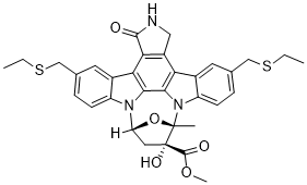
| SMILES |
CCSCC1=CC2=C(C=C1)N3[C@H]4C[C@@]([C@](O4)(N5C6=C(C=C(C=C6)CSCC)C7=C8CNC(=O)C8=C2C3=C75)C)(C(=O)OC)O
|
|
| InChi Key |
SCMLRESZJCKCTC-KMYQRJGFSA-N
|
|
| InChi Code |
InChI=1S/C33H33N3O5S2/c1-5-42-15-17-7-9-22-19(11-17)26-27-21(14-34-30(27)37)25-20-12-18(16-43-6-2)8-10-23(20)36-29(25)28(26)35(22)24-13-33(39,31(38)40-4)32(36,3)41-24/h7-12,24,39H,5-6,13-16H2,1-4H3,(H,34,37)/t24-,32+,33+/m1/s1
|
|
| 化学名 |
|
|
| 别名 |
|
|
| HS Tariff Code |
2934.99.9001
|
|
| 存储方式 |
Powder -20°C 3 years 4°C 2 years In solvent -80°C 6 months -20°C 1 month |
|
| 运输条件 |
Room temperature (This product is stable at ambient temperature for a few days during ordinary shipping and time spent in Customs)
|
| 溶解度 (体外实验) |
|
|||
|---|---|---|---|---|
| 溶解度 (体内实验) |
注意: 如下所列的是一些常用的体内动物实验溶解配方,主要用于溶解难溶或不溶于水的产品(水溶度<1 mg/mL)。 建议您先取少量样品进行尝试,如该配方可行,再根据实验需求增加样品量。
注射用配方
注射用配方1: DMSO : Tween 80: Saline = 10 : 5 : 85 (如: 100 μL DMSO → 50 μL Tween 80 → 850 μL Saline)(IP/IV/IM/SC等) *生理盐水/Saline的制备:将0.9g氯化钠/NaCl溶解在100 mL ddH ₂ O中,得到澄清溶液。 注射用配方 2: DMSO : PEG300 :Tween 80 : Saline = 10 : 40 : 5 : 45 (如: 100 μL DMSO → 400 μL PEG300 → 50 μL Tween 80 → 450 μL Saline) 注射用配方 3: DMSO : Corn oil = 10 : 90 (如: 100 μL DMSO → 900 μL Corn oil) 示例: 以注射用配方 3 (DMSO : Corn oil = 10 : 90) 为例说明, 如果要配制 1 mL 2.5 mg/mL的工作液, 您可以取 100 μL 25 mg/mL 澄清的 DMSO 储备液,加到 900 μL Corn oil/玉米油中, 混合均匀。 View More
注射用配方 4: DMSO : 20% SBE-β-CD in Saline = 10 : 90 [如:100 μL DMSO → 900 μL (20% SBE-β-CD in Saline)] 口服配方
口服配方 1: 悬浮于0.5% CMC Na (羧甲基纤维素钠) 口服配方 2: 悬浮于0.5% Carboxymethyl cellulose (羧甲基纤维素) 示例: 以口服配方 1 (悬浮于 0.5% CMC Na)为例说明, 如果要配制 100 mL 2.5 mg/mL 的工作液, 您可以先取0.5g CMC Na并将其溶解于100mL ddH2O中,得到0.5%CMC-Na澄清溶液;然后将250 mg待测化合物加到100 mL前述 0.5%CMC Na溶液中,得到悬浮液。 View More
口服配方 3: 溶解于 PEG400 (聚乙二醇400) 请根据您的实验动物和给药方式选择适当的溶解配方/方案: 1、请先配制澄清的储备液(如:用DMSO配置50 或 100 mg/mL母液(储备液)); 2、取适量母液,按从左到右的顺序依次添加助溶剂,澄清后再加入下一助溶剂。以 下列配方为例说明 (注意此配方只用于说明,并不一定代表此产品 的实际溶解配方): 10% DMSO → 40% PEG300 → 5% Tween-80 → 45% ddH2O (或 saline); 假设最终工作液的体积为 1 mL, 浓度为5 mg/mL: 取 100 μL 50 mg/mL 的澄清 DMSO 储备液加到 400 μL PEG300 中,混合均匀/澄清;向上述体系中加入50 μL Tween-80,混合均匀/澄清;然后继续加入450 μL ddH2O (或 saline)定容至 1 mL; 3、溶剂前显示的百分比是指该溶剂在最终溶液/工作液中的体积所占比例; 4、 如产品在配制过程中出现沉淀/析出,可通过加热(≤50℃)或超声的方式助溶; 5、为保证最佳实验结果,工作液请现配现用! 6、如不确定怎么将母液配置成体内动物实验的工作液,请查看说明书或联系我们; 7、 以上所有助溶剂都可在 Invivochem.cn网站购买。 |
计算结果:
工作液浓度: mg/mL;
DMSO母液配制方法: mg 药物溶于 μL DMSO溶液(母液浓度 mg/mL)。如该浓度超过该批次药物DMSO溶解度,请首先与我们联系。
体内配方配制方法:取 μL DMSO母液,加入 μL PEG300,混匀澄清后加入μL Tween 80,混匀澄清后加入 μL ddH2O,混匀澄清。
(1) 请确保溶液澄清之后,再加入下一种溶剂 (助溶剂) 。可利用涡旋、超声或水浴加热等方法助溶;
(2) 一定要按顺序加入溶剂 (助溶剂) 。
CEP-1347induces the differentiation of cancer stem cells into non-cancer stem cells.Oncotarget.2017 Oct 24;8(55):94872-94882. |
|---|
CEP-1347treatment deprives cancer stem cells of their sphere forming ability.Cells treated without (Control) or withCEP-1347(300 nM for PANC-1 CSLC, 200 nM for the others) for 6 days were subjected to a sphere formation assay in the absence ofCEP-1347.Oncotarget.2017 Oct 24;8(55):94872-94882. |
Systemically administeredCEP-1347selectively targets and inhibits tumor-initiating cancer stem cells within tumors in tumor-bearing mice.Oncotarget.2017 Oct 24;8(55):94872-94882. |
CEP-1347inhibits the tumor-initiating capacity of cancer stem cells.Oncotarget.2017 Oct 24;8(55):94872-94882. |
|---|
Short-term systemicCEP-1347treatment inhibits tumor formation by glioma stem cells in an orthotopic brain tumor model.Mice implanted intracranially with GS-Y03 cells underwent a daily i.p. injection ofCEP-1347(1.5 mg/kg/day) for 10 days.Oncotarget.2017 Oct 24;8(55):94872-94882. |
InvivoChem的所有产品仅用于作科学研究,不面向患者销售
Copyright 2020 InvivoChem LLC | All Rights Reserved 粤ICP备20063088号-1


 COA
COA


 463611831
