| 规格 | 价格 | |
|---|---|---|
| 500mg | ||
| 1g | ||
| Other Sizes |
| 靶点 |
Ki: 220/300 nM (ROCK-I/II)[1]
|
|---|---|
| 体外研究 (In Vitro) |
Y-27632具有以剂量依赖方式诱导ADSCs神经元样分化的效力。此外,Y-27632诱导的分化在停药后恢复。用Y-27632处理的ADSCs表达神经元标志物,如NSE、MAP-2和巢蛋白,而未处理的ADSC不表达这些标志物。结论:选择性ROCK抑制剂Y-27632可以增强ADSCs的神经元样分化,表明Y-27632可用于诱导ADSCs向神经元分化,促进ADSCs在组织工程中的临床应用[2]。
Y-27632 在 BrdU 标记出现时的浓度抑制延迟增加了 100 倍,并且延迟持续时间延长。在分别用 10 和 100 μM Y-27632 处理的 Swiss 3T3 细胞中,观察到约 1 和 4 小时的延迟 [1]。 Y-27632 在神经元口腔中促进脂肪组织源性干细胞 (ADSC) 的生长。与 1.0 和 2.5 μM Y-27632 诱导组相比,5.0 μM Y-27632 诱导组具有最多数量的神经样细胞 [2]。在上皮细胞中,肌动蛋白会重塑并上调细胞因子[8]。
Y-27632[(+)-(R)-反式-4-(1-氨基乙基)-N-(4-吡啶基)环己烷甲酰胺+++二盐酸盐]被广泛用作Rho相关卷曲卷曲形成蛋白丝氨酸/苏氨酸激酶(ROCK)家族蛋白激酶的特异性抑制剂。本研究考察了Y-27632和相关化合物Y-30141[(+)-(R)-反式-4-(1-氨基乙基)-N-(1H-吡咯并[2,3-b]吡啶-4-基)环己酰胺二盐酸盐]的抑制机制和作用特征。Y-27632和Y-30141在体外抑制了ROCK-I和ROCK-II的激酶活性,这种抑制作用被ATP以竞争方式逆转。这表明这些化合物通过与催化位点结合来抑制激酶。通过K(i)值确定的它们对ROCK激酶的亲和力比另外两种Rho效应激酶(柠檬酸激酶和蛋白激酶PKN)的亲和力高至少20至30倍。[(3)H]Y-30141以温度、时间和可饱和的方式被细胞吸收,这种吸收与未标记的Y-27632竞争。没有发现集中的积累,这表明摄取是载体介导的促进扩散。Y-27632在10微M时消除了瑞士3T3细胞中的应力纤维,但在此浓度下,细胞周期和胞质分裂的G(1)-S相变几乎不受影响。Y-30141在抑制激酶活性和应激纤维形成方面的效力是Y-27632的10倍,并且在10微M时导致G(1)-S转换和胞质分裂抑制的显著延迟。[1] Y-27632具有以剂量依赖方式诱导ADSCs神经元样分化的效力。此外,Y-27632诱导的分化在停药后恢复。用Y-27632处理的ADSCs表达神经元标志物,如NSE、MAP-2和巢蛋白,而未处理的ADSC不表达这些标志物。 结论:选择性ROCK抑制剂Y-27632可以增强ADSCs的神经元样分化,表明Y-27632可用于诱导ADSCs向神经元分化,促进ADSCs在组织工程中的临床应用 对人类诱导多能干细胞(hIPSC)分化的强有力控制对于实现其针对患者的治疗潜力至关重要。在这里,我们展示了小分子Rho相关蛋白激酶(ROCK)抑制剂Y-27632的一种新应用,以谱系特异性方式显著影响hIPSC的分化。Y-27632在hIPSC上的应用导致肌动蛋白捆绑减少,集落形成以浓度和时间依赖的方式中断。细胞和集落形态的这种变化与细胞-细胞连接蛋白E-钙粘蛋白的表达降低有关,与Y-27632暴露量的增加成正比。有趣的是,多能性标记物如NANOG和OCT4的基因和蛋白质表达在暴露于Y-27632长达36小时后没有下调。同时,上皮间充质(EMT)过渡标志物随着Y-27632的暴露而上调。随着Y-27632暴露时间的延长,细胞中的这些EMT样变化导致随后向中胚层谱系分化的效率显著提高。相比之下,当细胞在长期暴露于Y-27632后经历外胚层分化时,观察到抑制作用。总的来说,这些结果提出了一种新的方法,通过简单应用Y-27632来启动hIPSC以调节其分化潜力。[5] c-kit+CSC在CSC培养基中培养3-5天,然后单独用0至10μM Y-27632、0至1.0μM Dox或Y-27632和Dox处理48小时。检测细胞活力、毒性、增殖、形态、迁移、Caspase-3活性、凋亡相关关键蛋白和c-kit+的表达水平。结果显示,单独用Y-27632处理48小时后,c-kit+表达、增殖、Caspase-3活性或凋亡没有发生太大变化;然而,细胞活力显著增加,细胞迁移得到促进。这些效应可能涉及ROCK/Actin通路。相比之下,单独使用Dox治疗48小时会显著增加Caspase-3活性,导致细胞死亡。尽管单独使用Y-27632在基础条件下不影响凋亡相关关键因子(p-Akt、Akt、Bcl-2、Bcl-xl、Bax、切割的Caspase-3和Caspase-3)的表达水平,但它显著抑制了Dox诱导的切割的Caspase3的增加,并减少了Dox处理下的细胞死亡。 结论:我们得出结论,用Y-27632预处理人CSCs可显著降低Dox诱导的细胞死亡,并可能涉及切割的Caspase-3和ROCK/Actin通路。Y-27632的有益作用可应用于基于干细胞的治疗,以提高移植后的细胞存活率,或作为Dox治疗的癌症患者的心脏保护剂。[6] RhoA/ROCK信号通路的激活已被证明有助于胚胎和神经干细胞的解离诱导凋亡。我们之前已经证明,在40个Lin(-)Sca-1(+)CD49f(高)(LSC)前列腺基底上皮细胞中,大约有1个具有干细胞自我更新和多谱系分化的能力。我们在这里表明,在体外前列腺集落试验中,用ROCK激酶抑制剂Y-27632处理LSC细胞可将其克隆效率提高8倍。Y-27632治疗允许前列腺集落细胞有效地重新生长,否则不会发生这种情况。Y-27632还提高了前列腺球试验和分离前列腺细胞再生试验中前列腺干细胞的克隆效率。克隆效率的提高是由于抑制了RhoA/ROCK活化介导的前列腺干细胞的解离诱导凋亡。前列腺上皮细胞与细胞外基质的分离增加了PTEN活性并减弱了AKT活性。单独的Y-27632处理足以抑制细胞解离诱导的PTEN活性激活。然而,这并没有提高克隆效率,因为Y-27632处理在类似程度上增加了野生型和Pten-null前列腺细胞的球体形成单位。最后,敲低两种ROCK激酶的表达略微提高了前列腺集落细胞的重铺效率,证实了它们在Y-27632介导的克隆效率提高中起着重要作用。我们的研究表明,根据目前采用的检测方法,具有干/祖细胞活性的前列腺细胞的数量可能被低估,支持解离诱导的凋亡是具有上皮表型的胚胎和体细胞干细胞的共同特征,并强调了环境线索对干细胞维持的重要性。[7] 在用胶原蛋白(COL)或LPA刺激细胞之前,在0、0.03、0.3、3或10微M的ROCK抑制剂Y27632存在下培养整片禽胚胎角膜上皮。通过Caspase-3活性测定评估细胞凋亡,并用膜联蛋白V结合进行可视化。ROCK抑制剂以剂量依赖的方式阻断了肌动蛋白皮质垫的重建,破坏了基底细胞侧膜,并增加了凋亡标志物膜联蛋白V。此外,使用体外半胱氨酸天冬氨酸蛋白酶-3活性测定来确定,用10微M Y-27632处理的上皮细胞中的半胱氨酸天冬氨酰蛋白酶-3活性高于没有基底膜的上皮细胞或用纤维连接蛋白、COL或LPA刺激的上皮细胞。总之,ECM分子降低了凋亡标志物,抑制了ROCK通路,阻断了ECM刺激的肌动蛋白皮质垫的重建,增加了胚胎角膜上皮细胞的凋亡[8]。 |
| 体内研究 (In Vivo) |
与盐水组相比,Y-27632(5 和 10 mg/kg)显着延长了肌阵挛发作的发作时间。当给予 Y-27632(5 和 10 mg/kg)时,阵挛性惊厥的发作与盐水组相比显着延迟 [3]。与对照组(SS 组)相比,用二甲基亚硝胺(DMN)治疗的大鼠的肝脏重量和体重(DMN-S 组)显着减少。通过口服 Y27632 (30 mg/kg),大鼠(DMN-Y 组)基本上免受 DMN 引起的体重和肝脏重量损失[4]。
Y27632治疗显著降低了DMN诱导的肝纤维化的发生率,降低了肝脏中胶原蛋白和羟脯氨酸含量以及α-SMA表达。[3] Y27632可防止DMN引起的身体和肝脏重量减轻[3] 口服Y27632对腹腔注射和不注射DMN的大鼠体重和肝脏重量的影响如图1和表1所示。与对照组动物(S-S组)相比,DMN治疗导致大鼠体重和肝脏重量显著降低(DMN-S组)。口服Y27632基本上阻止了DMN诱导的大鼠体重和肝脏减轻(DMN-Y组)。S-S/S-Y组和DMN-Y组之间没有显著差异。第四周结束时,DMN-S组和DMN-Y组的血清ALT水平没有显著差异。 Y27632降低肝脏胶原蛋白和羟脯氨酸含量[3] 胶原测定证实,Y27632治疗组(DMN-Y组)的肝胶原沉积显著减少(DMN-S为808.5±122.4μg/100mg,而DMN-Y为601.3±67.7μg/100mg,P<0.05)(图3A)。肝纤维化也通过测量肝羟脯氨酸来定量。在这里,发现DMN治疗组(DMN-S)的羟脯氨酸含量显著高于DMN和Y27632治疗组(DMP-Y)(DMN-S为5.95±1.41μmol/100mg,而DMN-Y为2.98±0.91μmol/100mg)(图3B)。此外,DMN未治疗组(S-S、S-Y)和DMN和Y27632治疗组(DMN-Y)的胶原蛋白和羟脯氨酸含量没有显著差异。这些结果表明,Y27632治疗几乎完全预防了DMN诱导的肝纤维化。 Y27632抑制I型胶原mRNA表达[3] 为了评估Y27632处理对I型胶原mRNA转录的抑制水平,我们通过半定量RT-PCR检测了DMN处理肝脏中α1(I)胶原mRNA的水平。在对照组(S-S组)中,即使只有0.02 fg的竞争对手,在α1(I)胶原蛋白的竞争性RT-PCR中也检测到主要竞争对手特异性带和较弱的α1(Ⅰ)胶原蛋白特异性带(图4A,泳道4)。相比之下,在DMN-S组中,DMN治疗(在没有Y27632的情况下)使α1(I)胶原基因的转录比基础表达(S-S)增强了100多倍,即使在存在2fg竞争对手的情况下,也会产生一条代表α1(Ⅰ)胶原产物的条带(图4B,泳道2)。与DMN治疗相比(在没有Y27632的情况下),Y27632治疗导致了更强烈的竞争带;在0.02fg竞争对手存在的情况下,α1(I)胶原PCR产物的强度与竞争对手相同(图4C,泳道4)。这些结果表明,Y27632治疗可将肝脏中α1(I)胶原mRNA的表达抑制到DMN诱导表达的约10%水平。 法舒地尔和Y-27632对急性PTZ注射诱导的肌阵挛发作的影响[4] 法舒地尔和Y-27632对肌阵挛发作的影响如图1所示。剂量为25 mg kg−1,法舒地尔显著延长了单剂量PTZ(65 mg)诱导的肌阵挛发作时间 与生理盐水组相比(p<0.05)。然而,在5的剂量下,它没有改变发病时间 mg kg−1。此外,在5和10的剂量下 mg 与生理盐水组相比,Y-27632显著延长了肌阵挛发作时间(P<0.05)。 法舒地尔和Y-27632对急性PTZ注射诱发的阵挛性惊厥发作的影响[4] 法舒地尔和Y-27632对阵挛性惊厥发作的影响如图1所示。剂量为25 mg kg−1,法舒地尔显著延长了单剂量PTZ(65 mg)诱导的阵挛性惊厥的发作时间 与生理盐水组相比(p<0.05)。然而,在5的剂量下,它没有改变发病时间 mg kg−1。此外,在5和10的剂量下 mg 与生理盐水组相比,Y-27632显著延长了阵挛性惊厥的发作时间(P<0.05)。 法舒地尔和Y-27632对急性PTZ注射诱导的强直性后肢伸展的影响[4] PTZ治疗组中,十分之七的小鼠进行了强直性后肢伸展,随后死亡。然而,两种剂量的法舒地尔都能防止强直性后肢伸展和死亡的发生(数据未显示)。与法舒地尔类似,两种剂量的Y-27632也能防止强直性后肢伸展和死亡的发生(数据未显示)。 法舒地尔和Y-27632对MES系列强直性后肢伸展持续时间和翻正反射恢复潜伏期的影响[4] 两种法舒地尔(25mg 千克-1)和Y-27632(5和10 mg kg−1)与生理盐水治疗组相比,显著缩短了单次应用MES诱导的强直性后肢伸展的持续时间(图2,P<0.05)。此外,与生理盐水组相比,Rho激酶抑制剂法舒地尔和Y-27632也显著降低了翻正反射的恢复潜伏期(图2,P<0.05)。 急性单次给予法舒地尔或Y-27632对PTZ点燃小鼠肌阵挛发作和阵挛性惊厥的影响[4] 法舒地尔(25mg 千克-1)和Y-27632(5和10 mg kg−1)与生理盐水组相比,改变了肌阵挛抽搐和阵挛性惊厥的发作时间(图3)。 反复服用法舒地尔或Y-27632对PTZ点燃发展的影响[4] 反复服用法舒地尔或Y-27632对PTZ点燃发展的影响如图4所示。通过组间相互作用注射PTZ对生理盐水有显著影响,25 mg kg−1法舒地尔和5 mg Y-27632组(P<0.05)。如图4所示,法舒地尔不能阻止PTZ点燃的发展,但与生理盐水组相比,它通过降低平均发作阶段,在PTZ点燃发展的第2、3和4天产生了显著影响(P<0.05)。与法舒地尔不同,Y-27632更有效,与生理盐水组相比,从第2天开始,通过降低平均发作阶段,对PTZ点燃的发展有显著影响(P<0.05)。 法舒地尔和Y-27632对旋转杆性能的影响[4] 法舒地尔(25mg 千克-1)和Y-27632(5和10 mg kg−1)在20℃时,通过旋转杆的性能评估电机协调性发生了变化 下午3点(表1)。 |
| 酶活实验 |
重组ROCK-I、ROCK-II、PKN或柠檬酸激酶通过使用Lipofectamine转染在HeLa细胞中表达为Myc标记蛋白,并通过使用与G蛋白琼脂糖偶联的9E10单克隆抗Myc抗体从细胞裂解物中沉淀出来。在30°C下,在不存在或存在不同浓度Y-27632或Y-30141的情况下,将回收的免疫复合物与不同浓度的[32P]ATP和10 mg组蛋白2作为底物在总体积为30μL的激酶缓冲液中孵育30分钟,该缓冲液含有50 mM HEPES NaOH、pH 7.4、10 mM MgCl2、5 mM MnCl2、0.02%Briji 35和2 mM二硫苏糖醇。在30°C下,在含有50 mM Tris-HCl、pH 7.5、0.5 mM CaCl2、5 mM乙酸镁、25μg/mL磷脂酰丝氨酸、50 ng/mL 12-O-十四烷酰佛波醇-13-乙酸酯和0.001%亮肽的激酶缓冲液中,在不存在或存在不同浓度Y-27632或Y-30141的情况下,将PKCa与5μM[32P]ATP和200μg/mL组蛋白2作为底物孵育10分钟,总体积为30μL。通过加入10μL的43 Laemmli样品缓冲液终止培养。煮沸5分钟后,将混合物在16%凝胶上进行SDS-聚丙烯酰胺凝胶电泳。凝胶用考马斯亮蓝染色,然后干燥。切除与组蛋白2型相对应的条带,并测量放射性[1]。
|
| 细胞实验 |
Y-27632是Rho相关卷曲激酶(ROCK)的特异性抑制剂,已被证明可以促进多种细胞类型的存活和诱导分化。然而,Y-27632对成人脂肪组织来源干细胞(ADSCs)的影响尚不清楚。本研究旨在探讨Y-27632对ADSCs神经元样分化的影响。
方法:从接受整形手术的女性中分离出ADSCs并进行培养。用不同剂量的Y-27632处理ADSCs,并在显微镜下观察形态学变化。通过免疫细胞化学和蛋白质印迹分析检测Y-27632处理的ADSCs中巢蛋白、神经元特异性烯醇化酶(NSE)和微管相关蛋白-2(MAP-2)的表达[2]。
HeLa细胞以每3.5cm培养皿3×10⁴个细胞的密度铺板。在10mM胸苷存在下,将细胞在含有10%FBS的DMEM中培养16小时。用含有10%FBS地DMEM洗涤细胞后,再培养8小时,然后加入40ng/mL的诺考达唑。在诺考达唑治疗11.5小时后,加入不同浓度的Y-27632(0-300μM)、Y-30141或载体,并将细胞再孵育30分钟[1]。
从接受整形手术的女性中分离出ADSCs并进行培养。用不同剂量的Y-27632处理ADSCs,并在显微镜下观察形态学变化。通过免疫细胞化学和蛋白质印迹分析检测Y-27632处理的ADSCs中巢蛋白、神经元特异性烯醇化酶(NSE)和微管相关蛋白-2(MAP-2)的表达。[2] 细胞分离和培养[3] 如前所述,分离并培养未接受DMN或Y-27632的雄性Wistar大鼠肝脏的HSC。本研究中描述的实验是在第二和第四系列通道之间的HSC上进行的。在一些实验中,HSC在第一次传代之前使用。 α-SMA的蛋白质印迹分析[3] HSC在6cm塑料组织培养板中以1×10⁶个细胞/ml的密度用DMEM(20%FCS)培养。更换培养基后,HSC在有或没有Y-27632的情况下得以维持。全细胞裂解物的制备和蛋白质印迹分析如前所述进行。 免疫细胞化学[3] HSC在有或没有Y-27632(30μM)的情况下维持2天。免疫细胞化学如前所述。载玻片用FITC偶联的山羊抗小鼠IgG进行α-SMA染色。 为了提高初始细胞存活率,将hIPSC在ROCK抑制剂Y-27632存在下以10μM接种12小时,然后将培养基换成常规维持培养基mTeSR™1再接种12小时以获得典型的hIPSC细胞/集落形态。在接下来的预培养期间,细胞暴露于不同浓度和暴露时间的Y-27632中,随后进行基因和蛋白质分析。[5] 钙黄绿素AM细胞活力测定[6] 在96孔板中培养2天后,用Y-27632(0.0、0.1、1.0和10.0μM)或阿霉素(0.0、0.2、0.4、0.6、0.8和1.0μM)处理细胞48小时,然后用Ham's F12洗涤,并在37°C的黑暗中用2μM钙黄绿素AM孵育30分钟。荧光显微镜和Spectra Max平板阅读器都用于获得染色后的原始荧光图像或相对荧光强度(RFI)。钙黄绿素阳性细胞(绿色)对应于荧光图像上的活细胞。来自平板读数器的RFI用于计算药物治疗组与对照组的比率或倍数变化。 Caspase-3活性分析[6] QuantiFluo Caspase-3试剂盒用于测定Caspase-3活性。简而言之,在用Y-27632(0.0、0.1、1.0、10.0μM)、Dox(0.0、0.2、0.4、0.6、0.8、1μM)或Y-27632(0.0、0.1%、1.0、10.0M)加Dox(0.6μM)处理2天后,收集了贴壁和漂浮(死亡)细胞。将细胞颗粒重新悬浮在冰上的200μl裂解缓冲液(50mM HEPES,100mM NaCl,0.5%Triton X-100,pH7.2)中30分钟,然后将细胞悬浮液在-80°C下快速冷冻直至使用。使用Bradford试剂测定蛋白质含量。为了测量Caspase-3活性,将含有20μg总蛋白的细胞提取物等分试样在含有20mM HEPES(pH7.4)、0.1%CHAPS、5mM DTT、2mM EDTA和20μM Caspase底物N-乙酰基-Asp-Glu-Val-Asp-7氨基-4-甲基香豆素(Ac-DEVD-AMC)的反应缓冲液中在室温(RT)下孵育2小时。使用96孔Spectra-Max荧光板阅读器在Ex 360nm和Em 460nm下测量释放的AMC作为RFI。 膜联蛋白V和丙啶(PI)的凋亡和坏死检测[6] 在12孔板中培养的CSCs用10μMY-27632或Y-27632加0.6μM Dox再处理2天。收集贴壁细胞和漂浮(死亡)细胞,并使用Annexin V试剂盒进行凋亡和坏死分析。简而言之,用冷PBS洗涤细胞两次,在室温下用100ul Annexin V-FITC缓冲液(10mM HEPES、150mM NaCl、5mM KCl、1mM MgCl2、2.5mM CaCl2,补充5μg/ml PI、5μg/ml Hoechst 33342和1:40稀释的Annexin V-FITC,pH7.4)在黑暗中孵育15分钟。用1X结合缓冲液(10M HEPES、150M NaCl、5mM KCl、1mM MgCl2、2.5M CaCl2、pH7.4)洗涤两次后,使用Cytospin 4离心机将细胞在载玻片上进行细胞纺丝。荧光图像用EVOS显微镜 拍摄或用BD FACSAIR流式细胞仪运行。当膜联蛋白V-FITC与碘化丙啶(PI)(一种重要的染料)结合使用时,它能够区分凋亡细胞(膜联蛋白-V-FITC阳性,PI阴性)和坏死/晚期凋亡(膜联素-VITC阳性,P1阳性)细胞。 使用C-kit抗体进行免疫细胞化学染色[6] 在10μMY-27632存在下培养4天后,用4%PFA固定细胞,然后在室温下用1.5%BSA和0.2%吐温的PBS溶液封闭。将1:100的第一c-kit+抗体在4°c下孵育过夜,然后将第二抗体(1:1000的Alex Fluor 488)和赫斯特在室温下共同孵育2小时。使用EVOS显微镜获得荧光图像。根据用Hoechst染色的细胞核总数计算C-kit+细胞。每张图像大约计数100-200个细胞核,每个实验取6张图像的平均值进行统计分析。 使用EdU试剂盒进行细胞增殖检测[6] 根据制造商的方案,使用Click-iT EdU Alexa Fluor 488试剂盒测定CSC增殖。简而言之,在12个孔组中用Y-27632治疗2天后,将10μM终浓度的EdU(5-乙炔基-2'-脱氧尿苷)加入对照(0μM Y27632)或治疗(10μM Y-27632)孔中,一式三份。在37°C的培养箱中额外培养16小时后,用温热的PBS洗涤细胞,并在室温下用4%PFA和0.5%Triton X-100固定/渗透20分钟。用250μl Click-iT反应混合物(1X Click-iT反应缓冲液、CuSO4、Alexa Fluor 488叠氮化物和反应缓冲液添加剂)将细胞连续在黑暗中保持30分钟。用3%BSA和PBS洗涤后,用DAPI对细胞核进行复染,并使用EVOS荧光显微镜拍摄荧光图像。计算DAPI染色的细胞核总数中EdU阳性细胞的百分比。每幅图像约计数200个细胞核,每项实验取五幅图像的平均值进行统计分析。 |
| 动物实验 |
溶于DMSO,并用生理盐水稀释(大鼠);0.9% NaCl(小鼠);30 mg/kg/天(大鼠);0-10 mg/kg(小鼠);大鼠口服,小鼠腹腔注射。
自发性或诱导性高血压雄性Wistar大鼠;患有艾氏腹水癌的瑞士白化小鼠 首次注射DMN后,每日口服给予Y-27632,剂量为30 mg/kg,持续4周。通过图像分析以及肝脏中胶原蛋白和羟脯氨酸含量的测定来评估纤维化程度。同时评估了肝脏和原代培养的肝星状细胞(HSC)中α-平滑肌肌动蛋白(α-SMA)的表达。采用半定量RT-PCR评估肝脏中I型胶原mRNA的表达。[3] 在用戊四唑(PTZ,65 mg kg(-1))或最大电休克(MES,50 Hz,50 mA,0.4 s)刺激的小鼠中,研究了Y-27632(5-10 mg kg(-1))和法舒地尔(5-25 mg kg(-1))对肌阵挛、阵挛性惊厥和强直性惊厥持续时间、强直性后肢伸展和强直性惊厥指数百分比以及翻正反射恢复潜伏期的影响。这些抑制剂也在PTZ(35 mg kg(-1),持续11天)诱导的点燃模型中进行了测试。在点燃小鼠的脑匀浆中测定了RhoA蛋白的膜和胞质水平。[4] 动物处理[3] DMN(1 g/ml)用生理盐水稀释10倍(终浓度1%),并按所述方法,每周前3天腹腔注射(ip)10 mg/kg/天的DMN,持续4周。Y-27632溶于生理盐水(终浓度2%),从首次注射DMN当天开始,每天口服一次,剂量为30 mg/kg,持续4周。选择30 mg/kg的剂量是基于先前的一项报道,该剂量在几种大鼠模型中纠正了高血压且无毒性。20只大鼠被随机分为4个实验组(每组n=5),如下所示:(1)SS组(腹腔注射生理盐水并口服生理盐水); (2)SY组(腹腔注射生理盐水并口服Y27632);(3)DMN-S组(腹腔注射DMN并口服生理盐水);(4)DMN-Y组(腹腔注射DMN并口服Y27632)。每周称量大鼠体重。在第四周结束时处死大鼠并取出肝脏。此外,在处死大鼠前立即采集血样。 急性戊四唑(PTZ)实验[4] 一组动物单次注射PTZ(65 mg kg−1),以研究两种Rho激酶抑制剂法舒地尔和Y-27632是否会改变PTZ诱发癫痫发作的时间。在PTZ注射前30分钟腹腔注射法舒地尔、Y-27632或生理盐水。随后对每只小鼠进行15分钟的观察,以测量首次肌阵挛抽搐、首次阵挛性惊厥以及强直性后肢伸展的发生时间。部分动物在强直性后肢伸展后死亡,这是急性戊四唑(PTZ)注射的预期结果。观察期结束后,所有动物均使用氟烷麻醉处死。 PTZ点燃实验[4] 另一组小鼠进行了PTZ点燃实验。每周一、周三和周五,小鼠腹腔注射亚惊厥剂量的PTZ(35 mg kg−1),共注射11次。每次PTZ注射后,观察小鼠30分钟,记录惊厥活动的发生情况,并使用Fischer和Kittner(1998)的评分系统进行分类,具体如下所述。 30分钟后,小鼠分别腹腔注射法舒地尔(25 mg kg−1)或Y-27632(5 mg kg−1),然后放回笼中,直至下次注射。法舒地尔和Y-27632的对照组小鼠注射生理盐水。出现5期惊厥的动物被认为已完全点燃,不再进行后续测试。癫痫发作分期标准如下: 0期:无惊厥活动; 1期:耳部和面部抽搐,点头; 2期:肌阵挛; 3期:前肢阵挛伴完全后肢站立; 4期:全身阵挛性惊厥伴翻正反射消失,后肢站立、跳跃和跌倒;和 第5阶段:阵挛-强直性惊厥伴强直性后肢伸展。 另一组点燃小鼠分别注射单剂量法舒地尔(25 mg kg−1)或Y-27632(5–10 mg kg−1),以测试急性给予这些Rho激酶抑制剂是否会改变点燃小鼠的癫痫易感性。点燃实验结束后,所有动物均通过颈椎脱臼处死,并通过蛋白质印迹法检测脑内RhoA易位。 MES实验[4] 为了研究Rho激酶抑制剂法舒地尔和Y-27632对MES诱导的强直性后肢伸展的可能影响,一组小鼠通过耳夹电极接受电击。在电击前 30 分钟注射法舒地尔、Y-27632 或生理盐水。电击后立即将每只小鼠放入有机玻璃箱中,观察 10 分钟内后肢强直伸展的持续时间和翻正反射的恢复潜伏期。观察期结束后,所有动物均采用氟烷麻醉处死。 转棒运动能力测试[4] 另一组小鼠分别接受25 mg kg−1的法舒地尔或10 mg kg−1的Y-27632,并进行转棒运动能力测试,以观察Rho激酶抑制剂是否引起运动协调障碍。 Y-27632(5或10 mg kg−1)和戊四唑(35或65 mg kg−1)溶于0.9%氯化钠溶液(生理盐水)中,并以每10 g体重0.1 mL的剂量腹腔注射。对照组动物注射生理盐水。 |
| 参考文献 |
|
| 其他信息 |
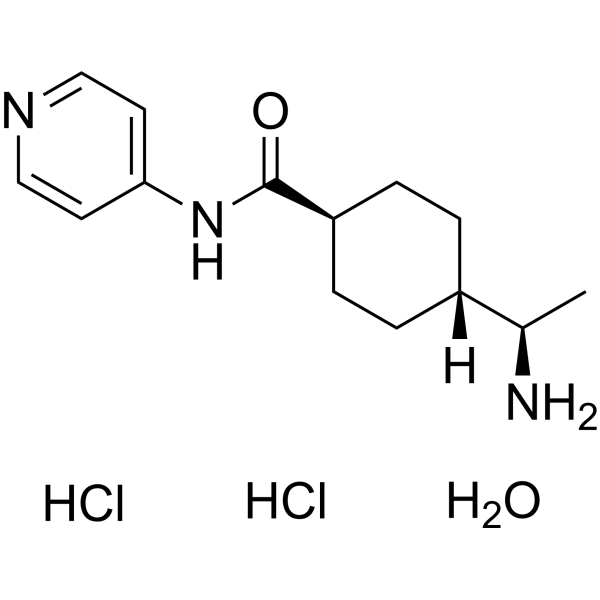
鸡胚角膜上皮是一种独特的组织,30多年来一直被用作体外上皮片状器官培养模型(Hay和Revel [1969]《发育中的禽类角膜的精细结构》。瑞士巴塞尔:S. Karger AG)。该组织被用于证实上皮细胞能够产生细胞外基质(ECM)蛋白,例如胶原蛋白和蛋白聚糖(Dodson和Hay [1971]《实验细胞研究》65:215-220;Meier和Hay [1973]《发育生物学》35:318-331;Linsenmayer等人[1977]《美国国家科学院院刊》74:39-43;Hendrix等人[1982]《眼科与视觉科学研究》22:359-375)。这一历史模型也被用于证实细胞外基质(ECM)蛋白能够刺激肌动蛋白重组并增加胶原蛋白合成(Sugrue 和 Hay [1981] J Cell Biol 91:45-54;Sugrue 和 Hay [1982] Dev Biol 92:97-106;Sugrue 和 Hay [1986] J Cell Biol 102:1907-1916)。我们的实验室利用该模型建立了参与 ECM 刺激的肌动蛋白重组的信号转导通路(Svoboda 等 [1999] Anat Rec 254:348-359;Chu 等 [2000] Invest Ophthalmol Vis Sci 41:3374-3382;Reenstra 等 [2002] Invest Ophthalmol Vis Sci 43:3181-3189)。本研究旨在探讨细胞外基质(ECM)在角膜上皮细胞存活中的作用,以及Rho相关激酶(p160 ROCK、ROCK-1、ROCK-2,简称ROCK)在ECM和溶血磷脂酸(LPA)介导的肌动蛋白重组中的作用。将禽类胚胎角膜上皮整片组织在ROCK抑制剂Y27632(浓度分别为0、0.03、0.3、3或10 μM)存在下进行培养,之后分别用胶原蛋白(COL)或LPA刺激细胞。通过Caspase-3活性检测评估细胞凋亡,并用膜联蛋白V结合进行可视化。 ROCK抑制剂以剂量依赖的方式阻断肌动蛋白皮质垫的重塑,破坏基底细胞侧膜,并增加凋亡标志物膜联蛋白V的表达。此外,体外caspase-3活性测定表明,与去除基底膜的上皮细胞或经纤连蛋白、胶原蛋白(COL)或溶血磷脂酸(LPA)刺激的上皮细胞相比,经10 μM Y-27632处理的上皮细胞中caspase-3活性更高。总之,细胞外基质(ECM)分子降低了凋亡标志物的表达,而抑制ROCK通路则阻断了ECM刺激的肌动蛋白皮质垫重塑,并增加了胚胎角膜上皮细胞的凋亡。[1]Y-27632是一种单羧酸酰胺,其结构为反式-[(1R)-1-氨基乙基]环己烷甲酰胺,其中氨基羰基的一个氮原子被吡啶环取代。研究表明,Y-27632 对 Rho 相关蛋白激酶 (ROCK) 具有抑制活性。它是一种 EC 2.7.11.1(非特异性丝氨酸/苏氨酸蛋白激酶)抑制剂。它是一种单羧酸酰胺,属于吡啶类化合物,也是一种伯氨基化合物。
Y-27632 是一种 Rho 相关蛋白激酶抑制剂。它通过抑制钙敏化来影响平滑肌舒张。 背景:p160ROCK 是 Rho 的直接靶点,介导 Rho 诱导的黏着斑和应力纤维的组装。我们之前报道过,Rho 信号通路参与体外肝星状细胞 (HSC) 的激活。本研究旨在验证p160ROCK特异性抑制剂(Y27632)能否预防二甲基亚硝胺(DMN)诱导的大鼠实验性肝纤维化。方法:在首次注射DMN后,每日口服Y27632(30 mg/kg),持续4周。采用图像分析和肝脏胶原蛋白及羟脯氨酸含量测定评估肝纤维化的程度。同时检测肝脏及原代培养的肝星状细胞(HSC)中α-平滑肌肌动蛋白(α-SMA)的表达。采用半定量RT-PCR检测肝脏中I型胶原mRNA的表达。结果:Y27632治疗显著降低了DMN诱导的肝纤维化的发生率,并降低了肝脏中胶原蛋白、羟脯氨酸含量以及α-SMA的表达。体外实验也发现 HSC 中 α-SMA 的表达受到抑制。结论:这些发现表明,Rho-ROCK 通路抑制剂可能对肝纤维化具有治疗作用。[3] 在本研究中,我们报道了一个此前未被探索的领域,其中 Y-27632 被鉴定为一种有效的小分子,能够诱导 hIPSC 选择性地向中内胚层谱系分化。Y-27632 通过有效调节细胞骨架和细胞间连接蛋白,诱导 hIPSC 发生类似 EMT 的变化,从而使细胞易于向中内胚层谱系分化。同时,这种对肌动蛋白和 E-钙黏蛋白组织的破坏导致外胚层分化受到抑制。这些结果提出了一种增强人多能干细胞向中内胚层靶细胞类型定向分化的新方法。 [5] 总之,本研究表明,Y-27632预处理可显著降低Caspase-3活性,并保护CSCs免受Dox诱导的细胞凋亡。其潜在机制尚未完全阐明,可能涉及ROCK与Caspase-3、ROCK与LIMK/ADF/Cofilin或ROCK与p-MCL之间的直接和/或间接相互作用(图8)。这些相互作用的总体结果或平衡可能导致对人CSCs的抗凋亡作用。尽管仍有许多问题尚未解答,但Y-27632值得在动物模型中基于干细胞的治疗中进行进一步评估,最终结果可能适用于人体临床试验。[6] 背景:近期使用c-kit+人源心脏干细胞(CSCs)的临床试验在提高心脏功能和改善生活质量方面显示出令人鼓舞的结果。然而,CSCs的效率较低,可能是由于移植后细胞存活率和植入率有限。 Rho相关蛋白激酶(ROCK)抑制剂Y-27632显著提高了多种细胞类型(包括干细胞)的存活率、黏附性和迁移能力,提示ROCK介导的细胞凋亡通路可能具有普遍特征,该特征也可能存在于人类癌症干细胞(CSC)中。本研究旨在验证Y-27632预处理人类CSC是否能保护细胞免受阿霉素(Dox)诱导的细胞凋亡。[6] |
| 分子式 |
C14H25CL2N3O2
|
|---|---|
| 分子量 |
338.27
|
| 精确质量 |
337.132
|
| 元素分析 |
C, 49.71; H, 7.45; Cl, 20.96; N, 12.42; O, 9.46
|
| CAS号 |
331752-47-7
|
| 相关CAS号 |
146986-50-7; 129830-38-2 (2HCl); 331752-47-7 (HCl hydrate); 138381-45-0 (racemate HCl); 310898-86-3 (recamate free base)
|
| PubChem CID |
9797929
|
| 外观&性状 |
Typically exists as solid at room temperature
|
| 熔点 |
180-230°C
|
| LogP |
4.486
|
| tPSA |
77.24
|
| 氢键供体(HBD)数目 |
5
|
| 氢键受体(HBA)数目 |
4
|
| 可旋转键数目(RBC) |
3
|
| 重原子数目 |
21
|
| 分子复杂度/Complexity |
268
|
| 定义原子立体中心数目 |
1
|
| SMILES |
C[C@H](C1CCC(CC1)C(=O)NC2=CC=NC=C2)N.O.Cl.Cl
|
| InChi Key |
BLQHLDXMMWHXIT-UYRCGDKNSA-N
|
| InChi Code |
InChI=1S/C14H21N3O.2ClH.H2O/c1-10(15)11-2-4-12(5-3-11)14(18)17-13-6-8-16-9-7-13;;;/h6-12H,2-5,15H2,1H3,(H,16,17,18);2*1H;1H2/t10-,11?,12?;;;/m1.../s1
|
| 化学名 |
4-[(1R)-1-aminoethyl]-N-pyridin-4-ylcyclohexane-1-carboxamide;hydrate;dihydrochloride
|
| 别名 |
331752-47-7; Y-27632 dihydrochloride monohydrate; Y-27632 Dihydrochloride Hydrate; Y-27632 (hydrochloride hydrate); (R)-(+)-trans-4-(1-Aminoethyl)-N-(4-Pyridyl)cyclohexanecarboxamide dihydrochloride monohydrate; 4-[(1R)-1-aminoethyl]-N-pyridin-4-ylcyclohexane-1-carboxamide;hydrate;dihydrochloride; trans-4-[(R)-1-Aminoethyl]-N-(pyridin-4-yl)cyclohexanecarboxamide dihydrochloride hydrate; Y 27632 Dihydrochloride Hydrate;Y27632 Dihydrochloride Hydrate;
|
| HS Tariff Code |
2934.99.9001
|
| 存储方式 |
Powder -20°C 3 years 4°C 2 years In solvent -80°C 6 months -20°C 1 month |
| 运输条件 |
Room temperature (This product is stable at ambient temperature for a few days during ordinary shipping and time spent in Customs)
|
| 溶解度 (体外实验) |
May dissolve in DMSO (in most cases), if not, try other solvents such as H2O, Ethanol, or DMF with a minute amount of products to avoid loss of samples
|
|---|---|
| 溶解度 (体内实验) |
注意: 如下所列的是一些常用的体内动物实验溶解配方,主要用于溶解难溶或不溶于水的产品(水溶度<1 mg/mL)。 建议您先取少量样品进行尝试,如该配方可行,再根据实验需求增加样品量。
注射用配方
注射用配方1: DMSO : Tween 80: Saline = 10 : 5 : 85 (如: 100 μL DMSO → 50 μL Tween 80 → 850 μL Saline)(IP/IV/IM/SC等) *生理盐水/Saline的制备:将0.9g氯化钠/NaCl溶解在100 mL ddH ₂ O中,得到澄清溶液。 注射用配方 2: DMSO : PEG300 :Tween 80 : Saline = 10 : 40 : 5 : 45 (如: 100 μL DMSO → 400 μL PEG300 → 50 μL Tween 80 → 450 μL Saline) 注射用配方 3: DMSO : Corn oil = 10 : 90 (如: 100 μL DMSO → 900 μL Corn oil) 示例: 以注射用配方 3 (DMSO : Corn oil = 10 : 90) 为例说明, 如果要配制 1 mL 2.5 mg/mL的工作液, 您可以取 100 μL 25 mg/mL 澄清的 DMSO 储备液,加到 900 μL Corn oil/玉米油中, 混合均匀。 View More
注射用配方 4: DMSO : 20% SBE-β-CD in Saline = 10 : 90 [如:100 μL DMSO → 900 μL (20% SBE-β-CD in Saline)] 口服配方
口服配方 1: 悬浮于0.5% CMC Na (羧甲基纤维素钠) 口服配方 2: 悬浮于0.5% Carboxymethyl cellulose (羧甲基纤维素) 示例: 以口服配方 1 (悬浮于 0.5% CMC Na)为例说明, 如果要配制 100 mL 2.5 mg/mL 的工作液, 您可以先取0.5g CMC Na并将其溶解于100mL ddH2O中,得到0.5%CMC-Na澄清溶液;然后将250 mg待测化合物加到100 mL前述 0.5%CMC Na溶液中,得到悬浮液。 View More
口服配方 3: 溶解于 PEG400 (聚乙二醇400) 请根据您的实验动物和给药方式选择适当的溶解配方/方案: 1、请先配制澄清的储备液(如:用DMSO配置50 或 100 mg/mL母液(储备液)); 2、取适量母液,按从左到右的顺序依次添加助溶剂,澄清后再加入下一助溶剂。以 下列配方为例说明 (注意此配方只用于说明,并不一定代表此产品 的实际溶解配方): 10% DMSO → 40% PEG300 → 5% Tween-80 → 45% ddH2O (或 saline); 假设最终工作液的体积为 1 mL, 浓度为5 mg/mL: 取 100 μL 50 mg/mL 的澄清 DMSO 储备液加到 400 μL PEG300 中,混合均匀/澄清;向上述体系中加入50 μL Tween-80,混合均匀/澄清;然后继续加入450 μL ddH2O (或 saline)定容至 1 mL; 3、溶剂前显示的百分比是指该溶剂在最终溶液/工作液中的体积所占比例; 4、 如产品在配制过程中出现沉淀/析出,可通过加热(≤50℃)或超声的方式助溶; 5、为保证最佳实验结果,工作液请现配现用! 6、如不确定怎么将母液配置成体内动物实验的工作液,请查看说明书或联系我们; 7、 以上所有助溶剂都可在 Invivochem.cn网站购买。 |
| 制备储备液 | 1 mg | 5 mg | 10 mg | |
| 1 mM | 2.9562 mL | 14.7811 mL | 29.5622 mL | |
| 5 mM | 0.5912 mL | 2.9562 mL | 5.9124 mL | |
| 10 mM | 0.2956 mL | 1.4781 mL | 2.9562 mL |
1、根据实验需要选择合适的溶剂配制储备液 (母液):对于大多数产品,InvivoChem推荐用DMSO配置母液 (比如:5、10、20mM或者10、20、50 mg/mL浓度),个别水溶性高的产品可直接溶于水。产品在DMSO 、水或其他溶剂中的具体溶解度详见上”溶解度 (体外)”部分;
2、如果您找不到您想要的溶解度信息,或者很难将产品溶解在溶液中,请联系我们;
3、建议使用下列计算器进行相关计算(摩尔浓度计算器、稀释计算器、分子量计算器、重组计算器等);
4、母液配好之后,将其分装到常规用量,并储存在-20°C或-80°C,尽量减少反复冻融循环。
计算结果:
工作液浓度: mg/mL;
DMSO母液配制方法: mg 药物溶于 μL DMSO溶液(母液浓度 mg/mL)。如该浓度超过该批次药物DMSO溶解度,请首先与我们联系。
体内配方配制方法:取 μL DMSO母液,加入 μL PEG300,混匀澄清后加入μL Tween 80,混匀澄清后加入 μL ddH2O,混匀澄清。
(1) 请确保溶液澄清之后,再加入下一种溶剂 (助溶剂) 。可利用涡旋、超声或水浴加热等方法助溶;
(2) 一定要按顺序加入溶剂 (助溶剂) 。
 BBO-11818
 RMC-5127
 Degarelix acetate hydrate
 DDO-2728
InvivoChem的所有产品仅用于作科学研究,不面向患者销售
Copyright 2020 InvivoChem LLC | All Rights Reserved 粤ICP备20063088号-1


 463611831
