| 规格 | 价格 | 库存 | 数量 |
|---|---|---|---|
| 100mg |
|
||
| 250mg |
|
||
| 500mg |
|
||
| Other Sizes |
|
| 体外研究 (In Vitro) |
Cl 链霉素可降低通道活性和 Ca2+ 进入的有效部分[3]。
|
|---|---|
| 药代性质 (ADME/PK) |
吸收、分布和排泄
由于口服吸收不良,包括链霉素在内的氨基糖苷类药物通常采用肠外给药。链霉素可肌注,在某些情况下也可静脉注射。肌注1克链霉素后,1小时内血清浓度峰值可达25-50微克/毫升。 静脉或肌注后,约50%的链霉素在24小时内经尿液排出。 肌注1克硫酸链霉素后,1小时内血清浓度峰值可达25至50微克/毫升,5至6小时后缓慢下降至约50%。除脑组织外,所有器官组织中均能检测到明显的链霉素浓度。在胸腔积液和结核性空洞中均发现了大量的链霉素。链霉素可通过胎盘,脐带血中的血清浓度与母体血清浓度相似。少量链霉素会通过乳汁、唾液和汗液排出。 链霉素不经胃肠道吸收。 肌注后链霉素吸收迅速。肾功能正常的成年人肌注单剂量1克链霉素后,血清链霉素浓度在1小时内达到峰值,范围为25-50微克/毫升;给药后5-6小时血清浓度下降50%。 一项针对早产儿的研究表明,肌注10-11毫克/公斤链霉素后,2小时内血清平均浓度达到峰值约29微克/毫升; 12 小时血清浓度平均为 11 μg/mL。 有关链霉素(共 16 项)的更多吸收、分布和排泄(完整)数据,请访问 HSDB 记录页面。 代谢/代谢物 氨基糖苷类抗生素不经代谢,主要通过肾小球滤过以原形经尿液排出。/氨基糖苷类/ 生物半衰期 链霉素的血清半衰期估计为 2.5 小时。 肾功能正常的成年人,链霉素的血浆消除半衰期通常为 2-3 小时;据报道,严重肾功能损害的成年人,其血浆消除半衰期可达 110 小时。据报道,早产儿和新生儿的链霉素血浆消除半衰期为 4-10 小时。据报道,肝肾功能受损患者的血浆消除半衰期比单纯肾功能受损患者更长。 |
| 毒性/毒理 (Toxicokinetics/TK) |
肝毒性
链霉素的静脉和肌肉注射治疗与血清碱性磷酸酶轻度升高(无症状)有关,但治疗很少影响氨基转移酶水平或胆红素,且一旦停用链霉素,这些变化通常会迅速恢复。仅有零星病例报告显示,链霉素治疗可导致急性肝损伤伴黄疸,且这些病例均与其他肝毒性更强的抗结核药物(如异烟肼、吡嗪酰胺和利福平)联合使用。在药物性肝病和急性肝衰竭的大型病例系列研究中,并未提及链霉素和氨基糖苷类抗生素;因此,链霉素引起的肝损伤即使发生,也极其罕见。 可能性评分:E(不太可能是临床上明显的肝损伤原因)。 妊娠和哺乳期影响 ◉ 哺乳期用药概述 与其他氨基糖苷类抗生素类似,链霉素很少分泌到母乳中。新生儿似乎会吸收少量氨基糖苷类抗生素,但其血清浓度远低于治疗新生儿感染时达到的浓度,因此不太可能出现链霉素的全身性影响。预计较大婴儿对链霉素的吸收量会更少。应监测婴儿胃肠道菌群可能受到的影响,例如腹泻、念珠菌病(如鹅口疮、尿布疹)或罕见的便血,便血可能提示抗生素相关性结肠炎。 ◉ 对母乳喂养婴儿的影响 截至修订日期,未找到相关的已发表信息。 ◉ 对泌乳和母乳的影响 一项观察性研究发现链霉素不会抑制泌乳。 |
| 参考文献 | |
| 其他信息 |
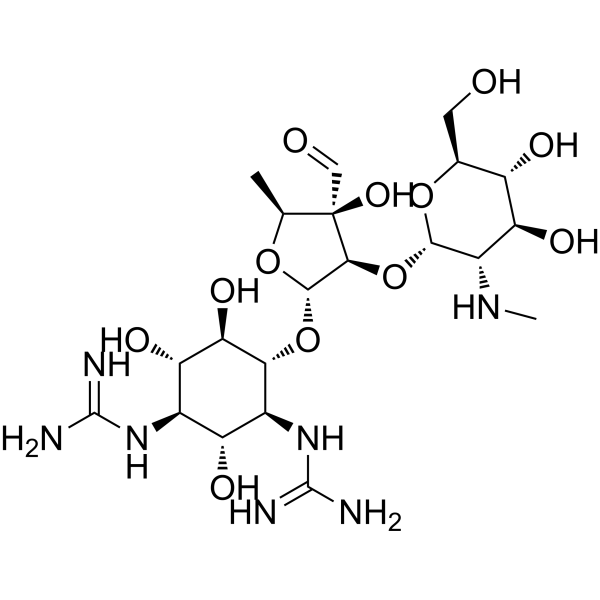
链霉素是一种氨基环醇糖苷,由链霉肽在4位连接一个二糖基组成。它是链霉素类药物的母体化合物,具有抗菌、抗微生物、抗病毒、蛋白质合成抑制剂、细菌代谢产物和抗真菌农药等多种功能。它是一种抗生素、抗真菌药物、抗生素杀菌剂,也是链霉素家族的成员。其功能与链霉肽相关,是链霉素(3+)的共轭碱。
链霉素是一种源自灰色链霉菌的抗生素,是20世纪40年代发现并应用于临床的第一个氨基糖苷类抗生素。塞尔曼·瓦克斯曼和阿尔伯特·沙茨因发现链霉素及其抗菌活性而荣获诺贝尔生理学或医学奖。虽然链霉素是第一个被证实对结核分枝杆菌有效的抗生素,但由于耐药性的出现,它已不再被广泛使用,现在主要用于多重耐药结核病的辅助治疗。 链霉素是一种氨基糖苷类抗菌药和抗分枝杆菌药。 链霉素是一种广谱氨基糖苷类抗生素,通常用于治疗活动性结核病,但总是与其他抗结核药物联合使用。链霉素通常与已知具有肝毒性的药物联合使用,其在肝损伤中的作用难以评估,但大多数信息表明链霉素不具有肝毒性。 据报道,链霉素存在于大丝状菌(Lyngbya majuscula)、千里光属(Senecio)和其他一些有相关数据的生物体中。 链霉素是一种氨基糖苷类抗生素,来源于灰色链霉菌(Streptomyces griseus),具有抗菌活性。链霉素与细菌30S核糖体亚基内的16S rRNA和S12蛋白不可逆地结合。因此,该药物会干扰mRNA与细菌核糖体之间起始复合物的组装,从而抑制蛋白质合成的起始。此外,链霉素还会诱导mRNA模板的错读,导致翻译移码,从而导致翻译提前终止。这最终会导致细菌细胞死亡。 链霉素是一种由土壤放线菌灰色链霉菌(Streptomyces griseus)产生的抗生素。它的作用机制是抑制蛋白质合成过程中的起始和延伸过程。 另见:硫酸链霉素(有盐形式);泛酸链霉素(其活性成分);盐酸链霉素(其活性成分)。 药物适应症 虽然链霉素是首个用于治疗结核分枝杆菌感染的抗生素,但由于耐药性和毒性,现在它主要作为二线药物使用。链霉素也可用于治疗由敏感需氧菌菌株引起的多种其他感染,尤其是在其他毒性较小的药物无效的情况下。例如:鼠疫耶尔森菌、土拉弗朗西斯菌、布鲁氏菌、肉芽肿棒状杆菌(引起腹股沟肉芽肿、杜氏肉芽肿)、杜克雷嗜血杆菌(引起软下疳)、流感嗜血杆菌(引起呼吸道感染、心内膜感染和脑膜感染,需与其他抗菌药物联合使用)、肺炎克雷伯菌(引起肺炎,需与其他抗菌药物联合使用)、大肠杆菌、变形杆菌、产气杆菌、克雷伯菌等。肺炎链球菌和粪肠球菌可引起尿路感染,草绿色链球菌和粪肠球菌(在心内膜感染中,需与青霉素联合使用)可引起革兰氏阴性杆菌菌血症(需与其他抗菌药物联合使用)。 作用机制 氨基糖苷类药物进入细胞主要分为三个阶段。第一个“离子结合阶段”发生在多聚阳离子氨基糖苷类药物通过静电作用与细菌细胞膜的带负电荷的成分结合时,例如革兰氏阴性菌外膜中的脂多糖和磷脂,以及革兰氏阳性菌细胞膜中的磷壁酸和磷脂。这种结合导致二价阳离子被置换,膜通透性增加,从而使氨基糖苷类药物得以进入细胞。氨基糖苷类药物进入细胞质的第二个“能量依赖性阶段I”依赖于质子动力势,并允许少量氨基糖苷类药物到达其主要的细胞内靶点——细菌30S核糖体。这最终导致蛋白质翻译错误和细胞质膜破坏。最后,在“能量依赖性阶段II”中,观察到浓度依赖性的杀菌作用。由于细胞质膜受损,氨基糖苷类药物在细胞内迅速积累,蛋白质翻译错误和合成抑制作用被放大。因此,氨基糖苷类药物既通过破坏细胞膜发挥即时杀菌作用,也通过抑制蛋白质合成发挥延迟杀菌作用;观察到的实验数据和数学模型均支持这种双机制模型。抑制蛋白质合成是氨基糖苷类药物发挥疗效的关键组成部分。结构和细胞生物学研究表明,氨基糖苷类抗生素与16S rRNA的44号螺旋(h44)结合,该螺旋位于30S核糖体亚基A位点附近,从而改变h44和h45之间的相互作用。这种结合还会使h44上的两个重要残基A1492和A1493发生位移,模拟A位点密码子-反密码子配对成功时发生的正常构象变化。总体而言,氨基糖苷类抗生素的结合会产生多种负面影响,包括抑制翻译、起始、延伸和核糖体循环。最近的证据表明,后一种影响是由于50S核糖体亚基23S rRNA的h69上存在一个隐蔽的第二个结合位点。此外,氨基糖苷类抗生素通过稳定模拟正确密码子-反密码子配对的构象,促进易错翻译。翻译错误的蛋白质可以整合到细胞膜中,从而导致上述损伤。氨基糖苷类抗生素的主要细胞内作用位点是30S核糖体亚基,该亚基由21种蛋白质和一条16S RNA分子组成。至少有三种蛋白质,或许还有16S核糖体RNA,参与链霉素的结合,这些分子的改变会显著影响链霉素的结合及其后续作用。例如,在一种核糖体蛋白(S12)的第42位氨基酸中,赖氨酸被天冬酰胺取代,即可阻止药物结合;由此产生的突变体对链霉素完全耐药。另一种突变体,其该位置的氨基酸为谷氨酰胺,则依赖于链霉素。 在蛋白质合成过程中,核糖体选择反密码子与小核糖体亚基A位点上的信使RNA密码子相匹配的氨酰转移RNA。氨基糖苷类抗生素链霉素通过结合在密码子识别位点附近来干扰解码过程。本文利用X射线晶体衍射技术,研究了链霉素对嗜热栖热菌30S核糖体亚基解码位点的影响,该亚基与同源或近同源的反密码子茎环类似物和信使RNA形成复合物。我们的晶体结构显示,链霉素诱导了16S核糖体RNA的显著局部畸变,包括直接参与密码子识别的关键碱基A1492和A1493。与动力学数据一致,我们观察到链霉素能够稳定近同源反密码子茎环类似物复合物,同时破坏同源反密码子茎环类似物复合物的稳定性。这些数据揭示了链霉素如何破坏同源反密码子茎环类似物的识别,却又如何增强近同源反密码子茎环类似物的识别。 链霉素是一种广泛用于治疗微生物感染的抗生素。其主要作用机制是通过与核糖体结合抑制翻译……在早期对该抗生素的研究中,人们观察到一种神秘的链霉素诱导的钾离子外流现象,该现象先于细胞活力的下降;据推测,这种外流改变了电化学梯度,使链霉素更容易进入细胞质。在此,我们使用高通量筛选方法寻找靶向大电导机械敏感通道(MscL)的化合物,并在筛选结果中发现了二氢链霉素。此外,我们发现 MscL 不仅是先前报道的链霉素诱导钾离子外流所必需的,而且在电生理学研究中还能直接增强 MscL 的活性。数据表明,调控 MscL 通道是二氢链霉素的一种新作用机制,并且 MscL 的大孔可能为药物进入细胞提供了一种机制。 ……氨基糖苷类抗生素是氨基环醇类抗生素,它们通过与 16S rRNA 结合并破坏细菌细胞膜的完整性来抑制蛋白质合成,从而杀死细菌。氨基糖苷类耐药机制包括:(a) 通过 N-乙酰化、腺苷酰化或 O-磷酸化使氨基糖苷失活;(b) 通过改变外膜通透性、降低内膜转运、主动外排和药物滞留等途径降低细胞内氨基糖苷浓度;(c) 通过突变改变 30S 核糖体亚基靶点;以及 (d) 氨基糖苷结合位点的甲基化。……/氨基糖苷类/ |
| 分子式 |
C21H39N7O12
|
|---|---|
| 分子量 |
581.57
|
| 精确质量 |
581.265
|
| CAS号 |
57-92-1
|
| 相关CAS号 |
Streptomycin sulfate;3810-74-0;Penicillin G;61-33-6
|
| PubChem CID |
19649
|
| 外观&性状 |
White to off-white solid powder
|
| 密度 |
2.0±0.1 g/cm3
|
| 沸点 |
872.9±75.0 °C at 760 mmHg
|
| 熔点 |
MW: 1457.383. Powder. MP: aproximately 230 °C /Streptomycin sulfate; 3810-74-0/
|
| 闪点 |
481.7±37.1 °C
|
| 蒸汽压 |
0.0±0.6 mmHg at 25°C
|
| 折射率 |
1.762
|
| LogP |
-2.53
|
| tPSA |
331.43
|
| 氢键供体(HBD)数目 |
12
|
| 氢键受体(HBA)数目 |
15
|
| 可旋转键数目(RBC) |
9
|
| 重原子数目 |
40
|
| 分子复杂度/Complexity |
940
|
| 定义原子立体中心数目 |
15
|
| SMILES |
C[C@H]1[C@@]([C@H]([C@@H](O1)O[C@@H]2[C@H]([C@@H]([C@H]([C@@H]([C@H]2O)O)N=C(N)N)O)N=C(N)N)O[C@H]3[C@H]([C@@H]([C@H]([C@@H](O3)CO)O)O)NC)(C=O)O
|
| InChi Key |
UCSJYZPVAKXKNQ-HZYVHMACSA-N
|
| InChi Code |
InChI=1S/C21H39N7O12/c1-5-21(36,4-30)16(40-17-9(26-2)13(34)10(31)6(3-29)38-17)18(37-5)39-15-8(28-20(24)25)11(32)7(27-19(22)23)12(33)14(15)35/h4-18,26,29,31-36H,3H2,1-2H3,(H4,22,23,27)(H4,24,25,28)/t5-,6-,7+,8-,9-,10-,11+,12-,13-,14+,15+,16-,17-,18-,21+/m0/s1
|
| 化学名 |
2-[(1R,2R,3S,4R,5R,6S)-3-(diaminomethylideneamino)-4-[(2R,3R,4R,5S)-3-[(2S,3S,4S,5R,6S)-4,5-dihydroxy-6-(hydroxymethyl)-3-(methylamino)oxan-2-yl]oxy-4-formyl-4-hydroxy-5-methyloxolan-2-yl]oxy-2,5,6-trihydroxycyclohexyl]guanidine
|
| HS Tariff Code |
2934.99.9001
|
| 存储方式 |
Powder -20°C 3 years 4°C 2 years In solvent -80°C 6 months -20°C 1 month |
| 运输条件 |
Room temperature (This product is stable at ambient temperature for a few days during ordinary shipping and time spent in Customs)
|
| 溶解度 (体外实验) |
H2O : ~125 mg/mL (~214.94 mM)
|
|---|---|
| 溶解度 (体内实验) |
注意: 如下所列的是一些常用的体内动物实验溶解配方,主要用于溶解难溶或不溶于水的产品(水溶度<1 mg/mL)。 建议您先取少量样品进行尝试,如该配方可行,再根据实验需求增加样品量。
注射用配方
注射用配方1: DMSO : Tween 80: Saline = 10 : 5 : 85 (如: 100 μL DMSO → 50 μL Tween 80 → 850 μL Saline)(IP/IV/IM/SC等) *生理盐水/Saline的制备:将0.9g氯化钠/NaCl溶解在100 mL ddH ₂ O中,得到澄清溶液。 注射用配方 2: DMSO : PEG300 :Tween 80 : Saline = 10 : 40 : 5 : 45 (如: 100 μL DMSO → 400 μL PEG300 → 50 μL Tween 80 → 450 μL Saline) 注射用配方 3: DMSO : Corn oil = 10 : 90 (如: 100 μL DMSO → 900 μL Corn oil) 示例: 以注射用配方 3 (DMSO : Corn oil = 10 : 90) 为例说明, 如果要配制 1 mL 2.5 mg/mL的工作液, 您可以取 100 μL 25 mg/mL 澄清的 DMSO 储备液,加到 900 μL Corn oil/玉米油中, 混合均匀。 View More
注射用配方 4: DMSO : 20% SBE-β-CD in Saline = 10 : 90 [如:100 μL DMSO → 900 μL (20% SBE-β-CD in Saline)] 口服配方
口服配方 1: 悬浮于0.5% CMC Na (羧甲基纤维素钠) 口服配方 2: 悬浮于0.5% Carboxymethyl cellulose (羧甲基纤维素) 示例: 以口服配方 1 (悬浮于 0.5% CMC Na)为例说明, 如果要配制 100 mL 2.5 mg/mL 的工作液, 您可以先取0.5g CMC Na并将其溶解于100mL ddH2O中,得到0.5%CMC-Na澄清溶液;然后将250 mg待测化合物加到100 mL前述 0.5%CMC Na溶液中,得到悬浮液。 View More
口服配方 3: 溶解于 PEG400 (聚乙二醇400) 请根据您的实验动物和给药方式选择适当的溶解配方/方案: 1、请先配制澄清的储备液(如:用DMSO配置50 或 100 mg/mL母液(储备液)); 2、取适量母液,按从左到右的顺序依次添加助溶剂,澄清后再加入下一助溶剂。以 下列配方为例说明 (注意此配方只用于说明,并不一定代表此产品 的实际溶解配方): 10% DMSO → 40% PEG300 → 5% Tween-80 → 45% ddH2O (或 saline); 假设最终工作液的体积为 1 mL, 浓度为5 mg/mL: 取 100 μL 50 mg/mL 的澄清 DMSO 储备液加到 400 μL PEG300 中,混合均匀/澄清;向上述体系中加入50 μL Tween-80,混合均匀/澄清;然后继续加入450 μL ddH2O (或 saline)定容至 1 mL; 3、溶剂前显示的百分比是指该溶剂在最终溶液/工作液中的体积所占比例; 4、 如产品在配制过程中出现沉淀/析出,可通过加热(≤50℃)或超声的方式助溶; 5、为保证最佳实验结果,工作液请现配现用! 6、如不确定怎么将母液配置成体内动物实验的工作液,请查看说明书或联系我们; 7、 以上所有助溶剂都可在 Invivochem.cn网站购买。 |
| 制备储备液 | 1 mg | 5 mg | 10 mg | |
| 1 mM | 1.7195 mL | 8.5974 mL | 17.1948 mL | |
| 5 mM | 0.3439 mL | 1.7195 mL | 3.4390 mL | |
| 10 mM | 0.1719 mL | 0.8597 mL | 1.7195 mL |
1、根据实验需要选择合适的溶剂配制储备液 (母液):对于大多数产品,InvivoChem推荐用DMSO配置母液 (比如:5、10、20mM或者10、20、50 mg/mL浓度),个别水溶性高的产品可直接溶于水。产品在DMSO 、水或其他溶剂中的具体溶解度详见上”溶解度 (体外)”部分;
2、如果您找不到您想要的溶解度信息,或者很难将产品溶解在溶液中,请联系我们;
3、建议使用下列计算器进行相关计算(摩尔浓度计算器、稀释计算器、分子量计算器、重组计算器等);
4、母液配好之后,将其分装到常规用量,并储存在-20°C或-80°C,尽量减少反复冻融循环。
计算结果:
工作液浓度: mg/mL;
DMSO母液配制方法: mg 药物溶于 μL DMSO溶液(母液浓度 mg/mL)。如该浓度超过该批次药物DMSO溶解度,请首先与我们联系。
体内配方配制方法:取 μL DMSO母液,加入 μL PEG300,混匀澄清后加入μL Tween 80,混匀澄清后加入 μL ddH2O,混匀澄清。
(1) 请确保溶液澄清之后,再加入下一种溶剂 (助溶剂) 。可利用涡旋、超声或水浴加热等方法助溶;
(2) 一定要按顺序加入溶剂 (助溶剂) 。
 AOH-1996
 hMAO-B-IN-3
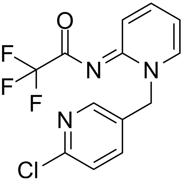 Flupyrimin
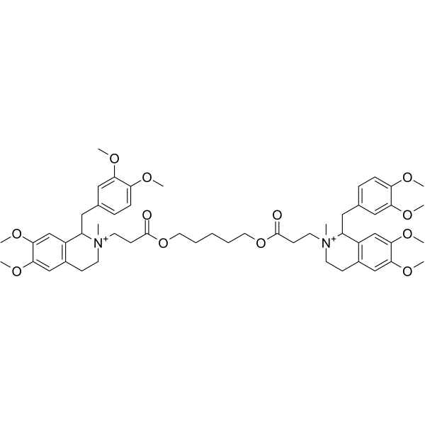 阿曲库铵
阿曲库铵
InvivoChem的所有产品仅用于作科学研究,不面向患者销售
Copyright 2020 InvivoChem LLC | All Rights Reserved 粤ICP备20063088号-1


 463611831
