| 规格 | 价格 | 库存 | 数量 |
|---|---|---|---|
| 100mg |
|
||
| 250mg |
|
||
| 500mg |
|
||
| 1g |
|
||
| 5g |
|
||
| 10g |
|
||
| Other Sizes |
|

| 靶点 |
Metabolite; ER; steroid hormone
Estrogen Receptor α (ERα): Estradiol valerate (hydrolyzes to estradiol in vivo) binds human ERα with high affinity, Ki = 0.2 nM (competitive binding assay in [1]); in rat hippocampal tissue, Ki = 0.3 nM [1] - Estrogen Receptor β (ERβ): Estradiol valerate binds human ERβ with moderate affinity, Ki = 0.8 nM; in zebrafish liver tissue, Ki = 1.1 nM (aquatic toxicity assay in [3]) [3] |
|---|---|
| 体外研究 (In Vitro) |
根据 MCF-7 细胞中 Ser225 磷酸化增强的情况确定,雌二醇 (10 nM) 可快速激活鞘氨醇激酶同工酶 SphK1。 Estradiol (20 nM) 刺激 MCF-7 细胞快速释放 1-磷酸鞘氨醇 (S1P) 和二氢-S1P。 SphK1和雌激素受体α主要负责S1P和二氢S1P的形成。使用 siRNA 或药物抑制剂下调 ABCC1 或 ABCG2 可减少雌二醇 (10 nM) 介导的 MCF-7 细胞中 S1P 或二氢-S1P 的释放。 Estradiol (10 nM) 抑制 MCF-7 人乳腺癌细胞中由雌激素受体 α 介导的 miR-21 表达。雌二醇 (10 nM) 通过抑制 miR-21 表达来激活 MCF-7 细胞中的多个 miR-21 靶基因报告基因活性。雌二醇 (10 nM) 会增加 MCF-7 细胞中蛋白质中内源 miR-21 靶基因的表达,但不会增加 RNA 水平。
1. 海马神经元神经保护活性([1][2]): - 原代大鼠海马神经元:戊酸雌二醇(1–100 nM)处理48小时促进神经突生长:10 nM使神经突长度增加45%(免疫荧光,β-微管蛋白III染色),突触素I(synapsin I)表达增加35%(蛋白质印迹法)[1]。 - 谷氨酸诱导神经毒性:戊酸雌二醇(50 nM)使海马神经元凋亡减少60%(Annexin V-FITC染色),抗凋亡蛋白Bcl-2上调2.3倍[2] 2. 水生细胞毒性([3]): - 斑马鱼肝细胞(ZFL):戊酸雌二醇(0.1–10 μM)暴露72小时呈浓度依赖激活ER:1 μM使ER靶基因卵黄蛋白原(Vtg)mRNA上调8倍(实时PCR),Vtg蛋白上调6.5倍(ELISA);细胞毒性IC50=8.2 μM(MTT实验)[3]。 - 虹鳟鱼生殖细胞(RTG-2):戊酸雌二醇(5 μM)使ERα核转位增加70%(免疫细胞化学),对细胞周期分布无影响[3] |
| 体内研究 (In Vivo) |
雌二醇(80 μg/kg/天,皮下注射)显着降低卵巢切除的 C57BL/6J 小鼠腹膜细胞和巨噬细胞的绝对数量,其特征是 F4/80 和 CD11b 双重阳性染色。在卵巢切除的 C57BL/6J 小鼠中,雌二醇(80 μg/kg/天,皮下注射)通过抑制 PI3K 活性,增强 TGC 诱导的巨噬细胞 LPS 诱导的促炎细胞因子的表达。雌二醇的促炎作用可通过下调巯基乙酸引发的巨噬细胞中雌激素受体 α 的活性而消除。
β-雌二醇17-戊酸酯(EV)是一种合成雌激素,在激素替代治疗药物中广泛与其他类固醇激素联合使用,并在天然水中检测到。尽管EV被认为是一种雌激素化学物质,但仍然缺乏关于日本青鳉(Oryzias latipes)在胚胎-幼虫、幼年和成年阶段接触EV对鱼类发育和生殖毒性的数据。在生命早期,将青鳉受精卵暴露在1、10、100和1000 ng/L EV中15天,孵化的幼鱼继续暴露在相同浓度范围内15天。结果表明,暴露于10ng/L或以上会对孵化率和孵化时间产生不利影响,当暴露于10ng/mL或以上时,孵化的雌性数量是雄性的两倍。当孵化的鱼继续暴露在1、10和100 ng/L的EV中40天后,雄性和雌性的肝体指数(HSI)都增加了,雌性的性腺指数(GSI)降低了,雄性增加了。在暴露于1 ng/L及以上的鱼类中发现了性别逆转。定量实时RT-PCR显示,在所有浓度下,雌性肝脏中雌激素受体α(ER-α)和卵黄原蛋白-I(VTG-I)的mRNA水平均显著下调,而雄性肝脏中卵黄原素-I(VTG-I)的信使核糖核酸水平均显著上调。这些发现表明,EV是雄性和雌性鱼类的生殖毒物和雌激素化学物质。[3] 1. 去卵巢大鼠神经元调控([1][2]): - 动物模型:250–300 g雌性SD大鼠行双侧卵巢切除术(OVX),随机分为OVX对照、戊酸雌二醇10 μg/kg/天、50 μg/kg/天组。 - 结果([1]):50 μg/kg/天(皮下注射,21天)使海马CA1区神经元密度增加30%(尼氏染色),胆碱乙酰转移酶(ChAT)活性增加40%(酶活测定)。 - 结果([2]):10 μg/kg/天改善空间记忆(Morris水迷宫:逃避潜伏期减少35%),海马脑源性神经营养因子(BDNF)mRNA上调2.1倍[2] 2. 水生生物毒性([3]): - 斑马鱼(Danio rerio):暴露于戊酸雌二醇(0.01–1 μg/L)28天: - 0.1 μg/L:诱导雌性次级性征(卵巢成熟加速20%),雄性肝脏Vtg增加5倍(较对照)。 - 1 μg/L:雄性性腺重量减少35%,精子活力降低50%(光学显微镜)[3] |
| 酶活实验 |
1. ERα竞争结合实验([1]):
1. ER制备:人重组ERα(配体结合域LBD)在大肠杆菌中表达,镍螯合层析纯化;大鼠海马组织匀浆,100,000×g离心60分钟获得胞质ERα。 2. 反应体系:200 μL体系含50 mM Tris-HCl(pH7.4)、10%甘油、0.5 nM [³H]-雌二醇、100 ng ERα及戊酸雌二醇(0.01–10 nM,冷竞争剂)。 3. 孵育与分离:4°C孵育2小时;葡聚糖包被活性炭(1%活性炭、0.1%葡聚糖)去除未结合[³H]-雌二醇,3000×g离心10分钟。 4. 检测:液体闪烁计数器检测上清放射性,Cheng-Prusoff方程计算Ki值[1] 2. 斑马鱼肝ERβ结合实验([3]): 1. ERβ制备:斑马鱼肝组织在0.05 M磷酸盐缓冲液(pH7.4)中匀浆,120,000×g离心90分钟分离胞质ERβ。 2. 反应体系:300 μL体系含0.3 nM [³H]-雌二醇、150 μg胞质ERβ及戊酸雌二醇(0.1–20 nM)。 3. 孵育与检测:4°C孵育18小时;活性炭处理分离结合/游离配体,检测放射性,Ki=1.1 nM [3] |
| 细胞实验 |
先前的研究表明,雌二醇在海马CA1锥体细胞上诱导新的树突棘和突触。我们评估了雌二醇诱导的树突棘对CA1锥体细胞内在和突触电生理特性的影响。从用雌二醇或油载体处理的去卵巢大鼠制备海马切片。记录CA1锥体细胞,并注射生物细胞素以观察脊柱。然后使用线性回归分析测试每个细胞的树突棘密度和电生理参数的相关性。我们发现脊柱密度与输入阻力之间存在负相关关系;然而,没有其他测量的内在特性与树突棘密度显著相关。谷氨酸受体放射自显影显示,雌二醇诱导的NMDA受体结合增加,但AMPA受体结合不增加。然后,我们使用输入/输出(I/O)曲线(EPSP斜率与刺激强度)来确定CA1锥体细胞对突触输入的敏感性是否与树突棘密度相关。与雌二醇对AMPA受体结合缺乏影响一致,我们观察到在标准记录条件下产生的I/O曲线斜率与脊柱密度之间没有关系,其中AMPA受体主导EPSP。然而,记录药物分离的NMDA受体介导的EPSP成分揭示了I/O斜率与脊柱密度之间的显著相关性。这些结果表明,与雌二醇诱导的脊柱/突触密度和NMDA受体结合的增加并行,雌二醇治疗增加了CA1锥体细胞对NMDA受体介导的突触输入的敏感性;此外,对NMDA受体介导的突触输入的敏感性与树突棘密度密切相关[1]。
1. 海马神经元培养实验([1]): - 细胞分离:解剖E18大鼠胚胎海马,胰蛋白酶消化,接种于多聚L-赖氨酸包被板(5×10⁴细胞/孔),Neurobasal培养基(含2% B27)培养。 - 药物处理:接种24小时后加入戊酸雌二醇(1–100 nM),培养48小时;对照组加入0.1%乙醇。 - 检测: 1. 神经突生长:抗β-微管蛋白III抗体免疫染色,ImageJ定量神经突长度。 2. 突触素I:蛋白质印迹法检测(β-肌动蛋白为内参)[1] 2. 斑马鱼肝细胞实验([3]): - 细胞培养:ZFL细胞接种于6孔板(2×10⁵细胞/孔),L-15培养基(含10% FBS),28°C培养(无CO₂)。 - 药物处理:戊酸雌二醇(0.1–10 μM)暴露72小时;对照组加入0.01% DMSO。 - 检测: 1. 活力:MTT实验(570 nm吸光度)计算IC50。 2. Vtg表达:实时PCR(Vtg mRNA)和ELISA(Vtg蛋白)[3] |
| 动物实验 |
80 μg/kg/天,皮下注射 小鼠 我们发现,成年雌性大鼠海马CA1区放射层突触密度对雌二醇水平变化敏感,并随着5天动情周期中卵巢类固醇激素水平的变化而自然波动。在雌二醇水平较低和雌二醇水平较高的情况下,突触密度均较低,而雌二醇水平较高则与突触密度较高相关。这些突触变化发生得非常迅速,在动情周期的动情前期和动情期之间约24小时内,我们观察到海马突触密度下降了32%。随后,突触密度似乎会在几天内恢复到动情前期的水平。据我们所知,这是首次证实这种短期类固醇介导的突触可塑性在成年哺乳动物大脑中自然发生。[1]
成对的种鱼在实验室中饲养。在自然受精后数小时内,小心地从种鱼(约40条雌鱼)的腹侧收集自然产下的卵。用手指轻轻滚动卵块即可获得卵。将卵置于0.9%的过氧化氢溶液中消毒10分钟(Marking等人,1994;Sun等人,2007),然后使用解剖显微镜检查受精情况。根据初步浓度范围研究的结果(数据未显示),将胚胎暴露于浓度分别为 1、10、100 和 1000 ng/L 的 β-雌二醇-17-戊酸酯 (EV) 溶液(稀释水,活性炭除氯自来水)中,持续 15 天。此外,实验设计中还设置了稀释水对照组 (DWC) 和溶剂对照组 (SC)。SC 组和所有 EV 暴露组均含有 0.1 ml/L DMSO 和 1% 亚甲蓝,而 DWC 组仅含有 1% 亚甲蓝。将处理组和对照组的胚胎随机分配到不同的处理组,置于盛有 100 mL 测试溶液的玻璃培养皿中(每皿 30 个胚胎)。每个浓度和对照组均设置三个重复。胚胎在 25 ± 1 °C 的温度下,以 16:8 小时的光暗周期进行培养。每24小时更换80%的测试溶液。每天观察胚胎两次,并移除死亡胚胎(通过亚甲蓝染色鉴定)。记录孵化率、孵化时间和肉眼可见的异常情况。[3] 1. 卵巢切除大鼠神经保护方案 ([1][2]): - 动物选择:8周龄雌性Sprague-Dawley大鼠(250-300 g),每组n=6(假手术组、卵巢切除对照组、戊酸雌二醇10/50 μg/kg组)。 - 模型建立:卵巢切除组进行双侧卵巢切除术;假手术组暴露卵巢但不切除。 - 药物制备:将戊酸雌二醇溶于芝麻油中,配制成1/5 μg/mL的溶液。 - 给药途径:皮下注射(10 mL/kg),每日一次,持续21天;假手术/卵巢切除对照组注射芝麻油。 - 检测:处死大鼠;解剖海马,进行尼氏染色(神经元密度)和BDNF mRNA检测;采集血液用于雌二醇水平测定(放射免疫分析法)[1][2] 2. 斑马鱼毒性试验方案 ([3]): - 动物选择:受精后7天(dpf)的斑马鱼幼鱼,每组n=30(对照组,0.01/0.1/1 μg/L 戊酸雌二醇)。 - 药物配制:戊酸雌二醇溶于乙醇,用复水稀释至目标浓度(乙醇<0.001%)。 - 给药途径:静态暴露28天;每48小时更换一次水。 - 检测方法:对斑马鱼实施安乐死;称量性腺重量,进行组织学染色(H&E染色);收集肝脏用于卵黄蛋白原(Vtg)蛋白检测(ELISA)[3] |
| 药代性质 (ADME/PK) |
吸收、分布和排泄
肌注:当与芳基和烷基结合用于肠外给药时,油性制剂的吸收速度减慢,作用持续时间延长,因此单次肌注戊酸雌二醇或环戊丙酸雌二醇可在数周内被吸收。Natazia:口服戊酸雌二醇后,在肠黏膜吸收过程中或在肝脏首过过程中,会裂解为17β-雌二醇和戊酸。这会产生雌二醇及其代谢物,如雌酮和其他代谢物。在28天序贯给药方案的第1天,空腹单次服用一片含3毫克戊酸雌二醇的片剂后,血清雌二醇浓度峰值达到73.3 pg/mL,中位时间为约6小时(范围:1.5-12小时),雌二醇浓度曲线下面积[AUC(0-24h)]为1301 pg·h/mL。 雌二醇、雌酮和雌三醇与葡萄糖醛酸苷和硫酸盐结合物一起经尿液排出。 代谢/代谢物 外源性雌激素的代谢机制与内源性雌激素相同。雌激素部分经细胞色素P450代谢。 1. 啮齿动物药代动力学 ([1][2]): - 吸收:大鼠皮下注射戊酸雌二醇 (50 μg/kg):12 小时血浆雌二醇峰浓度 (Cmax) = 85 pg/mL;生物利用度 = 92%(与静脉注射雌二醇相比)。 - 代谢:在血浆中迅速水解为雌二醇(水解半衰期 = 45 分钟);雌二醇在肝脏中代谢为雌酮(半衰期 = 6 小时)[1]。 - 分布:在脑(海马:血浆浓度的 2.5 倍)和子宫(血浆浓度的 5 倍)中高度蓄积[2] |
| 毒性/毒理 (Toxicokinetics/TK) |
妊娠期和哺乳期影响
◉ 哺乳期用药概述 尚未对戊酸雌二醇在哺乳期进行研究。注射用戊酸雌二醇通常与睾酮联合使用,用于抑制泌乳。一般来说,希望哺乳的母亲应避免使用注射用戊酸雌二醇,尤其是在产后约6周乳汁分泌尚未完全建立之前开始使用。乳汁分泌减少可能发生在雌激素暴露的最初几天。 在美国,口服戊酸雌二醇仅存在于含有地诺孕素的复方口服避孕药中。根据现有证据,专家意见认为,哺乳期妇女应优先选择非激素避孕方法,并且哺乳期妇女,尤其是在产后最初4周内,应优先选择仅含孕激素的避孕药而非复方口服避孕药。更多信息,请参阅题为“口服复方避孕药”的记录。 ◉ 对母乳喂养婴儿的影响 截至修订日期,未找到相关的已发表信息。 ◉ 对泌乳和母乳的影响 戊酸雌二醇注射液曾用于抑制泌乳,通常与睾酮联合使用。 一项回顾性队列研究比较了371名在青春期接受高剂量雌激素(每日3毫克己烯雌酚或150微克炔雌醇)以降低成年身高的女性与409名未接受雌激素的女性。两组间母乳喂养持续时间无差异,表明青春期高剂量雌激素对后期母乳喂养无影响。 1. 啮齿动物毒性 ([1][2]): - 子宫效应:戊酸雌二醇 50 μg/kg/天 (21 天) 使大鼠子宫湿重增加 2.3 倍(与卵巢切除对照组相比);未见子宫内膜增生(H&E 染色)[1]。 - 肝脏安全性:血清 ALT/AST 水平未发生变化;肝脏组织学正常 [2] 2. 水生动物毒性 ([3]): - 急性毒性:斑马鱼 96 小时 LC50 = 8.5 μg/L;浓度 <1 μg/L 时无死亡。 - 内分泌干扰:0.1 μg/L 可诱导雄性斑马鱼雌性化(雌雄同体形成率 = 30%);1 μg/L 可使繁殖力降低 40% [3] |
| 参考文献 |
[1]. J Neurosci.1997 Mar 1;17(5):1848-59.
[2]. J Neurosci.1992 Jul;12(7):2549-54. [3]. Aquat Toxicol. 2013 Jun 15:134-135:128-34. |
| 其他信息 |
药效学
雌激素通过强效激动雌激素受体 (ER) 发挥其在全身的作用,雌激素受体分布于包括乳腺、子宫、卵巢、皮肤、前列腺、骨骼、脂肪和大脑在内的多种组织中。雌二醇可与雌激素受体的两种亚型结合:雌激素受体α (ERα) 和雌激素受体β (ERβ)。雌二醇也是G蛋白偶联雌激素受体 (GPER) 的强效激动剂,GPER 近期被认为是雌二醇快速细胞效应的主要介质。 1. 药物背景 ([1][2]): 戊酸雌二醇是雌二醇的长效酯类前药,临床上用于绝经后妇女的激素替代疗法 (HRT)。它也是神经科学中研究雌激素介导的神经保护作用的工具化合物[1][2] 2. 作用机制 ([1][3]): - 神经保护:水解为雌二醇,激活海马中的ERα/β,上调BDNF和突触素I,促进神经元存活和突触可塑性[1]。 - 水生内分泌干扰:与鱼类ERβ结合,诱导雄性鱼类产生Vtg(雌性特异性蛋白),干扰生殖发育[3] 3. 治疗和实验用途 ([1][2][3]): - 临床:通过皮下注射(10–50 μg/kg/周)治疗绝经后症状(潮热、骨质疏松症)[1]。 - 研究:用于啮齿动物神经生物学(卵巢激素缺乏模型)和水生毒理学(内分泌干扰物筛选)[2][3] |
| 分子式 |
C23H32O3
|
|
|---|---|---|
| 分子量 |
356.5
|
|
| 精确质量 |
356.235
|
|
| 元素分析 |
C, 77.49; H, 9.05; O, 13.46
|
|
| CAS号 |
979-32-8
|
|
| 相关CAS号 |
Estradiol valerate;979-32-8; Alpha-Estradiol;57-91-0;Estradiol (Standard);50-28-2;Estradiol-d3;79037-37-9;Estradiol-d4;66789-03-5;Estradiol-d5;221093-45-4;Estradiol-13C2;82938-05-4;Estradiol (cypionate);313-06-4;Estradiol benzoate;50-50-0;Estradiol enanthate;4956-37-0;Estradiol hemihydrate;35380-71-3;Estradiol-d2;53866-33-4;Estradiol-13C6;Estradiol-d2-1;3188-46-3;rel-Estradiol-13C6; 979-32-8 (valerate); 113-38-2 (dipropionate); 57-63-6 (ethinyl); 172377-52-5 (sulfamate); 3571-53-7 (undecylate)
|
|
| PubChem CID |
13791
|
|
| 外观&性状 |
White to off-white solid powder
|
|
| 密度 |
1.1±0.1 g/cm3
|
|
| 沸点 |
486.2±45.0 °C at 760 mmHg
|
|
| 熔点 |
144°C
|
|
| 闪点 |
191.1±21.5 °C
|
|
| 蒸汽压 |
0.0±1.3 mmHg at 25°C
|
|
| 折射率 |
1.568
|
|
| LogP |
6.62
|
|
| tPSA |
46.53
|
|
| 氢键供体(HBD)数目 |
1
|
|
| 氢键受体(HBA)数目 |
3
|
|
| 可旋转键数目(RBC) |
5
|
|
| 重原子数目 |
26
|
|
| 分子复杂度/Complexity |
518
|
|
| 定义原子立体中心数目 |
5
|
|
| SMILES |
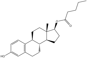
CCCCC(=O)O[C@H]1CC[C@@H]2[C@@]1(CC[C@H]3[C@H]2CCC4=C3C=CC(=C4)O)C
|
|
| InChi Key |
RSEPBGGWRJCQGY-RBRWEJTLSA-N
|
|
| InChi Code |
InChI=1S/C23H32O3/c1-3-4-5-22(25)26-21-11-10-20-19-8-6-15-14-16(24)7-9-17(15)18(19)12-13-23(20,21)2/h7,9,14,18-21,24H,3-6,8,10-13H2,1-2H3/t18-,19-,20+,21+,23+/m1/s1
|
|
| 化学名 |
(17β)-3-hydroxyestra-1,3,5(10)-trien-17-yl valerate
|
|
| 别名 |
|
|
| HS Tariff Code |
2934.99.9001
|
|
| 存储方式 |
Powder -20°C 3 years 4°C 2 years In solvent -80°C 6 months -20°C 1 month |
|
| 运输条件 |
Room temperature (This product is stable at ambient temperature for a few days during ordinary shipping and time spent in Customs)
|
| 溶解度 (体外实验) |
|
|||
|---|---|---|---|---|
| 溶解度 (体内实验) |
注意: 如下所列的是一些常用的体内动物实验溶解配方,主要用于溶解难溶或不溶于水的产品(水溶度<1 mg/mL)。 建议您先取少量样品进行尝试,如该配方可行,再根据实验需求增加样品量。
注射用配方
注射用配方1: DMSO : Tween 80: Saline = 10 : 5 : 85 (如: 100 μL DMSO → 50 μL Tween 80 → 850 μL Saline)(IP/IV/IM/SC等) *生理盐水/Saline的制备:将0.9g氯化钠/NaCl溶解在100 mL ddH ₂ O中,得到澄清溶液。 注射用配方 2: DMSO : PEG300 :Tween 80 : Saline = 10 : 40 : 5 : 45 (如: 100 μL DMSO → 400 μL PEG300 → 50 μL Tween 80 → 450 μL Saline) 注射用配方 3: DMSO : Corn oil = 10 : 90 (如: 100 μL DMSO → 900 μL Corn oil) 示例: 以注射用配方 3 (DMSO : Corn oil = 10 : 90) 为例说明, 如果要配制 1 mL 2.5 mg/mL的工作液, 您可以取 100 μL 25 mg/mL 澄清的 DMSO 储备液,加到 900 μL Corn oil/玉米油中, 混合均匀。 View More
注射用配方 4: DMSO : 20% SBE-β-CD in Saline = 10 : 90 [如:100 μL DMSO → 900 μL (20% SBE-β-CD in Saline)] 口服配方
口服配方 1: 悬浮于0.5% CMC Na (羧甲基纤维素钠) 口服配方 2: 悬浮于0.5% Carboxymethyl cellulose (羧甲基纤维素) 示例: 以口服配方 1 (悬浮于 0.5% CMC Na)为例说明, 如果要配制 100 mL 2.5 mg/mL 的工作液, 您可以先取0.5g CMC Na并将其溶解于100mL ddH2O中,得到0.5%CMC-Na澄清溶液;然后将250 mg待测化合物加到100 mL前述 0.5%CMC Na溶液中,得到悬浮液。 View More
口服配方 3: 溶解于 PEG400 (聚乙二醇400) 请根据您的实验动物和给药方式选择适当的溶解配方/方案: 1、请先配制澄清的储备液(如:用DMSO配置50 或 100 mg/mL母液(储备液)); 2、取适量母液,按从左到右的顺序依次添加助溶剂,澄清后再加入下一助溶剂。以 下列配方为例说明 (注意此配方只用于说明,并不一定代表此产品 的实际溶解配方): 10% DMSO → 40% PEG300 → 5% Tween-80 → 45% ddH2O (或 saline); 假设最终工作液的体积为 1 mL, 浓度为5 mg/mL: 取 100 μL 50 mg/mL 的澄清 DMSO 储备液加到 400 μL PEG300 中,混合均匀/澄清;向上述体系中加入50 μL Tween-80,混合均匀/澄清;然后继续加入450 μL ddH2O (或 saline)定容至 1 mL; 3、溶剂前显示的百分比是指该溶剂在最终溶液/工作液中的体积所占比例; 4、 如产品在配制过程中出现沉淀/析出,可通过加热(≤50℃)或超声的方式助溶; 5、为保证最佳实验结果,工作液请现配现用! 6、如不确定怎么将母液配置成体内动物实验的工作液,请查看说明书或联系我们; 7、 以上所有助溶剂都可在 Invivochem.cn网站购买。 |
| 制备储备液 | 1 mg | 5 mg | 10 mg | |
| 1 mM | 2.8050 mL | 14.0252 mL | 28.0505 mL | |
| 5 mM | 0.5610 mL | 2.8050 mL | 5.6101 mL | |
| 10 mM | 0.2805 mL | 1.4025 mL | 2.8050 mL |
1、根据实验需要选择合适的溶剂配制储备液 (母液):对于大多数产品,InvivoChem推荐用DMSO配置母液 (比如:5、10、20mM或者10、20、50 mg/mL浓度),个别水溶性高的产品可直接溶于水。产品在DMSO 、水或其他溶剂中的具体溶解度详见上”溶解度 (体外)”部分;
2、如果您找不到您想要的溶解度信息,或者很难将产品溶解在溶液中,请联系我们;
3、建议使用下列计算器进行相关计算(摩尔浓度计算器、稀释计算器、分子量计算器、重组计算器等);
4、母液配好之后,将其分装到常规用量,并储存在-20°C或-80°C,尽量减少反复冻融循环。
计算结果:
工作液浓度: mg/mL;
DMSO母液配制方法: mg 药物溶于 μL DMSO溶液(母液浓度 mg/mL)。如该浓度超过该批次药物DMSO溶解度,请首先与我们联系。
体内配方配制方法:取 μL DMSO母液,加入 μL PEG300,混匀澄清后加入μL Tween 80,混匀澄清后加入 μL ddH2O,混匀澄清。
(1) 请确保溶液澄清之后,再加入下一种溶剂 (助溶剂) 。可利用涡旋、超声或水浴加热等方法助溶;
(2) 一定要按顺序加入溶剂 (助溶剂) 。
 ET receptor antagonist 1
 ERα degrader 6
 hFSH-β-(33-53) (TFA)
 Bisphenol AF-d4 (BPAF-d4; 4,4'-(Perfluoropropane-2,2-diyl)diphenol-d4)
InvivoChem的所有产品仅用于作科学研究,不面向患者销售
Copyright 2020 InvivoChem LLC | All Rights Reserved 粤ICP备20063088号-1


 COA
COA
 463611831
